Alle indlæg af urtehave_com

Treatment(a-1)

SportNetDoc

Epistaxis: a review of hospitalized patients.

Huang CL, Shu CH. Zhonghua Yi Xue Za Zhi (Taipei) 2002 Feb;65(2):74-8

BACKGROUND: Epistaxis is a common disease. That is usually self-limited and controlled by extemal compression. However, posterior epistaxis is occasionally complicated and needs hospitalization. The aim of this study is to analyze the etiology of epistaxis that requires hospitalization, and present the results of the treatments. METHODS: From June 1997 to May 1999, 44 patients admitted under diagnosis of epistaxis were retrospectively investigated. Among these patients, 7 were admitted twice for epistaxis. The demographics, administrative history, post-history and associated diseases, treatment and complications were analyzed. Noninterventional treatment comprised of nasal packing and local electrocauterization; interventional treatment included surgery and embolization. RESULTS: The mean age of the patients enrolled was 53.4 years. Men outnumbered women in a rate of 3:1. The mean length for hospital stays was 8.1 days for noninterventionally-treated patients, 11.8 days for interventionally-treated patients. Emergency room visit prior to admission was noted in 68.6% of the patients. Hypertension, nasopharyngeal carcinoma (NPC) after radiotherapy, a history of nasal operations and smoking were four major associated diseases. The failure rate to control the epistaxis was 26.7% for noninterventional treatments and 16.7% for interventional treatments. The complication rate was 2% for the entire study, and the mortality rate was 7.8%. All the mortal patients were post-irradiation NPC patients with internal carotid artery bleeding. CONCLUSIONS: Most epistaxis patients can be managed in a noninterventional manner. Interventional treatment is only warranted in those whose epistaxis persists after adequate noninterventional treatment. For internal carotid artery epistaxis in NPC patients, embolization should be performed as soon as possible.

KONDITION

step2

Training ladder for:
TENDINITIS AT THE ISCHIATIC BONE
(ENTESOPATIA TUBER ISCHIADICUM)

STEP 2

KONDITION
Unlimited: Cycling. Swimming. Jogging.

UDSPÆNDING
(15 min)

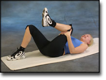
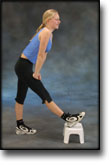
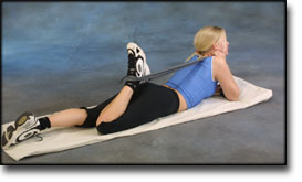
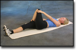
Lie on your back. Draw the injured leg up towards your head so that the muscles in the back of the thigh become increasingly stretched. Perform the exercise with outstretched as well as bent knee. Hold the position for 20 seconds and relax for 20 seconds before repeating. The exercise can also be performed standing with the injured leg outstretched on a chair while the upper body is bent slightly forwards.

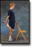
Stand with support from the back of a chair or the wall. Using your hand, bend the knee and draw the foot up and your knee slightly backwards so that the muscles in the front of the thigh become increasingly stretched. Hold the position for 20 seconds and relax for 20 seconds before repeating. The exercise can also be done lying down. If you lie on your stomach you can draw the foot up by using a towel.

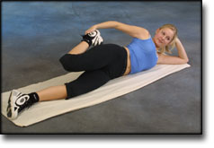
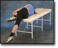
Lie on your side on a table. Bend one leg up under your body and let the other hang over the edge of the table so that the muscles in the outer side of the thigh become increasingly stretched. Hold the position for 20 seconds and relax for 20 seconds before repeating. The exercise can also be done standing by placing the outstretched injured leg behind the good leg at the same time as bending over the injured leg.

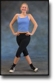
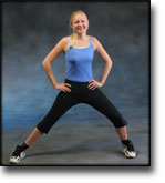
Stand with one leg outstretched and the other slightly bent. Thrust your weight to the side over the bent leg so that the inner side of the opposite thigh becomes increasingly stretched. Hold the position for 20 seconds and relax for 20 seconds before repeating.

Lie on your back with one leg outstretched and the other bent with the foot on the other side of the outstretched leg. Draw the knee up towards the opposite shoulder so that the buttocks become increasingly stretched. Hold the position for 20 seconds and relax for 20 seconds before repeating.

KOORDINATION
(5 min)

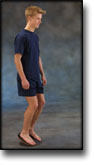
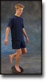
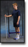
Seesaw. Balance on two legs, possibly using a hand as support against the wall, balancing subsequently on one leg without support. Look straight ahead and keep knees bent.

STYRKE
(40 min)

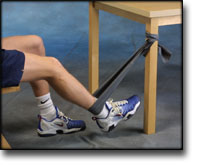
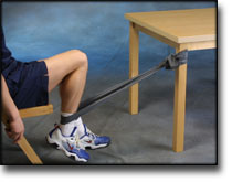
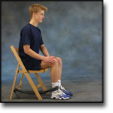
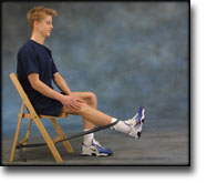
Sit on a chair with elastic around the ankle, facing the elastic. Lift the leg and slowly bend and stretch the knee.

Sit on a chair with elastic attached to the ankle. Raise the leg and slowly stretch and bend the knee.

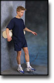
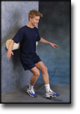
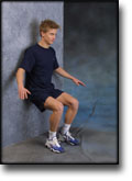
Stand with your back against a wall with a ball or firm round cushion between the wall and your back. Slowly go down to bend your knee 90 degrees before slowly rising up again.

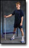
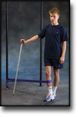
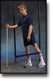
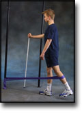
Stand on the healthy leg with the elastic around the inside of the injured leg. Move the injured leg from side to side in a slow smooth movement. Moving the position of the elastic lower down the leg can increase the load.

Stand on the healthy leg with the elastic around the outside of the injured leg. Move the injured leg from side to side in a slow smooth movement. Moving the position of the elastic lower down the leg can increase the load.

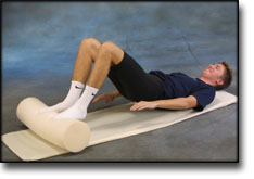
Lie on your back with a ball or firm round cushion under both feet. Roll the ball backwards and forwards in a steady pace while lifting your backside.

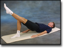
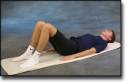
Lie on your back with bent knees. Lift one leg and stretch while at the same time lifting your hip from the floor.

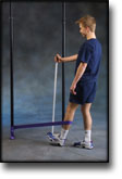
Stand with the elastic around the injured leg, facing towards the elastic. Move the leg backwards and slowly forwards. The elastic can be moved up and down the leg depending upon the strength of the knee – the stronger the knee, the lower the elastic should be.

Stretching is carried out in the following way: stretch the muscle group for 3-5 seconds. Relax for 3-5 seconds. The muscle group should subsequently be stretched for 20 seconds. The muscle is allowed to be tender, but must not hurt. Relax for 20 seconds, after which the procedure can be repeated. The time consumed for stretching, coordination and strength training can be altered depending on the training opportunities available and individual requirements.

Special(a-2)

SportNetDoc

Mouth guard protection for prevention of athletic injuries to teeth, mouth and jaw.

Mischkowski RA, Siessegger M, Zoller JE. Sportverletz Sportschaden 1999 Sep;13(3):65-7.

Sport is well known as a common cause of dental and oral injuries. Sports-related injuries account for 13% to 39% of all trauma cases in this area. The knowledge about prevention of dental and oral injuries among hobby and professional athletes can be regarded as not satisfactory yet. Mouth guards are considered as one of the most effective means for injury prevention. However, no statements or recommendations regarding use of mouth guards in sports have been published in German language literature yet. The following review article describes several types of available mouth guards and discuss their advantages and disadvantages. Guidelines concerning indications and use of mouth guards are presented.

Special(a-1)

SportNetDoc

Athletic mouth guards prevent orofacial injuries: a review.

Woodmansey KF. Gen Dent 1999 Jan-Feb;47(1):64-9; quiz 70-1.

Athletes who participate in competitive and recreational sports are at significant risk of orofacial injury. The use of mouth guards can reduce that risk substantially. The known incidence of orofacial trauma and benefits of athletic mouth guard use are reviewed. Dental health professionals are advised to advocate mouth guards for athlete-patients.

Symptoms(a-2)

SportNetDoc

Ocular sports injuries: the current picture.

Barr A, Baines PS, Desai P, MacEwen CJ. Br J Sports Med 2000 Dec;34(6):456-8.

OBJECTIVES.
To determine the recent incidence of eye injury due to sport in Scotland, identify any trend, and establish which sports are responsible for most injury? The type of injury and final visual outcome is also evaluated.

METHODS.
A prospective observational study of ocular injuries sustained during sport was performed over a one year period. Only patients requiring hospital admission were included. Data were collected on a standardised proforma and entered into a central database. Patients were followed up for at least three months.

RESULTS.
Of 416 patients admitted because of ocular injury, 52 (12.5%) resulted from playing a sport. Although all racquet sports together accounted for 47.5% of these injuries, football was the single most common sport associated with ocular trauma, being responsible for 32.5% of cases. The most common clinical finding was macroscopic hyphaema occurring in 87.5% of patients. Overall the final visual acuity was 6/6 in 92.5% of patients.

CONCLUSIONS.
The incidence of eye injury due to sport at 12.5% is lower than previously reported, suggesting a change in the pattern of ocular trauma. Football is the single most common cause of ocular injury from sport in Scotland, but the wearing of protective headgear would be difficult to instigate. The incidence of hyphaema in sport related ocular trauma (87.5%) is almost double that of all ocular injury (47.8%), so the potential for serious visual loss as the result of a sports injury should not be underrated. Ophthalmologists have a role in protecting this young population at risk by actively encouraging the design and use of protective eyewear.

KONDITION

step1

Training ladder for:
TENDINITIS AT THE ISCHIATIC BONE
(ENTESOPATIA TUBER ISCHIADICUM)

STEP 1

The indications of time after stretching, coordination training and strength training show the division of time for the respective type of training when training for a period of one hour. The time indications are therefore not a definition of the daily training needs, as the daily training is determined on an individual basis.

KONDITION
Unlimited: Cycling. Swimming. Running in deep water.

UDSPÆNDING
(15 min)

Lie on your back. Draw the injured leg up towards your head so that the muscles in the back of the thigh become increasingly stretched. Perform the exercise with outstretched as well as bent knee. Hold the position for 20 seconds and relax for 20 seconds before repeating. The exercise can also be performed standing with the injured leg outstretched on a chair while the upper body is bent slightly forwards.

Stand with support from the back of a chair or the wall. Using your hand, bend the knee and draw the foot up and your knee slightly backwards so that the muscles in the front of the thigh become increasingly stretched. Hold the position for 20 seconds and relax for 20 seconds before repeating. The exercise can also be done lying down. If you lie on your stomach you can draw the foot up by using a towel.

Lie on your side on a table. Bend one leg up under your body and let the other hang over the edge of the table so that the muscles in the outer side of the thigh become increasingly stretched. Hold the position for 20 seconds and relax for 20 seconds before repeating. The exercise can also be done standing by placing the outstretched injured leg behind the good leg at the same time as bending over the injured leg.

Stand with one leg outstretched and the other slightly bent. Thrust your weight to the side over the bent leg so that the inner side of the opposite thigh becomes increasingly stretched. Hold the position for 20 seconds and relax for 20 seconds before repeating.

Lie on your back with one leg outstretched and the other bent with the foot on the other side of the outstretched leg. Draw the knee up towards the opposite shoulder so that the buttocks become increasingly stretched. Hold the position for 20 seconds and relax for 20 seconds before repeating.

KOORDINATION
(5 min)

Seesaw. Balance on two legs, possibly using a hand as support against the wall, balancing subsequently on one leg without support. Look straight ahead and keep knees bent.

STYRKE
(40 min)

Sit on a chair with elastic attached to the ankle. Raise the leg and slowly stretch and bend the knee.

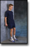
Stand with your back to the wall with your weight on both feet. Slowly go down and bend the knee to 90 degrees, and slowly rise again.

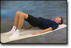
Lie on your back with bent legs. Lift your hip from the floor and hold the position for 5 seconds. Rest for 5 seconds before repeating.

Stand on the healthy leg with the elastic around the inside of the injured leg. Move the injured leg from side to side in a slow smooth movement. Moving the position of the elastic lower down the leg can increase the load.

Stand on the healthy leg with the elastic around the outside of the injured leg. Move the injured leg from side to side in a slow smooth movement. Moving the position of the elastic lower down the leg can increase the load.

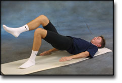
Lie on your back and bend the injured leg. Lift the healthy leg, and then raise your backside with the weight on the injured leg to be trained. Hold the position a few seconds and lower your backside again. Moving the injured leg further away from the body will increase the load.

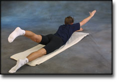
Lie on your stomach on the floor with your arms above your head and with outstretched legs. Lift right arm and left leg together, changing to lift left arm and right leg together.

Stretching is carried out in the following way: stretch the muscle group for 3-5 seconds. Relax for 3-5 seconds. The muscle group should subsequently be stretched for 20 seconds. The muscle is allowed to be tender, but must not hurt. Relax for 20 seconds, after which the procedure can be repeated. The time consumed for stretching, coordination and strength training can be altered depending on the training opportunities available and individual requirements.

Symptoms(a-1)

SportNetDoc

Eye injuries in athletics and recreation.

Napier SM, Baker RS, Sanford DG, Easterbrook M. Surv Ophthalmol 1996 Nov-Dec;41(3):229-44.

Sports-related eye injuries represent a significant eye health hazard worldwide. In the USA, it is estimated that eye injuries from sports account for more than 100,000 physician visits per year at the cost of greater than $175 million. The sports responsible for the greatest number of injuries are baseball, ice hockey, and racquet sports. In the USA there are over 189 million participants in these sports with a disproportionate number (one-third) of injuries involving the pediatric population. The most common injuries are abrasions and contusions, followed by more serious injuries, including hyphema. Substantial reduction in the incidence of eye injuries through prevention has been demonstrated in the sport of hockey which can serve as a model for other sports. Specific criteria for protective eyewear must be developed. Polycarbonate plastic lenses and frames which are sturdy and impact resistant provide optimal protection. Lensless goggles, street wear and spectacle correction glasses do not provide adequate eye protection. The ophthalmologist’s role in preventing sports-related eye injuries includes addressing particular sports participants needs, identifying one-eyed athletes, and informing patients of the need for protective eyewear.

Metatarsus ligament rupture

Diagnosis: METATARSUS LIGAMENT RUPTURE
(Ruptura traumatica ligamenti pedis)


Anatomy:
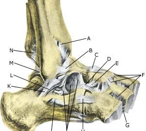
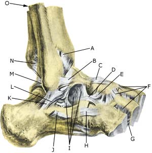
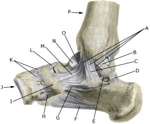
The foot bones comprise the 7 tarsal bones (ossa tarsi), the 5 metatarsal bones (ossa metatarsi) and the 14 bones in the toes (phalanx). Large or small ligaments (metarsus ligaments) run between all the bones, which stabilise the joints if twisted and running with directional change.

  1. Lig. tibiofibulare anterius
  2. Lig. talofibulare anterius
  3. Lig. talonaviculare
  4. Lig. calcaneocuboideum (Lig. bifurcatum)
  5. Lig. calcaneonaviculare (Lig. bifurcatum)
  6. Ligg. tarsi dorsalia
  7. Lig. cuneocuboideum interosseum
  8. Lig. calcaneocuboideum
  9. Lig. talocalcaneum interosseum
  10. Retinaculum mm. peroneorum inferius
  11. Lig. calcaneofibulare
  12. Lig. talocalcaneum laterale
  13. Lig. talofibulare posterius
  14. Lig. tibiofibulare posterius
  15. Fibula

OUTER ANKLE JOINT


  1. Ligamentum mediale/deltoideum
  2. Pars tibiotalaris posterior
  3. Pars tibiocalcanearis
  4. Retinaculum mm. flexorum (m. flexor hallucis longus)
  5. Lig. talocalcaneum mediale
  6. Lig plantare longum
  7. Lig. calcaneonaviculare plantare
  8. Lig. cuneonaviculare plantare
  9. Os naviculare
  10. Os cuneiforme mediale
  11. Ligg. cuneonavicularia dorsalia
  12. Lig talonaviculare
  13. Pars tibionavicularis
    (lig. deltoidei)
  14. Talus
  15. Pars tibiotalaris anterior
  16. Tibia

INNER ANKLE JOINT

Cause: A violent twist of the joint can in light cases result in a strain or sprain of the ligament, whereas sever cases can result in full or partial rupture or tear.

Symptoms: Pain in the ligament also occuring with load (twist) on the joint which the ligament stabilises.

Acute treatment: Click here.

Examination: Clinical examination is normally not required in light cases (strain/sprain) with only minimal tenderness, and no discomfort when walking. More pronounced pain will demand examination to exclude bone fracture, bone membrane tear (periosteal avulsion), inflammation of the tendon sheath, concentration of fluid in the joint (traumatic arthritis/synovitis). A normal medical (evt. clinical) examination is usually sufficient in order to make the diagnosis. X-ray will confirm or exclude any suspicion of fracture. Small bone membrane tears are best seen under ultrasound scanning.

Treatment: Treatment of uncomplicated metatarsus ligament rupture will normally be conservative (rehabilitation).

Bandage: In the rehabilitation phase, tape can be used when running on uneven surfaces or running with rapid directional change begins. The tape has, however, not the same important purpose as for ligament injuries on the large outer or inner ligaments in the ankle, (tape-instruction).

Complications: If there is not a steady improvement in the condition consideration must be given as to whether the diagnosis is correct, or if complications have arisen:

Muscular bleeding

Diagnosis: BLEEDING IN THE MUSCLE IN THE FOOT
(Haematoma)


Anatomy:
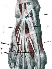
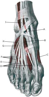
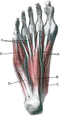
The muscles often comprise a muscle belly and a muscular tendon, which are attached to a bone. The muscle belly is found on the back of the foot, between forefoot bones and under the sole.

  1. M. extensor hallucis longus
  2. M. extensor hallucis brevis
  3. M. abductor hallucis
  4. Mm. interossei dorsales
  5. M. abductor digiti minimi
  6. M. extensor dititorum brevis
  7. Tendo m. peronei tertii
  8. M. extensor digitorum brevis
  9. M. extensor digitorum longus

MUSCLES IN THE BACK OF THE FOOT

  1. M. flexor hallucis brevis
  2. M. flexor digitorum brevis
  3. M. abductor hallucis
  4. M. abductor digiti minimi
  5. M. flexor digiti minimi brevis
  6. Mm. lumbricales

MUSCLES IN THE SOLE OF THE FOOT

Cause: The fleshy part of the muscles contains a great number of blood vessels which will often bring about bleeding if there is a rupture of the muscle fibres due to a tearing or blow.

Symptoms: Pain when pressure is applied, activating and stretching the damaged muscle. There is often a sense of the muscle being firm or filling out.

Examination: A medical examination is not required in slight cases. If satisfactory progress is not achieved a medical examination may be required in order to make the diagnosis. A normal medical examination is usually sufficient in order
to make the diagnosis, however, if there is any doubt concerning the
diagnosis an ultrasound scan can be performed.

Treatment: Bleeding on the muscles is treated with relief and rehabilitation. In instances where the bleeding is particularly excessive it may be necessary to perform draining.

Rehabilitation: Rehabilitation is totally dependent upon which muscle is injured. Running can normally be resumed as soon as the pain subsides. Until this is possible, the guidelines under rehabilitation, general should be followed.

Complications: If satisfactory progress is not achieved, an x-ray examination should be performed to exclude the possibility of:

Similarly, an ultrasound scan should be performed to exclude:

Cause-article4

SportNetDoc

Apophyseal injuries in the young athlete.

Peck DM. Am Fam Physician 1995 Jun;51(8):1891-5, 1897-8

Apophyseal injuries, which are unique in the adolescent athlete, cause inflammation at the site of a major tendinous insertion onto a growing bony prominence. These injuries typically occur in active adolescents between the ages of eight and 15 years and usually present as periarticular pain associated with growth, skeletal immaturity, repetitive microtrauma and muscle-tendon imbalance. Common apophyseal injuries, and their sites, include Sever’s disease (posterior calcaneus), Osgood-Schlatter disease (tibial tuberosity), Sindig-Larsen-Johansson syndrome (inferior patella), medial epicondylitis (humeral medial epicondyle) and apophysitis of the hip (iliac crest, ischial tuberosity). Conservative therapy, including rest, ice, compression, elevation, nonsteroidal anti-inflammatory agents, modification of the athlete’s activity level and exercises for increased flexibility and strengthening, is usually effective.