Kategoriarkiv: Tendinitis at the ischiatic bone

Sportsman’s hernia

SPORTSMANS HERNIA

Diagnosis: “SPORTSMAN’S HERNIA”


Anatomy:
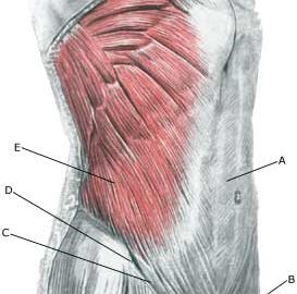
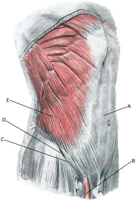
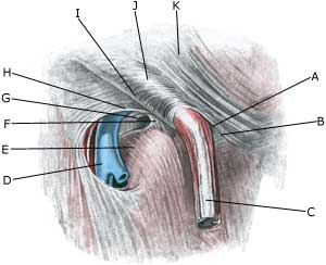
The various muscles of the abdominal wall are penetrated by the inguinal canal, which contains nerves (N ilioinguinalis and the genital branch of N genitofemoralis) and in men the spermatic cord (funilicus spermaticus). In women the inguinal canal instead contains a small fibrous ligament. Where the inguinal canal penetrates the abdominal wall weak spots arise (anulus inguinalis superficialis and anulus inguinalis profundus).

  1. M. recti abdominis
  2. Funiculus spermaticus
  3. Ligamentum inguinale
  4. Spina iliaca anterior superior
  5. M. obliquus externus abdominis

INGUINAL CANAL

  1. Anulus inguinalis superficialis
  2. Crus mediale
  3. Funiculus spermaticus
    et m. cremaster
  4. V. femoralis
  5. Hiatus saphenus
  6. Lig. lacunare
  7. Anulus femoralis
  8. Margo falciformis
    (cornu superius)
  9. Lig inguinale
  10. Fibrae intercrurales
  11. M. obliquus externus abdominis

RIGHT INGUINAL CANAL

Cause: The existence of “sportsman’s hernia” is debated amongst professionals (article 1). Generally it involves a inguinal hernia (direct hernia), that can not be detected with certainty neither before nor after surgery. Some consider the condition to be an early stage of an imminent inguinal hernia, while others believe it to be small ruptures in the muscles and tendons around the inguinal canal (article 2) (article 3).

Symptoms: Pain in the groin without any detectable swelling in the groin or any other explanation for the pain.

Examination: There are no examinations (X-ray, ultrasound, MRI-scan, scintigraphy), that can detect the sportsman’s hernia (article). Previously used attempts with X-ray contrast in the abdominal cavity (herniography) showed “sportsman’s hernia” in 49% of healthy subjects, which naturally renders the examination unusable.

Treatment: Before you choose to be operated for “sportsman’s hernia”, all non-operative possibilities should be attempted, including sufficient relief and rehabilitation of the most tender structures and muscles.

Training of the muscles around the groin, stomach and loin before possible surgery.

Complications: Since it may be difficult to make a correct diagnosis in athletes with long-term groin pain (article). A multidisciplinary approach, it should be supplemented with, amongst other things, ultrasound scan and consideration of X-ray scintigraphy and possibly MRI-scan. You should of course consider the correctness of the diagnosis and amongst other things consider the following:

Degenerative arthritis in the hip joint

RUPTURE OF THE POSTERIOR CRUCIATE LIGAMENT

Diagnosis: RUPTURE OF THE POSTERIOR CRUCIATE LIGAMENT
(Ruptura ligamentum cruciatum posterius)


Anatomy:
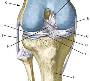
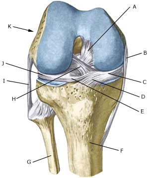
The bones in the knee joint comprise the thigh bone (femur), shin bone (tibia) and the knee cap (patella). There is furthermore a small joint between the shin bone and the calf bone (fibula). The knee joint is strengthened by a joint capsule which is in turn strengthened on the sides by an outer and an inner collateral ligament (ligamentum collaterale laterale (LCL) and ligamentum collaterale mediale (MCL)). Inside the knee there are two ligaments, the anterior and posterior cruciate ligaments (ligamentum cruciatum anterius and ligamentum cruciatum posterius).

  1. Ligamentum cruciatum posterius
  2. Ligamentum collaterale mediale/tibiale
  3. Meniscus medialis
  4. Insertio anterior
    menisci medialis
  5. Ligamentum transversum genus
  6. Tibiae
  7. Fibulae
  8. Ligamentum cruciatum anterius
  9. Ligamentum collaterale laterale/fibulare
  10. Meniscus lateralis
  11. Femur

KNEE JOINT FROM THE FRONT

Cause: Rupture of the posterior cruciate ligament usually occurs following a blow or kick direct on the front of the shin bone just below the knee (Photo).

Symptoms: Usually a snap can be heard or felt and continued sports activity must be aborted. The knee can swell within the first few hours, after which the knee can not bend completely. You can subsequently often sense that the leg gives way (knee failure).

Acute treatment: Click here.

Examination: If a partial or complete rupture of the cruciate ligament is suspected, you should seek medical attention (casualty ward) immediately, to obtain a diagnosis. The doctor can perform various tests on the knee (rear drawer looseness) to examine the stability of the knee. It should be noted that the looseness in the knee can often only be demonstrated after two weeks. It is often necessary to supplement the examination with a MR-scan, ultrasound scan (Ultrasonic image) (article), or arthroscopy to make the diagnosis with certainty.

Treatment: Treatment of a rupture of the posterior cruciate ligament usually comprises relief and rehabilitation. It is only in cases of pronounced looseness, or if the rupture is combined with other ligament ruptures, that surgery is recommended (article).

An intensive rehabilitation period of at least six months is to be expected.

Bandage: Hinge bandages (Don-Joy) can be utilised the first few weeks. Tape treatment of cruciate ligament ruptures in the knee has no sure effect.

Complications: If smooth progress is not achieved, it should be considered whether the diagnosis is correct or whether complications have arisen. Amongst others the following should be considered:

Special: Since there is a risk that the injury can cause permanent disability, the injury should be reported to your insurance company.

Stress fracture of the femoral neck

STRESS FRACTURE IN THE FEMORAL NECK

Diagnosis: STRESS FRACTURE IN THE FEMORAL NECK
(Stress fraktur)


Anatomy:
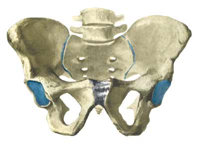
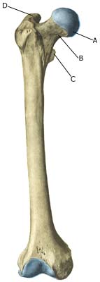
The femur and the hip bone form the hip joint.

  1. Caput femoris
  2. Collum femoris
  3. Trochanter minor
  4. Trochanter major

PELVIS AND THIGH BONE FROM THE FRONT


Cause: Repeated loads, especially when walking or running can cause cracks (stress fractures) in the femoral neck (collum femoris) (article-1) (article-2).

Symptoms: Pain in the hip when applying pressure (direct and indirect tenderness) and when under load (walking, running).

Examination: X-ray. Since many stress fractures cannot be seen early in the course of events, X-ray examination can be repeated after a few weeks. Scintigraphy, CT- and MRI and ultrasound scan can often diagnose stress fractures far earlier than X-rays (Ultrasonic image)
It is imperative for the result of the treatment that the diagnosis is made as early as possible (article).

Treatment: Relief. In some cases surgery is necessary (article).

Rehabilitation: The rehabilitation is completely dependent on the type of fracture and treatment (conservative or operative).
Also read rehabilitation, general.

Complications: If progress is not smooth, you should be re-examined to ensure that the fracture heals according to plan. In some cases a false joint can be formed (pseudoarthrosis), which requires surgical treatment.

Special: Shock absorbing shoes or inlays will reduce the load.

Rupture of the superficial hip flexor (ruptura musculus rectus femoris)

BRISTNING AF DEN OVERFLADISKE HOFTEBØJER

Diagnosis: BRUPTURE OF THE SUPERFICIAL HIP FLEXOR
(Ruptura musculus rectus femoris)


Anatomy:
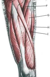
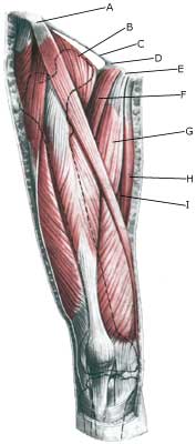
The superficial hip flexor (the forward straight thigh muscle, musculus quadriceps femoris) originate from the front edge of the hip (processus spinosus anterior inferior) and from the upper edge hip joint socket (acetabulum). The muscle is joined by three of the other thigh muscles and is attached in a common joint muscle tendon (quadriceps) on the upper edge of the kneecap (patella). The function of the superficial hip flexor is to stretch the knee and bend in the hip.

  1. Spina iliaca anterior superior
  2. M. iliopsoas
  3. Lig. inguinale
  4. Lig. lacunare
  5. Tuberculum pubicum
  6. M. pectineus
  7. M. adductor longus
  8. M. gracilis
  9. M. adductor magnus

THIGH FROM THE FRONT

Cause: When a muscle is subjected to loads (repeated smaller loads or one very powerful load), beyond the strength of the muscle (jumping, kicking), a rupture occurs. The rupture can be microscopic and due to repeated loads in continuing sports activity, many small loads can trigger a chronic inflammation or a rupture. The vast majority of cases are partial muscle ruptures.

Symptoms: In light cases a local tenderness is felt after the load (“muscle strain”, “imminent pulled muscle”, “tendinitis”). The symptoms can often decrease after a thorough warm-up, only to return when the sports activity has ceased. In severe cases a sudden shooting pain is felt in the muscle (“partial muscle rupture”, “pulled muscle”) and in the worst case a sudden snap is felt rendering the muscle unusable (“total muscle rupture”). With muscle injuries the following three symptoms are characteristic: pain when applying pressure, stretching and activation against resistance. In total ruptures a defect in the muscle can often be seen and felt, and a swelling is felt above or below the rupture (the contracted muscular belly and the bleeding).

Acute treatment: Click here.

Examination: In light cases with only minimal tenderness and no discomfort with ordinary walking, medical examination is not necessarily required. The extent of the tenderness is, however, not always a mark of the degree of the injury. In cases of more pronounced pain or tenderness, medical examination is required to ensure the correct diagnosis and treatment. A normal medical examination is usually sufficient in order to make the diagnosis, however, if there is any doubt concerning the diagnosis an ultrasound scan can be performed (Ultrasonic image). X-ray examination is recommended when it is suspected that the bone on which the muscle tendon fastens has been torn off.

Treatment: The treatment usually comprises relief and rehabilitation. Only in very rare cases is surgery indicated (e.g. total ruptures in the muscle tendon, close to the fastening). Even large ruptures in the femoral muscle can usually be rehabilitated without resulting in functional harm (but often cosmetic damage, with an irregular femoral muscle). If the condition concerns tendinitis where there has been no sensation of a “snap” in the muscle, and smooth improvement has not been achieved after relief and gradually increasing rehabilitation, treatment can be supplemented with rheumatic medicine (NSAID) oand possibly injection of corticosteroid in the area surrounding the inflamed part of the muscle attachment point. If it concerns ruptures, (“total or partial muscle rupture”) the injection of corticosteroid is not indicated.

Complications: If the treatment does not progress according to plan, it should be considered whether the diagnosis is correct or whether complication have arisen. In particular the following should be considered:

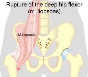
Rupture of the deep hip flexor (M iliopsoas)

RUPTURE OF THE DEEP HIP FLEXOR

Diagnosis: RUPTURE OF THE DEEP HIP FLEXOR
(Ruptura musculus iliopsoas)


Anatomy:
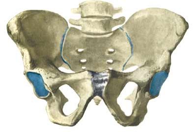
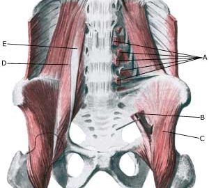
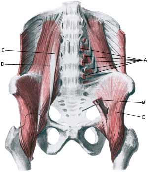
The deep hip flexor (M iliopsoas) consists of two muscles. The Psoas muscle originate from the lumbar vertebra and the Iliacus muscle from the inside of the hip bone. The two muscles fuse and are both fastened on the inside of the femur (trochanter minor). The iliopsoas is the strongest flexor muscle of the hip joint.

  1. Origines m. psoatis
  2. M. psoas major
  3. M. iliacus
  4. M. psoas major
  5. M. psoas minor

PELVIS FROM THE FRONT

Cause: When a muscle is subjected to a load beyond the strength of the muscle (jumping, kicking), a rupture occurs. The vast majority of ruptures are partial muscle ruptures.

Symptoms: In light cases a local tenderness is felt after the load (“sprained muscle”, “imminent pulled muscle”) e.g. kicking a ball with the instep. In severe cases a sudden shooting pain is felt in the muscle (“partial muscle rupture”, “pulled muscle”) and in the worst case a violent snap is felt, rendering the muscle unusable for e.g. walking up stairs (“total muscle rupture”). With muscle injuries the following three symptoms are characteristic: pain upon pressure, stretching and activation against resistance. In some cases the bleeding can be so great that it entraps the nerve to the bone (nervus femoralis) with increasing pains, reduction of power and symptoms into the leg (article).

Acute treatment: Click here.

Examination: In light cases with only minimal tenderness and no discomfort when walking normally (“sprained muscle”, “imminent pulled muscle”), medical examination is not necessarily required. The severity of the tenderness is, however, not always a measure of the extent of the injury. In case of more pronounced tenderness or pain medical examination is required to ensure the correct diagnosis and treatment. A normal medical examination is usually sufficient in order to make the diagnosis, however, if there is any doubt concerning the diagnosis an ultrasound scan can be performed. It is known that the larger the bleeding is assessed in the ultrasound scan, the longer the rehabilitation period (Ultrasonic image), (article).

Treatment: The treatment usually consists of relief and careful rehabilitation (article). Only in very rare cases is surgery indicated (e.g. in case of total ruptures or very heavy bleeding).

Complications: In case of lack of progress it should be considered if the diagnosis is correct or whether complications have arisen. In particular the following should be considered:

Tendonitis at the ischiatic bone.

Diagnosis: TENDINITIS AT THE ISCHIATIC BONE
(ENTESOPATIA TUBER ISCHIADICUM


Anatomy:
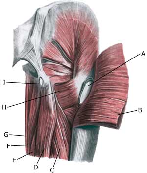
The large posterior thigh muscles (hamstring muscles) have a common muscle tendon fastening on the ischiatic bone (tuber ischiadicum). The posterior thigh muscles flex the knee and stretch the hip.

  1. Bursa trochanterica m. glutei maximi
  2. M. gluteus maximus
  3. M. biceps femoris (caput longum)
  4. M. semitendinosus
  5. M. semimembranosus
  6. M. adductor magnus
  7. M. gracilis
  8. M. quadratus femoris
  9. Bursa ischiadica m. glutei maximi

(Photo)

RIGHT GLUTEAL MUSCLES
FROM THE REAR

Cause: Inflammation of the tendon fastenings (tendinitis) at the ischiatic bone (tuber ischiadicum) occurs following repeated uniform (over)loads (e.g. running, sprinting) causing microscopic ruptures in the tendon, and especially at the tendon fastening. Tendinitis is a warning that the training performed is too strenuous for the muscles in question, and if the load is not reduced a rupture of the posterior thigh muscle fastening on the ischiatic bone (“pulled muscle”) may occur. This will result in a considerably prolonged rehabilitation period.

Symptoms: Pain in the ischiatic bone can occasionally radiate down into the rear of the thigh. The pain is aggravated when applying pressure on the bone (e.g. sitting position), stretching and activating the posterior thigh muscles (flexing the knee against resistance).

Acute treatment: Click here.

Examination: In slight cases with only minimal tenderness and no discomfort with walking, medical examination is not necessarily required. The extent of the tenderness is, however, not always a mark of the degree of the injury. In cases of more pronounced pain or tenderness, medical examination is required to ensure the correct diagnosis and treatment. The diagnosis is usually made on the basis of a normal medical examination, however, if there is any doubt concerning the diagnosis, this can be confirmed by ultrasound scanning or MR scanning (article).

Treatment: The treatment usually comprises relief, stretching and rehabilitation (article). If the rehabilitation does not progress satisfactorily, medicinal treatment in the form of rheumatic medicine (NSAID) can be considered, or corticosteroid injection in the area surrounding the inflamed tendon fastening on the ischiatic bone. As the injection of corticosteroid is always an element in the long term rehabilitation of a very serious, chronic injury, it is vital that the rehabilitation period lasts over several months in order to reduce the risk of a relapse or ruptures. The tendon can naturally not sustain maximal load after a long-term injury period and only a short-term rehabilitation period. If the diagnosis is made by use of ultrasound scanning, the injections are performed under guidance of ultrasound, and the rehabilitation is progressed in accordance with the guidelines mentioned, then the treatment involving corticosteroid injections has very few risks connected. If satisfactory progress is not made following the rehabilitation and medicinal treatment, surgical intervention can be considered. Long-term results of operations are often disappointing, despite publication of a minor series with good results (article).

Complications: If the treatment does not progress according to plan, it should be considered if the diagnosis is correct or whether complications have arisen. The following should in particular be considered:

complications-article3

Untitled Document

Legg-Calve-Perthes’ disease.

Wall EJ. Curr Opin Pediatr 1999 Feb;11(1):76-9.

The etiology, radiographic classification, and treatment of Legg-Calve-Perthes’ disease remain controversial. Several recent papers focus on these issues in an effort to provide guidance in the clinical care of Perthes’ disease. The research studied in this paper lends further support to the hypothesis of clotting abnormalities with vascular thrombosis, which seems to be the most likely etiology for Legg-Calve-Perthes’ disease. Several studies focus on use of magnetic resonance imaging for the early diagnosis and prognosis of Perthes’ disease. A few researchers whose work is featured in this paper add information on the treatment of Perthes’ disease, supporting surgical treatment for older patients with more severe disease and non-surgical treatment for younger patients with less extensive femoral head involvement.

complications-article2

Untitled Document

Diagnosis and treatment of slipped capital femoral epiphysis.

Reynolds RA. Curr Opin Pediatr 1999 Feb;11(1):80-3
.
Slipped capital femoral epiphysis remains a diagnostic problem despite numerous papers written on the subject. The most important factor in the diagnosis of slipped capital femoral epiphysis is suspicion by the practitioner. The history, physical examination, and radiographic imaging are important in the confirmation of the diagnosis. Imaging is the topic of 1998 with advances in the areas of ultrasound. Ultrasound may be better in experienced hands than plain radiography in the diagnosis of slipped capital femoral epiphysis. Magnetic resonance imaging is used for diagnosis of slipped capital femoral epiphysis and in the assessment of pre-slips. The magnetic resonance image can be oriented to a plane orthoganol to the plane of the physis to assess the width of the physis and to detect edema in the area of the physis.

complications-article1

Untitled Document

Hip pain in athletes.

Adkins SB 3rd, Figler RA. Am Fam Physician 2000 Apr 1;61(7):2109-18.

Hip pain in athletes involves a wide differential diagnosis. Adolescents and young adults are at particular risk for various apophyseal and epiphyseal injuries due to lack of ossification of these cartilaginous growth plates. Older athletes are more likely to present with tendinitis in these areas because their growth plates have closed. Several bursae in the hip area are prone to inflammation. The trochanteric bursa is the most commonly injured, and the lesion is easily identified by palpation of the area. Iliotibial band syndrome presents with similar lateral hip pain and may be identified by provocative testing (Ober’s test). A methodical physical examination that specifically tests the various muscle groups that move the hip joint can help determine a more specific diagnosis for the often vague complaint of hip pain. A number of hip conditions are more prevalent in athletes of certain ages. Transient synovitis is a common diagnosis in the very young, Legg-Calve-Perthes disease causes bony disruption of the femoral head in prepubescents, and slipped capital femoral epiphysis is seen most commonly in obese adolescent males. Femoral neck stress fractures are seen in adult athletes, especially those involved in endurance sports, and can progress to necrosis of the femoral head if not found early. Older athletes may be limited by degenerative joint disease but nonetheless should be encouraged to stay active.

NSAID

Untitled Document Untitled Document

NSAID (rheumatism medicine)

Use of NSAID is widespread in sport as a painkiller, and as treatment to subdue inflammation.

Indication. Over-load symptoms from tendons. A considerable number of scientific studies have been performed comprising NSAID treatment on acute tendon injuries. In the majority of studies, but not all, healing was achieved slightly quicker, and inflammation was slightly reduced compared with placebo treatment. Some studies have shown increased instability and reduced mobility in the joints after NSAID treatment.
Acute muscle injuries. There are only a handful of animal studies dealing with NSAID treatment of acute muscle injuries. Increased muscle strength has been proven, however, also reduced healing of damaged tissue.
Myositis ossifans (calcification in muscles after bleeding). One study shows that calcification in the muscles following a hip operation is reduced in patients who are treated with NSAID after the operation.
Chronic muscle and tendon injury. There is no conclusive scientific evidence supporting use of NSAID on chronic muscle or tendon injuries.

Side effects. Side effects from the abdomen and intestines (heartburn, gastric ulcer and sour eructation) are frequent following treatment with NSAID. The new rheumatism pills (“selective COX-2 inhibitors”, as for example, Vioxx) are by and large free of serious side effects from the abdomen and intestines. Serious side effects are rare, but allergic shock, kidney damage and bone marrow damage has been described. Only moderate side effects are seen following localised treatment with NSAID (allergy).

Contraindications. Allergy is on the whole the only contraindication for NSAID treatment in healthy athletes. Patients with gastric ulcer, high blood pressure, liver, heart and kidney illnesses should be cautious with NSAID treatment.

Administration. Tablet treatment is recommended. Some placebo controlled studies show that local NSAID as gel is better than placebo on acute injuries, despite the concentration of blood following localised treatment constituting less than 10% of the level after tablet treatment or after injection in the muscles. There are no scientific grounds for using injection methods. There are no studies which document the ideal point in time to start NSAID treatment, or the length of duration.

Discussion. There is no conclusive clarification as to whether inhibiting the acute inflammation is an absolute advantage. Pain and discomfort are in any event partially conditional upon the inflammation. By reducing the inflammation the symptoms are reduced, thereby allowing rehabilitation to start at an earlier stage. On the other hand, the inflamed cells are responsible for the decomposition of the tissue which has been destroyed, which is necessary for removal of dead muscle fibre and the like.

Conclusion. Several clinical studies have documented that treatment with NSAID has some effect on sports injuries. There are, however, still many unanswered questions preventing a sure, unequivocal indication for treatment with NSAID to be given. If systematic NSAID treatment is indicated, the new rheumatism pills (“selective COX-2 inhibitors” for example Vioxx), can be recommended. As mentioned above, NSAID treatment is merely a supplement to the base treatment which is “active rest” with increasing intensity in training within the pain threshold. If NSAID is misused as a painkiller to continue a potentially damaging sports activity, the treatment will indirectly increase the risk of the chronic injury. It is for this reason that all NSAID treatment on athletes must be administered by a physician with knowledge of the basic rehabilitation principles.