Alle indlæg af urtehave_com

Concentration of fluid in the joint (traumatic arthritis/synovitis)

Diagnosis: CONCENTRATION OF FLUID IN THE JOINT
(Traumatic arthritis / synovitis)


Anatomy:
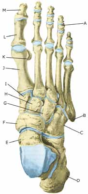
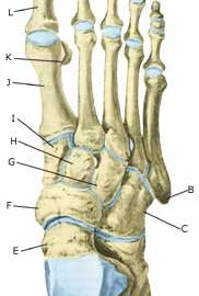
The joints in the foot comprise the large ankle joint (between the shin and the foot, articulatio talocruralis), as well as many smaller joints between the tarsal bones (ossa tarsi) and the metatarsal bones (ossa metatarsi), and the toe joints.

  1. Phalanx media
  2. Tuberositas ossis metatarsalis V
  3. Os cuboideum
  4. Calcaneus
  5. Talus
  6. Os naviculare
  7. Os cuneiforme laterale
  8. Os cuneiforme intermedium
  9. Os cuneiforme mediale
  10. Os metatarsalei
  11. Os sesamoideum
  12. Phalanx proximalis
  13. Phalanx distalis

THE FOOT FROM ABOVE

Cause: Inflammation of the synovial membrane can occur after a violent twist of a joint, which can cause the membrane to thicken and produce fluids resulting in the joint swelling up. The concentration of fluid in the joint (traumatic arthritis/synovitis) is often seen in connection with ligament injuries in the ankle joint area, Associated injuries in chronic lateral ankle instability, but is often overlooked.

Symptoms: Swelling of the joint. Pain with pressure on the joint lines, as well as with passive and active movement of the joint.

Acute treatment: Click here.

Examination: In pronounced cases the diagnosis is made from a normal medical examination, however, experience has shown that diagnosis is difficult and is often overlooked. Ultrasound scanning enables the diagnosis to be made from minor swelling, and swelling in joints that are otherwise difficult to examine.
(article), (Ultrasonic image-1), (Ultrasonic image-2).

Treatment: Treatment is primarily based on relieving the affected area (and treatment of other possible injuries that have developed at the same time). If swelling of the joint persists despite relieving the area as much as possible, a supplement of medicinal treatment in the form of rheumatic medicine (NSAID) can be administered. Alternatively, draining and evaluating the fluid can be performed, and injection of corticosteroid into the joint. Injection into the joint is best performed if ultrasound guided (article).

Rehabilitation: Rehabilitation is completely dependent upon which joint is injured, and which treatment has been administered. Until the swelling and the pain has subsided, the guidelines under rehabilitation, general should be followed.

Complications: If there is not a steady improvement in the condition consideration must be given as to whether the diagnosis is correct, or if complications have arisen:

Cause-article3

SportNetDoc

Hamstring strains in athletes: diagnosis and treatment.

Clanton TO, Coupe KJ. J Am Acad Orthop Surg 1998 Jul-Aug;6(4):237-48.

Hamstring strains are among the most common injuries (and reinjuries) in athletes. Studies combining electromyography with gait analysis have elucidated the timing of activity of the three muscles of the hamstring group; they function during the early-stance phase for knee support, during the late-stance phase for propulsion, and during midswing to control the momentum of the leg. Muscle injury, whether partial or complete, occurs at the myotendinous junction, where force is concentrated. The healing response begins with inflammation, associated edema, and localized hemorrhage. After an initial period of reduced tension, the healing muscle regains strength rapidly as long as reinjury does not occur. Although the use of anti-inflammatory medication is a keystone of treatment, a certain degree of inflammation is necessary for removing necrotic muscle fibers and rescaffolding to allow optimal recovery. The protocol of rest, ice, compression, and elevation is still the preferred first-aid approach. After a brief period of immobilization (usually less than 1 week for even the most severe strain), mobilization is begun to properly align the regenerating muscle fibers and limit the extent of connective tissue fibrosis. Concurrent pain-free stretching and strengthening exercises (beginning with isometrics and progressing to isotonics and isokinetics) are essential to regain flexibility and prevent further injury and inflammation. Readiness for return to competition can be assessed by isokinetic testing to confirm that muscle-strength imbalances have been corrected, the hamstring-quadriceps ratio is 50% to 60%, and the strength of the injured leg has been restored to within 10% of that of the unaffected leg. The only indication for surgery is a complete rupture at or near the origin from the ischial tuberosity or distally at its insertion (either soft-tissue avulsion with a large defect or bone avulsion with displacement by 2 cm).

Cause-article2

SportNetDoc

Hamstring injuries. Current trends in treatment and prevention.

Kujala UM, Orava S, Jarvinen M. Sports Med 1997 Jun;23(6):397-404.

Pre-exercise stretching and adequate warm-up are important in the prevention of hamstring injuries. A previous mild injury or fatigue may increase the risk of injury. Hamstring muscle tear is typically partial and takes place during eccentric exercise when the muscle develops tension while lengthening, but variation in injury mechanisms is possible. Diagnosis of typical hamstring muscle injury is usually based on typical injury mechanism and clinical findings of local pain and loss of function. Diagnosis of avulsion in the ischial tuberosity, with the need for longer immobilisation, and a complete rupture of the hamstring origin, in which immediate operative treatment is necessary, poses a challenge to the treating physician. X-rays, ultrasonography or magnetic resonance imaging (MRI) may be helpful in differential diagnostics. After first aid with rest, compression, cold and elevation, the treatment of hamstring muscle injury must be tailored to the grade of injury. Conservative treatment is based on a knowledge of the biological background of the healing process of the muscle. Experimental studies have shown that a short period of immobilisation is needed to accelerate formation of the granulation tissue matrix following injury. The length of the immobilisation is, however, dependent on the grade of injury and should be optimised so that the scar can bear the pulling forces operating on it without re-rupture. Mobilisation, on the other hand, is required in order to regain the original strength of the muscle and to achieve good final results in resorption of the connective tissue scar and re-capillarisation of the damaged area. Another important aim of mobilisation–especially in sports medical practice–is to avoid muscle atrophy and loss of strength and extensibility, which rapidly result from prolonged immobilisation. Complete ruptures with loss of function should be operated on, as should cases resistant to conservative therapy in which, in the late phase of repair, the scar and adhesions prevent the normal function of the hamstring muscle.

Inflammation of the tendon sheath

Diagnosis: INFLAMMATION OF THE TENDON SHEATH
(Tenosynovitis)


Anatomy:
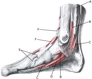
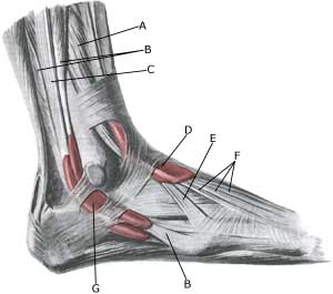
Tendons from the shin bone to the foot pass both inwardly, outwardly, in front and behind the ankle joint, and continue on the back of the foot and the sole to the toes. The tendons are during part of the course deposited in synovial sheaths.

  1. M. flexor digitorum longus
  2. Vagina synovialis tendinis m. tibialis posterioris
  3. Vagina synovialis tendinis m. flexoris hallucis longi
  4. Vagina tendinum m. flexoris digitorum pedis longi
  5. Tendo m. tibialis anterioris
  6. Vagina tendinis m. tibialis anterioris

INNER FOOT

  1. M. extensor digitorum longus
  2. M. peroneus brevis
  3. M. peroneus longus
  4. Vagina tendinum m. extensoris digitorum pedis longi
  5. M. peroneus tertius
  6. Tendines m. extensoris digitorum longi
  7. Vagina synovilis mm. peroneorum communis

OUTER FOOT

Cause: Inflammation of the tendon sheath (tenosynovitis) occurs either due to a mechanical irritation (e.g. a shoe tied too tightly, or from a kick), or often from a straining of the ligaments in the ankle joint. In some cases, a prolonged inflammation of the tendon sheath can cause a weakening, and in worst cases a rupture of the tendon. Inflammation of the tendon sheath is often seen as a result of ligament injuries (article), but is often unfortunately overlooked, (article). The tendon sheaths surrounding the ankle often communicate with the ankle joint itself, and accumulation of fluid in the tendon sheaths can therefore be a consequence of increased fluid in the ankle joint itself, concentration of fluid in the joint (traumatic arthritis / synovitis).

Symptoms: Inflammation of the tendon sheath causes pain along the tendon sheath itself, which is aggravated by stretching and activation of the muscle tendon.

Examination: In obvious cases the diagnosis can be established from a normal medical examination. The diagnosis is, however, easily determinable by use of ultrasound scanning, which can also be advantageously used in cases where steady improvement is not noticeable from relieving the affected area (Ultrasonic image).

Treatment: Treatment is primarily based on relieving the affected area and removal of the provoking factor, if such a factor is in evidence (i.e. tightly bound shoes). If satisfactory progress is not made, the treatment can be supplemented by medicinal treatment in the form of rheumatic medicine (NSAID) Alternatively, draining and evaluating fluid form the tendon sheath can be performed, and injection of corticosteroid into the tendon sheath. Injection into the tendon sheath is best performed if ultrasound guided.

Rehabilitation: Rehabilitation is completely dependent upon which tendon is involved, and whether the inflammation of the tendon sheath is a part of another injury (ligament rupture). The guidelines above (rehabilitation, general) should be followed. Strenuous exercises can be gradually resumed when the pain has decreased.

Complications: If there is not a steady improvement in the condition consideration must be given as to whether the diagnosis is correct, or if complications have arisen:

Special: Shock absorbing shoes or inner inlays will reduce the risk of various forms of inflammation of the tendon sheath. In the event of unsatisfactory progress, or relapse after successful rehabilitation, consideration must be given to performing an analysis of the patient’s running style to establish whether a correction of the running style should be recommended.

Tendon luxation of the outer ankle knuckle (peroneus luxation)

Diagnosis: TENDON LUXATION – OUTER ANKLE KNUCKLE
(Peroneus luxation)


Anatomy:
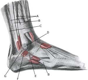
The tendons surrounding the ankle joint are connected to the bones in several places by connective tissue strings (retinaculum). Two peroneus muscles are positioned behind the outer ankle knuckle, which are used to stabilise and flex the ankle joint.


  1. M. peroneus brevis
  2. M. peroneus longus
  3. Tendo calcaneus (Achillis)
  4. Bursa subcutanea malleoli lateralis
  5. Retinaculum mm. peroneorum inferius
  6. Vagina synovialis mm. peroneorum communis
  7. Retinaculum mm. peroneorum superius

OUTER FOOT

Cause: Discomfort can be produced if a rupture of the connective tissue (retinaculum musculorum peroneorum superius & inferius) behind the outer ankle knuckle (malleolus lateralis) occurs. A rupture of the connective tissue (retinaculum) is relatively often combined with outer ligament injuries in the ankle joint.

Symptoms: Pain at the outer ankle knuckle (malleolus lateralis), where certain movements of the ankle joint can give rise to a painful sense of “slipping”.

Acute treatment: Click here.

Examination: When the painful movement of the ankle joint is performed, normal examination can detect the tendon slip over the outer ankle knuckle. A normal medical examination is usually sufficient in order
to make the diagnosis, however, if there is any doubt concerning the
diagnosis it can be made with certainty by use of ultrasound scanning while the ankle joint is in motion (dynamic ultrasound scanning). Ultrasound scanning will reveal whether there is bleeding in the acute stage. Inflammation of the tendon sheath can develop at a later stage.

Treatment: Treatment is primarily concentrated on providing rest from the painful activities (running). If steady progress from rest and rehabilitation is not achieved, and ultrasound scanning reveals inflammation of the tendon sheath, the treatment can be supplemented by medicinal treatment in the form of rheumatic medicine (NSAID) Alternatively, draining and evaluating fluid can be performed, and injection of corticosteroid into the tendon sheath. Injection into the tendon sheath is best performed if ultrasound guided. Surgical intervention can be attempted in certain cases, if resting, rehabilitation and medicinal treatment do not provide the desired result (article).

Complications: If there is not a steady improvement in the condition consideration must be given as to whether the diagnosis is correct, or if complications have arisen:

Possibly supplement with further examinations (x-ray, ultrasound scanning).

Bone membrane tear (periosteal avulsion)

Diagnosis: BONE MEMBRANE TEAR
(Perisotael avulsion)


Anatomy:
The foot bones comprise the 7 tarsal bones (ossa tarsi), the 5 metatarsal bones (ossa metatarsi) and the 14 bones in the toes (phalanx). Muscles or ligaments are attached or anchored to all the bones. The outer surface of the bone is called bone membrane (periosteum), (Ultrasonic image).

Cause: A violent twist can stretch the ligaments and tear a small piece of bone membrane (periosteum) from the ligament anchor point. In other cases, bone membrane tears can occur when the bones impact upon each other (for example if the foot is bent or flexed backwards with great force so that one of the tarsal bones (talus) impacts against the front edge of the shin bone). Bone membrane tears in the ankle joint area are common, and can be seen in most cases where the person has played football for many years (“football-ankle”). Bone membrane tears can occur on all the bones of the foot where tendons or ligaments are anchored.

Symptoms: Pain when applying pressure, and when stretching the tendon or ligament which is attached to the bone.

Acute treatment: Click here.

Examination: Normal clinical examination is often sufficient. Larger tears can be seen on an x-ray. Many lesser tears can be best seen via an ultrasound (Ultrasonic image) scan or MRI examination, (article).

Treatment: Relief from the pain inducing activities. Larger tears can require surgical operation. Lesser tears do not require treatment. Some cases can cause prolonged discomfort with pain which does not recede despite relief. This can be due to the tear causing inflammation in the tissue. In such cases, rheumatic medicine (NSAID) or injection of corticosteroid in the area surrounding the tear can be recommended.

Rehabilitation: Rehabilitation is totally dependent upon the type of tear, and the treatment (conservative or surgical).
Also read rehabilitation, general.

Complications: If there is not a steady improvement in the condition consideration must be given as to whether the diagnosis is correct, or if complications have arisen:

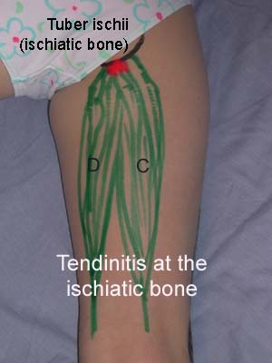
Cause-article1

SportNetDoc

Ischial tuberosity apophysitis and avulsion among athletes.

Kujala UM, Orava S, Karpakka J, Leppavuori J, Mattila K. Int J Sports Med 1997 Feb;18(2):149-55

Ischial tuberosity pain in athletes may be caused by several clinical entities, which include acute and old bony or periosteal avulsions and apophysitis. We studied the natural course of these injuries based on our clinical case series of fourteen patients with apophysitis and twenty-one with avulsion of the ischial tuberosity. Only patients with the diagnosis confirmed by X-ray finding were included. The clinical diagnostic criteria by ischial apophysitis consisted of gradually increasing functional and palpatory pain at the ischial tuberosity without any major trauma at the beginning of the symptoms. Typically there was asymmetry on plain radiographs of the ischial tuberosities in apophysitis; the involved apophyseal area became sclerotic, wider than the non-symptomatic apophysis, osteoporotic patches developed and the lower margin of the ischial tuberosity became irregular. The patients with avulsion reported an acute trauma at the beginning of the symptoms and an avulsion fragment was immediately after injury or later seen in plain radiographs. The mean age of the patients with apophysitis (14.1 yrs) was lower than that of the subjects with avulsions (18.9 yrs). Apophysitis of the ischial tuberosity usually healed well without complications. Avulsions often caused more prolonged pain with referral pain to the posterior parts of the thigh which often required operative interventions. A small bony or periosteal avulsion sometimes grew to a pseudotumor calcification. We recommend conservative treatment as the primary treatment modality for both ischial tuberosity apophysitis and avulsion fractures.

Inflammation of the bursa

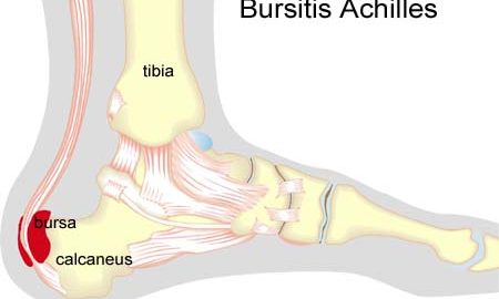
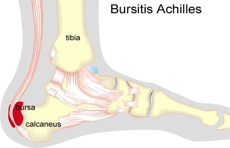
Diagnosis: INFLAMMATION OF THE BURSA AT THE ATTACHMENT OF THE ACHILLES TO THE HEEL BONE
(BURSITIS ACHILLES)


Anatomy:
The calf muscle (M Gastrocnemicus) is comprised of two muscle heads which gather in a wide tendinous ligament and continue in to the Achilles tendon. Another of the larger calf muscles (M Soleus) is attached to the front side of the Achilles tendon and thus forms a part of the Achilles tendon. The Achilles is attached to the heel bone (calcaneus). At the heel bone there is a bursa in front of the Achilles anchor point (bursae tendinis Achilles), as well as behind (bursae subcutanea calcanei). The bursa reduce the pressure against the heel bone.

Cause: Inflammation of the bursa in front of and behind the Achilles occurs with continued overload, where the bursa is squeezed against the heel bone (for example an ill-fitting heel cap on the shoe).

Symptoms: Pain when activating the Achilles tendon (running and jumping) and when applying pressure at the point of attachment of the tendon on the heel bone. Contrary to the tenderness occurring with inflammation of the Achilles tendon, the tenderness is localised to the point of attachment to the heel bone.

Acute treatment: Click here.

Examination: Medical examination is not necessarily required in light cases where the tenderness is minimal. In all cases where smooth improvement is not experienced, medical attention should be sought as soon as possible to exclude a (partial) rupture of the Achilles tendon or rupture of the soleus muscle. This situation is best determined by use of ultrasound scanning, as a number of injuries requiring treatment can easily be overlooked during a clinical examination (Ultrasonic image). Ultrasound scanning enables an evaluation of the extent of the change in the tendon; inflammation of the tendon (tendinitis), development of cicatricial tissue (tendinosis), calcification, inflammation of the tissue surrounding the tendon (peritendinitis), inflammation of the bursa (bursitis), as well as (partial) rupture (article).

Treatment: Treatment is primarily comprised of relief from the painful activity (running). It is important that shoes do not pinch the heel. If satisfactory progress is not made during the rehabilitation, medical treatment can be considered in the form of rheumatic medicine (NSAID) or injection of corticosteroid in the bursa. Injections should be performed under ultrasound guidance to ensure optimal effect and reduce the risk of injecting into the Achilles itself. If progress is not made neither through rehabilitation nor medicinal treatment, surgical treatment can be attempted (article-1), (article-2).

Bandage: In some cases a ring of felt (for example) can be taped around the tender bursa which will reduce the pressure from shoes. It is naturally important that the hole in the ring is positioned directly above the bursa.

Complications: If there is not a steady improvement in the condition an ultrasound scan should be performed to exclude:

KONDITION

STEP4

Training ladder for:
RUPTURE OF THE ACHILLES TENDON
(RUPTURA TRAUMATICA TENDINIS ACHILLES)

STEP 4

Stretching is carried out in the following way: stretch the muscle group for 3-5 seconds. Relax for 3-5 seconds. The muscle group should subsequently be stretched for 20 seconds. The muscle is allowed to be tender, but must not hurt. Relax for 20 seconds, after which the procedure can be repeated. The time consumed for stretching, coordination and strength training can be altered depending on the training opportunities available and individual requirements.

KONDITION
Unlimited: Cycling. Swimming. Running with increasing distance on a soft surface.

UDSPÆNDING
(5 min)

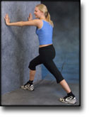
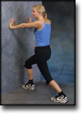
Stand with the injured leg stretched backwards with the toes facing front. Slowly bend the knee so that the calf muscles become increasingly stretched. Hold the position for 20 seconds and relax for 20 seconds before repeating.

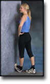
Stand with the injured leg outstretched with the ankle joint bent up against the wall. Press your abdomen against the wall so that the calf muscles become increasingly stretched. Hold the position for 20 seconds and relax for 20 seconds before repeating.

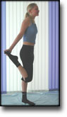
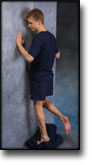
Stand on the good leg while gaining support on the wall with the hand on the same side. Bend the knee of the injured leg and draw the heel towards the buttocks. Take hold of the big toe side of the foot and draw slowly upwards so that the ankle joint is stretched to the maximum and drawn slightly outwards so that increased stretching is experienced on the inside of the shin bone. Hold the position for 20 seconds and relax for 20 seconds before repeating.

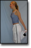
Stand on the good leg while gaining support from the wall with the opposite hand. Bend the knee of the injured leg and draw the heel towards the buttocks. Take hold of the little toe side of the foot and draw slowly upwards so that the ankle joint is stretched to the maximum and drawn over the good leg so that increased stretching is experienced on the outer side of the shin bone. Hold the position for 20 seconds and relax for 20 seconds before repeating.

KOORDINATION
(5 min)

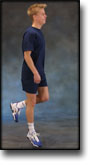
Stand on the leg to be trained. Take-off and land on the same leg.

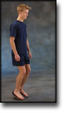
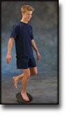
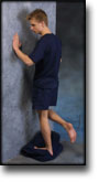
Seesaw. Balance on two legs, possibly using a hand as support against the wall, balancing subsequently on one leg without support. Look straight ahead and keep knees bent.

STYRKE
(50 min)

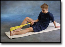
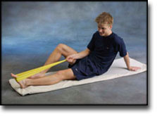
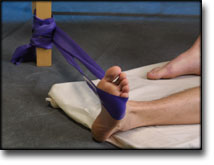
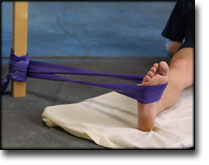
Sit on the floor. Tip the foot up and down with elastic under the forefoot so that the elastic becomes taut.

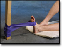
Sit on the floor. Tip the foot up and down with elastic on top of the forefoot so that the elastic becomes taut.

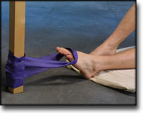
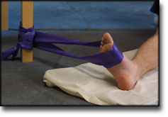
Sit on the floor. Tip the foot from side to side with elastic on the outer side of the foot, without moving the knee.

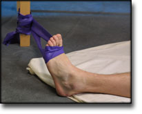
Sit on the floor. Tip the foot from side to side with elastic on the inner side of the foot, without moving the knee.

Stand on a soft surface. Rise slowly up on tiptoe and go down again.

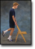
Stand behind a chair. Rise slowly up on tiptoe and go down again.

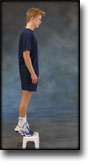
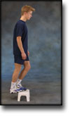
Stand with both forefeet on a stool or doorstep with the heel out over the edge. Slowly rise up on to your toes with your weight on the healthy leg. Go slowly down on the injured leg as far as you can go. Use the healthy leg to rise up on to your toes again. The exercise should be performed with stretched, as well as bent knee. Wearing a rucksack and gradually increasing the ballast in the rucksack can increase the load.

Stand with feet together. Using the ankle joint to take off, hop approx. 5 cm and land on both feet. The exercise should be done on one leg when you are able to do it without discomfort using both legs.

Stretching is carried out in the following way: stretch the muscle group for 3-5 seconds. Relax for 3-5 seconds. The muscle group should subsequently be stretched for 20 seconds. The muscle is allowed to be tender, but must not hurt. Relax for 20 seconds, after which the procedure can be repeated. The time consumed for stretching, coordination and strength training can be altered depending on the training opportunities available and individual requirements.