Kategoriarkiv: Buttock

Stress fracture in the femoral neck

STRESS FRACTURE IN THE FEMORAL NECK

Diagnosis: STRESS FRACTURE IN THE FEMORAL NECK
(Stress fraktur)


Anatomy:
The femur and the hip bone form the hip joint.


  1. Caput femoris
  2. Collum femoris
  3. Trochanter minor
  4. Trochanter major

PELVIS AND THIGH BONE FROM THE FRONT


Cause: Repeated loads, especially when walking or running can cause cracks (stress fractures) in the femoral neck (collum femoris) (article-1) (article-2).

Symptoms: Pain in the hip when applying pressure (direct and indirect tenderness) and when under load (walking, running).

Examination: X-ray. Since many stress fractures cannot be seen early in the course of events, X-ray examination can be repeated after a few weeks. Scintigraphy, CT- and MRI and ultrasound scan can often diagnose stress fractures far earlier than X-rays (Ultrasonic image).
It is imperative for the result of the treatment that the diagnosis is made as early as possible (article).

Treatment: Relief. In some cases surgery is necessary (article).

Rehabilitation: The rehabilitation is completely dependent on the type of fracture and treatment (conservative or operative).
Also read rehabilitation, general.

Complications: If progress is not smooth, you should be re-examined to ensure that the fracture heals according to plan. In some cases a false joint can be formed (pseudoarthrosis), which requires surgical treatment.

Special: Shock absorbing shoes or inlays will reduce the load.

Fluid accumulation in the joint

FLUID ACCUMULATION IN THE JOINT

Diagnosis: FLUID ACCUMULATION IN THE JOINT
(Synovitis / coxitis)


Anatomy:
The hip joint consists of the hip socket (acetabulum) and the femoral head (caput femoris). The articular surfaces are coated with a cartilage layer a few mm thick, which reduces the load on the articular surfaces.

Cause: Many repeated loads or one violent load can cause an inflammation of the synovial membrane (synovitis), fluid formation, swelling, restriction of movement and pain in the hip joint. The condition is relatively often seen in children (Ultrasonic image).

Symptoms: Pain in the joint upon movement with load. Often there will be movement restriction with rotation of the hip joint.

Examination: It will often be necessary to supplement the ordinary clinical examination with an ultrasound scan, where the fluid in the hip joint can easily be seen (Ultrasonic image).

Treatment: The treatment primarily comprises relief from the pain inducing activity until the swelling has abated. Rehabilitation, within the pain threshold, can subsequently be commenced. In cases of lack of progress with relief the treatment can be supplemented with a medical treatment in the form of rheumatic medicine (NSAID) or by drainage of the joint fluid and the injection of corticosteroid, which should be done with ultrasound guidance.

Complications: In particular you should consider a bacterial infection in the joint (pyarthron), which is determined by ultrasound guided draining of the joint fluid, the child hip diseases (epifysiolysis capitis femoris and Calvé-Legg-Perthes disease), where the joint head on the femur slips or collapses (X-ray examination will determine the diagnosis), arthritis or:

Special: There are two childrens’ hip diseases that should always be considered in children with hip pain.

  • SLIPPING OF THE FEMORAL HEAD IN THE GROWTH ZONE (EPIFYSIOLYSIS CAPITIS FEMORIS), which mainly affects boys age 11-16 years (article). There will often be limping, groin pain, but sometimes the pain is sensed in the knee. It is important in order to obtain a good result from the treatment to be examined by a doctor and have the diagnosis made as soon as possible (with X-rays).

  • CALVÉ-LEGG-PERTHES DISEASE is a disease which mainly affects boys age 3-11 years (article). The bone core in the femoral head is dissolved and flattened. There will often be limping, tiredness and pain in the groin, but once in a while the pain is sensed in the knee instead. It is important in order for a good result of the treatment to be examined by a doctor and have the diagnosis made as soon as possible (by means of X-rays or ultrasound).

Degenerative arthritis

DEGENERATIVE ARTHRITIS

Diagnosis: DEGENERATIVE ARTHRITIS
(Osteoarthritis)


Anatomy:
The hip joint consists of the hip socket and the femoral head (caput femoris). The articular surface is covered with a few mm thick cartilage layer, which reduces the load on the articular surfaces

Cause: In case of repeated loads, the cartilage primarily, and subsequently the bone below the cartilage, can be damaged (degenerative arthritis). The degenerative arthritis changes can in some cases cause an inflammation of the synovial membrane (synovitis) which causes fluid formation, swelling, movement restriction and pain in the hip joint.

Symptoms: Pain in the hip joint upon movement with load. There will often be movement restriction upon rotation in the hip joint.

Examination: Ordinary clinical examination is often sufficient to make the diagnosis. The examination can be supplemented with an X-ray examination. Ultrasound scan is the most suitable examination if you suspect a fluid accumulation in the hip joint.

Treatment: The treatment primarily comprises relief from the pain inducing activity until any swelling in the hip joint has decreased. Rehabilitation can subsequently be commenced with the primary goal to strengthen the muscles around the hip joint and preserve the joint mobility. There is no treatment that can restore the ruined cartilage (and bone). Cartilage transplants are not yet suitable for general degenerative arthritis changes. Upon swelling in the hip joint you can attempt to reduce the synovitis with rheumatic medicine (NSAID) or by attempting to drain the fluid and injecting corticosteroid, which should be conducted with ultrasound guidance to optimise the effect and minimize the risk. Pain without joint swelling is best treated with paracetamol. In cases of severe degenerative arthritis changes with pain when resting (at night) it may be necessary to replace the hip joint.

Complications: Degenerative arthritis which sits on the weight bearing parts of the joint is one of the most serious sports injuries, and often results in a termination of active sport. Cycling and swimming are significantly less stressful for the hip joint than running. In particular the following should be considered:

Special: Shock absorbing shoes or inlays will reduce the load.

Muscle infiltration in the buttock muscles

MUSKELINFILTRATION I EN AF BALDEMUSKLERNE

Diagnosis: MUSCLE INFILTRATION (MYALGIA) IN 
THE BUTTOCK MUSCLES

(PIRIFORMIS SYNDROM)


Anatomy:
The piriformis muscle is a small muscle which origin from the sacrum and fastens on the outer femoral bone projection (tronchanter major). The muscle assists in rotating the thigh outwards.

  1. Trochanter major
  2. Trochanter minor
  3. Femur
  4. M. obturatorius externus
  5. Tuber ischiadicum
  6. M. obturatorius internus
  7. Lig. sacrotuberale
  8. Lig. sacrospinale et. m coccygeus
  9. M. piriformis

M. PIRIFORMIS

Cause: If the muscle is over-loaded, it will become taut and tender. In some cases, the muscle can become so taut that it jams against the ischias nerve (nervus ischiadicus).

Symptoms: Pain deep in the buttock, with periodic radiation into the leg.

Examination: The diagnosis will be rendered probable under clinical examinations by demonstrating tenderness by applying pressure on the muscle deep within the buttock, as well as provoking pain by stretching and activating the muscle. There is no suitable method to provide a pictorial image of the muscle (MR scanning can be attempted in the event of suspicions of nerve impingement) (article).

Treatment: The treatment usually comprises stretching and subsequent strength training of the muscles surrounding the lower back and buttocks. It is only in very rare cases involving nerve impingement that surgery is indicated. Uncomfortable pain can be treated medicinally in the form of paracetamol, or possibly rheumatic medicine (NSAID) . If this treatment does not provide the desired relief, ultrasound guided injection of corticosteroid in the most tender part of the muscle can be attempted (article 1), (article 2).

Special: Shock absorbing shoes or inner inlays will reduce the risk of various forms of muscle infiltrations. In the event of unsatisfactory progress, or relapse after successful rehabilitation, consideration must be given to performing an analysis of the patient’s running style to establish whether a correction of the running style should be recommended.

Complications: If progress is not smooth, the correctness of the diagnosis should be considered or whether complications have arisen. The following should be considered in particular:

Bursitis

BURSITIS

Diagnosis: BURSITIS
(Inflammation of the bursa)


Anatomy:
There are numerous bursas around the hip joint, serving the purpose of reducing the pressure on muscles, tendons and ligaments where these lie close to a bone projection.

Cause: In case of repeated loads or blows, the bursa can produce additional fluid, swell and become inflamed and painful.

Symptoms: Pain when applying pressure on the bursa, which sometimes (but far from always) can feel swollen. Pain is aggravated upon activation of the muscle closest to the bursa.

Acute treatment: Click here.

Examination: In light cases with only minimal tenderness, medical examination is not necessarily required. In case of more pronounced pain or lack of progress, a medical examination should be carried out to ensure the correctness of the diagnosis and the commencement of any treatment. The diagnosis is most easily and quickly made with ultrasound (which allows simultaneous treatment).

Treatment: The treatment primarily comprises relief. If the direct cause of the complaint is known, it should of course be removed. The treatment can be supplemented by rheumatic medicine (NSAID) or injection of corticosteroid in the bursa, preceded by draining of this, which can advantageously be performed under ultrasound guidance.

Rehabilitation: The treatment is dependant upon which bursa is inflamed, but sports activity can usually be cautiously resumed when pain has diminished, especially if the provoking factor has been identified and removed.
Also read rehabilitation, general.

Complications: If progress is not smooth, the correctness of the diagnosis or whether complications have arisen should be considered:

Special: Shock absorbing shoes or inlays will reduce the load.

Bursitis at the outer femoral bone projection

BURSITIS AT THE OUTER FEMORAL BONE PROJECTION

Diagnosis: BURSITIS AT THE OUTER FEMORAL BONE PROJECTION
(Bursittis trochanterica)


Anatomy:
On the outside of the outer femoral bone projection (trochanter major) is a large bursa which reduces the load on muscles and tendons when these slide over the bone projection.

  1. M. piriformis
  2. M. gluteus minimus
  3. Bursa m. piriformis
  4. Bursae trochantericae m. glutei medii
  5. M. gluteus medius (resectus)
  6. Bursa trochanterica m. glutei maximi
  7. M. gluteus maximus
  8. Bursae intermusculares mm. gluteorum
  9. Tuberositas glutea
  10. Tractus iliotibialis
  11. M. biceps femoris
    (caput longum)
  12. M. biceps femoris
    (caput breve)
  13. M. adductor magnus
  14. B. subtendinea m. bicipitis femoris superior
  15. Tuber ischiadicum
  16. B. ischiadica m. glutei maximi
  17. Mm. gemilli sup. Et inf.
  18. B. ischiadica m. obturatorii interni

GLUTEAL MUSCLES FROM THE REAR

Cause: In case of repeated loads or blows the bursa can become inflamed, produce fluid, swell and become painful.

Symptoms: Pain when applying pressure on the thigh corresponding to the bursa, which sometimes (but far from always) can feel swollen. The pain can radiate down the thigh.

Acute treatment: Click here.

Examination: In light cases with only minimal tenderness medical examination is not necessarily required. In case of more pronounced pain or lack of progress, a medical examination should be carried out to ensure the diagnosis and commencement of any treatment. A normal medical examination is usually sufficient in order to make the diagnosis, however, if there is any doubt concerning the diagnosis an ultrasound scan can be performed (Ultrasonic image).

Treatment: The treatment primarily comprises relief. If the direct cause of the complaint is known, it should of course be removed. The treatment can be supplemented with rheumatic medicine (NSAID) or injection of corticosteroid in the bursa, which is best performed under the guidance of ultrasound.

Complications: If progress is not smooth, the correctness of the diagnosis should be considered or whether complications have arisen:

Special: Shock absorbing shoes or inlays will reduce the load. In case of lack of progress or recurrences after successful rehabilitation it can be considered to have a running style analysis, to evaluate whether correction of the running style is indicated.

Inflammation of the bursa at the ischiatic bone

Diagnosis: INFLAMMATION OF THE BURSA AT THE ISCHIATIC BONE
(BURSITTIS ISCHIADICA)


Anatomy:
The large posterior thigh muscles (hamstring muscles) have a common muscle tendon fastening on the ischiatic bone (tuber ischii). Below the tendon fastening there is a bursa which reduces the load on the tendon when the tendon slides against the bone. The posterior thigh muscles flex the knee and stretch the hip.


  1. Bursa trochanterica m. glutei maximi
  2. M. gluteus maximus
  3. M. biceps femoris (caput longum)
  4. M. semitendinosus
  5. M. semimembranosus
  6. M. adductor magnus
  7. M. gracilis
  8. M. quadratus femoris
  9. Bursa ischiadica m. glutei maximi

(Photo)

RIGHT GLUTEAL MUSCLES
FROM THE REAR

Cause: In case of repeated loads or blows the bursa can become inflamed, produce fluid, swell and become painful.

Symptoms: Pain upon applying pressure on the bursa (sitting position), which sometimes (but far from always) may feel swollen. Aggravated upon stretching and activation of the posterior thigh muscles (flexing of the knee against resistance) (article).

Acute treatment: Click here.

Examination: In slight cases with only minimal tenderness, medical examination is not necessarily required. In cases of more pronounced pain or lack of progress, a medical examination should be carried out to ensure a correct diagnosis and commencement of treatment. The diagnosis is usually made on the basis of a normal medical examination, however, if there is any doubt surrounding the diagnosis, it can easily and quickly be confirmed under an ultrasound scan.

Treatment: The treatment primarily consists of relief. The treatment can be supplemented with rheumatic medicine (NSAID) or the injection of corticosteroid in the bursa preceded by draining of the bursa, which can advantageously be done under ultrasound guidance.

Complications: If progress is not smooth, it should be considered if the diagnosis is correct or whether complications have arisen. The following should in particular be considered:

Rupture of the posterior thigh muscle fastening on the ischiatic bone

RUPTURE OF THE POSTERIOR THIGH MUSCLE FASTENING ON THE ISCHIATIC BONE

Diagnosis: RUPTURE OF THE POSTERIOR THIGH MUSCLE FASTENING ON THE ISCHIATIC BONE
(RUPTURA MUSCULI)


Anatomy:
The large posterior thigh muscles (hamstring muscles) have a common muscle tendon fastening on the ischiatic bone (tuber ischii). The posterior thigh muscles flex the knee and stretch the hip.


  1. Bursa trochanterica m. glutei maximi
  2. M. gluteus maximus
  3. M. biceps femoris (caput longum)
  4. M. semitendinosus
  5. M. semimembranosus
  6. M. adductor magnus
  7. M. gracilis
  8. M. quadratus femoris
  9. Bursa ischiadica m. glutei maximi

(Photo)

RIGHT GLUTEAL MUSCLES
FROM THE REAR

Cause: When a muscle is subjected to a load beyond the strength of the muscle (typically sprinting), a rupture occurs. The vast majority of ruptures are partial muscle ruptures. Many of the ruptures at the muscle fastening on the ischiatic bone (tuber ischiadicum) er preceded by lengthy tendinitis (entesopatia tuber ischiadicum) at the same location. In rare cases, the muscle fastening can tear a piece of the ischiatic bone off (especially seen in children as the growth zone on the ischiatic bone (apophysis) increases the risk of tears).

Symptoms: In slight cases a local tenderness is felt after being subjected to load (“sprained muscle”, “imminent pulled muscle”). In severe cases sudden shooting pains are felt in the muscle (“partial muscle rupture”, “pulled muscle”) and in the worst case a sudden snap is felt rendering the muscle unusable (“total muscle rupture”). The following three symptoms are characteristic in connection with muscle injuries: pain upon applying pressure, stretching and activation of the muscle (flexing knee) against resistance. With total ruptures a defect and a swelling (the contracted muscle belly and bleeding) can often be seen and felt in the posterior thigh muscle just below the ischiatic bone and below.

Acute treatment: Click here.

Examination: In very slight cases with only minimal tenderness and no discomfort when walking, medical examination is not necessarily required. The severity of the tenderness is however, not always a measure of the extent of the injury. In cases of more pronounced tenderness or pain, medical examination is required to ensure the diagnosis and treatment. The diagnosis is usually made following normal medical examination, however, if there is any doubt concerning the diagnosis, ultrasound scanning (or MRI scanning) can be performed, as these are the most suitable examinations to ensure the diagnosis (article). The larger the bleeding as assessed by ultrasound scanning, the longer the period needed to heal the injury. Tears on the ischiatic bone will usually be visible on x-rays.

Treatment: Treatment comprises relief and rehabilitation. The aim of the rehabilitation is to strengthen the posterior thigh muscles to enable the muscles to manage the loads which previously caused the rupture. It is only in very rare cases where there is a total rupture at the muscle fastening or tearing of a large piece of bone from the ischiatic bone that surgery is considered (article). Even large ruptures in the thigh muscles will usually be able to be healed and rehabilitated without giving functional disorder (but often cosmetic disfigurement with an irregular thigh muscle).

Complications: If steady progress is not experienced it should be considered if the diagnosis is correct or whether complications have arisen. The following should in particular be considered:

Tendinitis at the ischiatic bone

Diagnosis: TENDINITIS AT THE ISCHIATIC BONE
(ENTESOPATIA TUBER ISCHIADICUM


Anatomy:
The large posterior thigh muscles (hamstring muscles) have a common muscle tendon fastening on the ischiatic bone (tuber ischiadicum). The posterior thigh muscles flex the knee and stretch the hip.

  1. Bursa trochanterica m. glutei maximi
  2. M. gluteus maximus
  3. M. biceps femoris (caput longum)
  4. M. semitendinosus
  5. M. semimembranosus
  6. M. adductor magnus
  7. M. gracilis
  8. M. quadratus femoris
  9. Bursa ischiadica m. glutei maximi

(Photo)

RIGHT GLUTEAL MUSCLES
FROM THE REAR

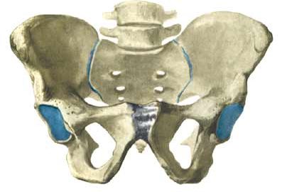
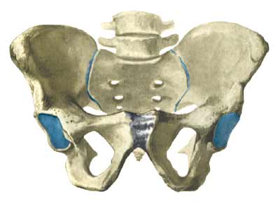
Cause: Inflammation of the tendon fastenings (tendinitis) at the ischiatic bone (tuber ischiadicum) occurs following repeated uniform (over)loads (e.g. running, sprinting) causing microscopic ruptures in the tendon, and especially at the tendon fastening. Tendinitis is a warning that the training performed is too strenuous for the muscles in question, and if the load is not reduced a rupture of the posterior thigh muscle fastening on the ischiatic bone (“pulled muscle”) may occur. This will result in a considerably prolonged rehabilitation period.

Symptoms: Pain in the ischiatic bone can occasionally radiate down into the rear of the thigh. The pain is aggravated when applying pressure on the bone (e.g. sitting position), stretching and activating the posterior thigh muscles (flexing the knee against resistance).

Acute treatment: Click here.

Examination: In slight cases with only minimal tenderness and no discomfort with walking, medical examination is not necessarily required. The extent of the tenderness is, however, not always a mark of the degree of the injury. In cases of more pronounced pain or tenderness, medical examination is required to ensure the correct diagnosis and treatment. The diagnosis is usually made on the basis of a normal medical examination, however, if there is any doubt concerning the diagnosis, this can be confirmed by ultrasound scanning or MR scanning (article).

Treatment: The treatment usually comprises relief, stretching and rehabilitation (article). If the rehabilitation does not progress satisfactorily, medicinal treatment in the form of rheumatic medicine (NSAID) can be considered, or corticosteroid injection in the area surrounding the inflamed tendon fastening on the ischiatic bone. As the injection of corticosteroid is always an element in the long term rehabilitation of a very serious, chronic injury, it is vital that the rehabilitation period lasts over several months in order to reduce the risk of a relapse or ruptures. The tendon can naturally not sustain maximal load after a long-term injury period and only a short-term rehabilitation period. If the diagnosis is made by use of ultrasound scanning, the injections are performed under guidance of ultrasound, and the rehabilitation is progressed in accordance with the guidelines mentioned, then the treatment involving corticosteroid injections has very few risks connected. If satisfactory progress is not made following the rehabilitation and medicinal treatment, surgical intervention can be considered. Long-term results of operations are often disappointing, despite publication of a minor series with good results (article).

Complications: If the treatment does not progress according to plan, it should be considered if the diagnosis is correct or whether complications have arisen. The following should in particular be considered: