Kategoriarkiv: Elbow

Disclocation of the elbow

DISCLOCATION OF THE ELBOW

Diagnosis: DISCLOCATION OF THE ELBOW
(LUXATIO ARTICULI CIBITI)


Anatomy:
The elbow joint is comprised of the upper arm bone (humerus), and one of the two forearm bones (ulna). The other forearm bone (radius) forms a joint with ulna (art. Radioulnaris proximalis). A joint capsule and several strengthening ligaments surround the elbow joint.

  1. Humerus
  2. Capsula articularis
  3. Epicondylus medialis
  4. Lig. collaterale ulnare
  5. Chorda obliqua
  6. Ulna
  7. Radius
  8. Tendo m. bicipitis brachii
  9. Lig. anulare radii
  10. Lig. collaterale radiale
  11. Epicondylus lateralis

Elbow joint

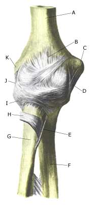
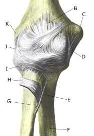
Cause: A dislocation of the elbow can occur following a direct fall on an outstretched arm. The dislocation can in some cases be complicated by a bone fracture, vascular damage or nerve damage.

Symptoms: Sudden insetting pain around the elbow, with pain-conditional restriction of mobility of the arm following a sudden, violent load (fall).

Acute treatment:
Click here.

Examination: Sudden, strong pain in the arm with restriction of movement following a fall should always lead to acute medical examination. Acute medical assistance should be sought due to the risk of damage to blood vessels and nerves. An X-ray examination will usually reveal the dislocation and rule out bone fracture.

Treatment: The dislocation can usually, in uncomplicated cases, be put in place without the need of surgery. Some recommend a short time where bandaging is used after the dislocation has been put into place. Surgery is often necessary in cases where complications arise from the dislocation in the form of bone fracture, vascular damage or nerve damage. Rehabilitation with exercises involving movement should be commenced as soon as possible, (article-1)(article-2).

Rehabilitation can commence shortly after the dislocation is put into place (and possible bandaging has been removed) in uncomplicated cases without bone fracture, vascular damage or nerve damage. Recommendations from your doctor must be taken into consideration in the rehabilitation program if the dislocation has involved complications and has possibly required surgery.
Also read rehabilitation, general.

Complications: Tears or ruptures around the elbow will in the vast majority of cases heal without complication. Some cases will experience persistent stiffness in the elbow, looseness of the elbow, calcification in the muscles surrounding the elbow and vascular or nerve damege. Dislocation of the elbow can in some cases be complicated by ligament injuries in the wrist, (article).

Fracture of the humeral shaft at the elbow

FRACTURE OF THE HUMERAL SHAFT AT THE ELBOW

Diagnosis: FRACTURE OF THE HUMERAL SHAFT AT THE ELBOW
(fractura supracondylaris humeri)


Anatomy:
The upper arm (humerus) consists of the capitulum, the neck (collum), the long tubular bone (corpus) and the articulated part in the elbow (epicondylus).

  1. Caput humeri
  2. Collum chirurgicum
  3. Epicondylus medialis
  4. Epicondylus lateralis
  5. Tuberculum minus
  6. Sulcus intertubercularis
  7. Tuberculum majus
  8. Collum anatomicum

UPPER ARM FROM THE FRONT

Cause: A fracture of the humerus can occur in cases of a direct fall on the outstretched arm. The fracture may occur anywhere on the humerus, but a fracture through the neck of the humerus (collum) and the middle of the long tubular bone (corpus) are the most common locations. However, in children especially a fracture of the upper arm just above the elbow is often seen (fractura supracondylaris humeri), which in rare cases can cause entrapment of blood vessels and nerves.

Symptoms: Sudden pain in the elbow region and pain induced constriction of movement of the arm after a fall.

Acute treatment: Click here.

Examination: Sudden, powerful pains in the arm with constriction of movement after a fall, should always lead to acute medical examination due to the risk of damage to the blood vessels and nerves. The fracture is usually visible on x-rays, and on the basis of the type of fracture, the correct treatment can be determined.

Treatment: The fracture can normally be set in place under an aenesthetic, followed by bandaging for a few weeks. Surgical fixation may be required for certain types of fractures.

Rehabilitation: When pain has decreased and the bandage has been removed fitness training in the form of cycling and running may be started along with retraining as specified under  rehabilitation, general. Children will often be able to resume sports activity within 2-3 months.

Complications: In the vast majority of cases the fracture heals without complications although in some instances a poor healing occurs affecting the blood and nerve supply to the arm, or development of muscle acute compartment syndrome (article).

Inflammation of the bursa

INFLAMMATION OF THE BURSA

Diagnosis: INFLAMMATION OF THE BURSA
(BURSITIS)


Anatomy:
There are numerous bursas surrounding the elbow, with the purpose of reducing the pressure on muscles and tendons, where these lie close to the bone.

Cause: In cases of repeated loads or blows the bursas can become inflamed, produce fluid, swell and become painful. One of the most frequent bursitis forms in the elbow region is inflammation of the bursa located between the biceps tendon and the fastening on the radius (tuberositas radii), (article).

Symptoms: Pain upon applying pressure on the bursa, which sometimes (but far from always) may feel swollen. Aggravated upon activation of the muscle located immediately above the bursa.

Acute treatment: Click here.

Examination: In light cases with only minimal tenderness, medical examination is not necessarily required. In cases of more pronounced pain or lack of progress, a medical examination should be carried out to ensure a correct diagnosis and treatment. The diagnosis is made most easily and quickly with an ultrasound scan.

Treatment: The treatment primarily consists of relief with removal of the provoking cause if known. The treatment can be supplemented with rheumatic medicine (NSAID) or the injection of corticosteroid in the bursa preceded by draining of the bursa, which can advantageously be performed under ultrasound guidance.

Rehabilitation: The treatment is completely dependant on which bursa is inflamed, but sports activity can usually be cautiously resumed once pain has decreased, particularly if it has been possible to remove the provoking cause.
See also rehabilitation, general.

Complications: If progress is not smooth, it should be considered if the diagnosis is correct or whether complications have arisen: In rare cases the bursa can become infected with bacteria. This is a serious condition where the bursa becomes red, warm and increasingly swollen and tender, and requires immediate medical examination and treatment. 
If there is no progress with relief, medical treatment rheumatic medicine (NSAID) and the ultrasound guided injection of corticosteroid
surgical removal of the bursa may be attempted.

Inflammation of the bursa at the elbow

INFLAMMATION OF THE BURSA AT THE ELBOW

Diagnosis: INFLAMMATION OF THE BURSA AT THE ELBOW
(BURSITIS OLECRANEI)


Anatomy:
There is a large bursa on the point of the elbow (olecranon) which protects the elbow bone against blows and pressure.

Cause: A blow or fall on the point of the elbow (football goal-keeper) can cause the bursa to become inflamed, swell and become tender.

Symptoms: Tenderness and swelling on the point of the elbow with pain conditioned constriction of movement of the arm following a strenuous load (fall). The pain is aggravated when the elbow is supported on a table top (the injury is also called “student’s elbow”).

Acute treatment: Click here.

Examination: The diagnosis is usually made on the basis of a normal medical examination, however, if any doubt surrounds the diagnosis an ultrasound scan can be performed which will clearly show the bursa (Ultrasonic image).

Treatment: Relief and protection from further blows. If no change for the better is experienced, the treatment can be supplemented with rheumatic medicine (NSAID) or injection of corticosteroid in the bursa preceded by draining of the bursa fluid (which can be sent for bacteriological examination) (article).

Rehabilitation: Normal training can generally be resumed taking care to avoid further blows to the elbow.
See also: rehabilitation, general.

Complications: If the bursa does not diminish following the treatment outlined, the fluid should be drained once again to rule out a bacterial infection in the bursa. If it proves impossible to make the bursa diminish despite repeated treatment, the bursa can be surgically removed, however this will often result in discomfort when resting the elbow on a table top or receiving blows of a similar nature.

Special: The injury can be partially guarded against by use of elbow protection (volley ball players, football goal-keepers).

Golf elbow

Diagnosis: GOLF ELBOW
(EPICONDYLITIS MEDIALIS)


Anatomy:
A large number of the muscles in the forearm that are designed to flex the wrist and fingers fasten on the inner bone projection on the elbow (epicondylus medialis).


  1. M. biceps brachii
  2. Epikondylus mediale
  3. Aponeurosis m. bicipitis brachii
  4. M. pronator teres
  5. M. flexor carpi radialis
  6. M. palmaris longus
  7. M. flexor digitorum superficialis
  8. M. flexor carpi ulnaris

FLEXORS OF THE FOREARM

Cause: The strength of the muscle fastening will be exceeded if subjected to repeated uniform (over)loads, causing microscopic ruptures in the tendon, and especially at the tendon fastening, resulting in an inflammation. This tendinitis is a warning that the training performed is too strenuous for the muscle tendons in question, and if the load is not reduced a chronic inflammation can arise which is problematic to treat. The condition is also called “golf elbow”, and is often a consequence of incorrect stroke technique but can be the result of a number of other causes.

Symptoms: Tenderness and pain in the area of the inner bone projection elbow (epicondylus medialis) on the elbow which is aggravated when activating the muscle group which fastens there (flexing of the wrist (flexion) against resistance and when stretching).

Acute treatment: Click here.

Examination: The diagnosis is usually made based on a normal medical examination, however, if there are any doubts surrounding the diagnosis an ultrasound scan can be performed which will often reveal the inflammatory changes at the muscle fastening (article). Novel use of laser doppler imaging for investigating epicondylitis. With prolonged discomfort a fraying of the bone membrane (“entesopati”) (Ultrasonic image) can be observed, as well as calcification of the soft parts which in places can have the characteristics of a calcaneal spur.

Treatment: Correction of stroke technique and adjustment of equipment are naturally vital elements for a successful rehabilitation. Treatment primarily comprises relief, stretching and strength training of the forearm muscles (article). If the discomfort does not abate, the treatment can be supplemented with medicinal treatment in the form of rheumatic medicine (NSAID) or injection of corticosteroid (article). Surgical treatment can be considered if there is no change for the better, however, the results are far from convincing.

Bandage: Some patients experience an improvement in the symptoms by applying tape (or a bandage) around the forearm just below the elbow (tape-instruction)

Complications: If satisfactory progress is not achieved it should be considered whether the diagnosis is correct or whether complications have arisen, which can amongst others be:

Tennis elbow

TENNIS ELBOW

Diagnosis: TENNIS ELBOW
(EPICONDYLITIS LATERALIS)


Anatomy:
A large number of the muscles in the forearm that are designed to flex the wrist backwards and stretch the fingers, fasten on the outer bone projection on the elbow.


  1. M. extensor carpi radialis brevis
  2. M. extensor digitorum
  3. M. extensor carpi ulnaris
  4. M. anconeus
  5. Olecranon
  6. Epicondylus lateralis

EXTENSORS OF THE FOREARM

Cause: The strength of the muscle fastening will be exceeded if subjected to repeated uniform (over)loads, causing microscopic ruptures in the tendon, and especially at the tendon fastening, resulting in an inflammation. This tendinitis is a warning that the training performed is too strenuous for the muscle tendons in question, and if the load is not reduced a chronic inflammation can arise which is problematic to treat. The condition is also called “tennis elbow”, and is often a consequence of incorrect stroke technique or unsuitable equipment (racket).

Symptoms: Tenderness and pain in the area of the outer bone projection on the elbow (epicondylus lateralis) which is aggravated when activating the muscle group which fastens there (backwards flexing of the wrist (extension) against resistance and when stretching).

Acute treatment: Click here.

Examination: The diagnosis is usually made based on a normal medical examination, however, if there are any doubts surrounding the diagnosis an ultrasound scan can be performed which will often reveal the inflammatory changes at the muscle fastening (article). With prolonged discomfort a fraying of the bone membrane (entesopatia) can be observed, as well as calcification of the soft parts which in places can have the characteristics of a calcaneal spur (Ultrasonic image).

Treatment: Correction of stroke technique and adjustment of equipment are naturally vital elements for a successful rehabilitation. Treatment primarily comprises relief, stretching and strength training of the forearm muscles (article). If the discomfort does not abate, the treatment can be supplemented with medicinal treatment in the form of rheumatic medicine (NSAID) or injection of corticosteroid (article). During recent years, different types of experimental treatment have been seen such as sclerosis injection (where injections are performed around the tendon with a drug to destroy the small blood vessels (and nerves) that infiltrate the sick tendons), and shock-wave (ultrasound treatment). However, there is no sure or clear documentation for the effect of these kinds of treatment. Surgical treatment can be considered if there is no change for the better, however, the results are far from convincing.

Bandage: Some patients experience an improvement in the symptoms by applying tape (or a bandage) around the forearm just below the elbow (tape-instruction).

Complications: If satisfactory progress is not achieved it should be considered whether the diagnosis is correct or whether complications have arisen, which can amongst others be: