Kategoriarkiv: Foot, front

Degenerative arthritis

SLIDGIGT

Diagnosis: DEGENERATIVE ARTHRITIS
(Osteoarthritis)


Anatomy:
The surfaces of the joints are lined with a cartilage covering of a few millimetre’s thickness which serves to reduce the load or strain on the joint surfaces.

  1. Phalanx media
  2. Tuberositas ossis metatarsalis V
  3. Os cuboideum
  4. Calcaneus
  5. Talus
  6. Os naviculare
  7. Os cuneiforme laterale
  8. Os cuneiforme intermedium
  9. Os cuneiforme mediale
  10. Os metatarsalei
  11. Os sesamoideum
  12. Phalanx proximalis
  13. Phalanx distalis


THE FOOT FROM ABOVE

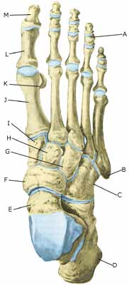
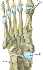
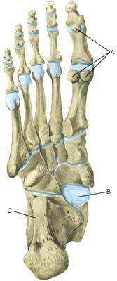
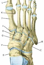
Cause: Degenerative arthritis occurs with repeated (over) load when first the cartilage takes damage, and then the bone under the cartilage. Degenerative arthritis can in some instances cause an irritation of the synovial membrane which will result in concentration of fluid, swelling, reduction in mobility and pain in the joints. Degenerative arthritis in the ankle joint is often seen after repeated ligament injuries (outer ankle joint ligaments, inner ankle joint ligaments), where cartilage lesions in the ankle joint have occurred at the same time.

Symptoms: Pain in the joint with movement under load. Occasionally swelling in the joint.

Examination: Normal clinical examination is often sufficient. However, it is also often necessary to perform an x-ray (or ultrasound scan or MRI examination) to make the diagnosis. Ultrasound scanning will often reveal inflammation surrounding new bone development at the joint surfaces.

Treatment: Treatment comprises relief from the painful activities until the swelling has gone down, after which training can commence with the primary aim to strengthen the muscles surrounding the joint and retain joint mobility. There is no treatment which can restore the damaged cartilage (and bone). Cartilage transplants are, as yet, not suitable for general degenerative arthritis. In cases of swelling in the joint, and with inflamed new bone development at the joint surfaces, inflammation of the synovial membrane can be attempted subdued by using rheumatic medicine (NSAID), or by draining the fluid and injecting corticosteroid. The injections can be performed to advantage by utilising an ultrasound guided method. Pain without swelling of the joints is best treated with paracetamol. In severe cases of degenerative arthritis where there is pain when resting (at night), it may be necessary to fix the joint by operation.

Rehabilitation: Rehabilitation is completely dependent upon the degree of the degenerative arthritis and in which joints it is located.
Also read rehabilitation, general.

Bandage: A supportive tape (Hollywood bandage) can be attempted to aid degenerative arthritis in small joints (toes) (tape-instruction). Tape provides no help to attacks in the ankle joint.

Complications: Degenerative arthritis which sits on the weight bearing parts of the joint is one of the most serious sports injuries, and often results in a termination of active sport. It is usually possible to continue sport activities with light strain on the joints (cycling, swimming), whereas it is advisable to participate in activities with great strains on the joint (running, ball games) with restraint. The diagnostic considerations in connection with degenerative arthritis include:

Special: Shoes with shock absorbing inlays will reduce the discomfort of degenerative arthritis.

Stiff toe syndrome

STIVHED AF STORETÅEN

Diagnosis: STIFF TOE SYNDROME
(Hallux rigidus functionalis)


Anatomy:
The flexing tendons of the big toe (musculus flexor hallucis longus & brevis) run under the sole of the foot and are attached to the bones of the big toe.

  1. M. flexor hallucis brevis
  2. M. flexor hallucis longus

MUSCLES IN THE SOLE OF THE FOOT

Cause: The stiffness is due to an irritation in the flexing tendons under the toe (M flexor hallucis). It occurs with repeated overloading of the flexing tendons (jumping, handball).

Symptoms: Pain in the metatarsophalangeal toe joint when walking. The pain is aggravated when the big toe is flexed backwards (extension). Often seen in girls in the 14-18 year age group.

Examination: Medical examination is not necessarily required in slight cases with minimal tenderness and no discomfort when walking. Examination is required in cases of more pronounced pain in order to confirm the diagnosis. The examination will show that the big toe can not be flexed upwards (extension), when the ankle joint is flexed 90 degrees, whilst the big toe can flex upwards when the ankle joint is stretched (flexion). In some instances it will be necessary to perform an x-ray or ultrasound scan, possibly supplemented with MRI examination (article).

Treatment: Treatment comprises relief with the aid of shoes with stiff soles (article). The treatment can be combined with rheumatic medicine (NSAID) (gel or crème). Furthermore, increased mobility of the joint is aimed at by active and passive movement of the big toe (hallux) within the pain threshold. Operations have been attempted, but have not yielded satisfactory results (article).  

Rehabilitation: Load or strain within the pain threshold is allowed. Shoes with stiff soles are recommended. The guidelines under rehabilitation, general should be followed. Passive and active movement of the big toe within the pain barrier.

Bandage: Taping which supports the metatarsophalangeal toe joint can be used to reduce the pain from sports activity and walking (tape-instruction). If this is not sufficient, special soles can be manufactured to support the big toe.

Complications: If there is not a steady improvement in the condition, medical assistance should be sought to determine whether the diagnosis is correct or whether complications have arisen:

Imflammation of the bursa

Diagnosis: INFLAMMATION OF THE BURSA
(Bursitis)


Anatomy:
There are numerous bursas around the foot for the purpose of reducing the pressure on the muscles, tendons and ligaments which lie close to bone projections. The bursas at the achilles tendon are those which most often give rise to symptoms.

Cause: The bursas can become inflamed, produce fluid, swell and become painful with repeated over-load or due to blows.

Symptoms: Pain when applying pressure to the bursa, which sometimes, but far from always, can give the impression of being swollen.

Acute treatment: Click here.

Examination: Medical examination is usually not required in light cases with only minimal tenderness. With more pronounced pain, or lack of improvement, medical examination should always be performed for confirm the diagnosis and commencement of treatment if required. The diagnosis is best made using ultrasound examination.

Treatment: Treatment is primarily concentrated on providing rest. If the provoking factor is known (i.e. tight shoes), this should naturally be corrected. Treatment can be supplemented with rheumatic medicine (NSAID) or injection of corticosteroid in the bursa preceded by draining, which can be best performed if ultrasound-guided.

Rehabilitation: Treatment is completely dependent upon which bursa is inflamed, but the sports activity can be cautiously resumed when the pain has diminished, especially if the provoking factor has been identified and removed.
Also read rehabilitation, general.

Bandage: In some cases a ring of felt (for example) can be taped around the tender bursa which will reduce the pressure from shoes. It is naturally important that the hole in the ring is positioned directly above the bursa.

Complications: If there is not a steady improvement in the condition consideration must be given as to whether the diagnosis is correct, or if complications have arisen:

In rare cases, the bursa can be infected with bacteria. This is a serious condition if the bursa becomes red, warm and increasingly swollen and tender. This condition requires immediate examination and treatment.

Fracture of the sesamoid bone under the toe

FRACTURE OF THE SESAMOID BONE UNDER THE TOE

Diagnosis: FRACTURE OF THE SESAMOID BONE 
UNDER THE TOE


Anatomy:
Sesamoid bones are located in several places in the foot, embedded in tendons and ligaments. The sesamoid bones protect the tendons and ligaments against over load. There are almost always two sesamoid bones embedded in the flexing tendon of the big toe (musculus flexor hallucis brevis) under the metatarsophalangeal joint of the big toe.

  1. Ossa sesamoidea
  2. Talus
  3. Calcaneus

THE FOOT FROM BELOW

Cause: Fracture of the seamoid bones can either occur acutely after sudden forceful strain/load, or in the form of a stress fracture subsequent to repeated, monotonous activity (running).

Symptoms: Pain just below the metatarsophalangeal joint of the big toe which is aggravated when the big toe is flexed backwards. 

Acute treatment: Click here.

Examination: Clinical examination is usually necessary in order to make the diagnosis. The fracture can often be seen on x-rays, MRI examination (article), or ultrasound scanning.

Treatment: Treatment of a fracture of the sesamoid bone comprises relief, possibly in the form of shoes with a fixed sole.

Rehabilitation: Load or strain within the pain threshold is allowed. Shoes with stiff soles are recommended. The guidelines under rehabilitation, general should be followed.

Complications: It is unfortunately often seen that a fracture of the sesamoid bone does not heal, and frequently results in prolonged discomfort. If there is not steady improvement in the condition, consideration must though be given as to whether the diagnosis is correct or whether complications have arisen:

Surgical intervention can be attempted in cases with persistent discomfort, where one of the most used methods entails removal of one part of the fractured sesam bone (article).

Bone mambrane tear

Diagnosis: BONE MEMBRANE TEAR
(Perisotael avulsion)


Anatomy:
The foot bones comprise the 7 tarsal bones (ossa tarsi), the 5 metatarsal bones (ossa metatarsi) and the 14 bones in the toes (phalanx). Muscles or ligaments are attached or anchored to all the bones. The outer surface of the bone is called bone membrane (periosteum), (Ultrasonic image).

Cause: A violent twist can stretch the ligaments and tear a small piece of bone membrane (periosteum) from the ligament anchor point. In other cases, bone membrane tears can occur when the bones impact upon each other (for example if the foot is bent or flexed backwards with great force so that one of the tarsal bones (talus) impacts against the front edge of the shin bone). Bone membrane tears in the ankle joint area are common, and can be seen in most cases where the person has played football for many years (“football-ankle”). Bone membrane tears can occur on all the bones of the foot where tendons or ligaments are anchored.

Symptoms: Pain when applying pressure, and when stretching the tendon or ligament which is attached to the bone.

Acute treatment: Click here.

Examination: Normal clinical examination is often sufficient. Larger tears can be seen on an x-ray. Many lesser tears can be best seen via an ultrasound (Ultrasonic image) scan or MRI examination, (article).

Treatment: Relief from the pain inducing activities. Larger tears can require surgical operation. Lesser tears do not require treatment. Some cases can cause prolonged discomfort with pain which does not recede despite relief. This can be due to the tear causing inflammation in the tissue. In such cases, rheumatic medicine (NSAID) or injection of corticosteroid in the area surrounding the tear can be recommended.

Rehabilitation: Rehabilitation is totally dependent upon the type of tear, and the treatment (conservative or surgical).
Also read rehabilitation, general.

Complications: If there is not a steady improvement in the condition consideration must be given as to whether the diagnosis is correct, or if complications have arisen:

Bleeding under toe nail

BLEEDING UNDER TOE NAIL

Diagnosis: BLEEDING UNDER TOE NAIL
(Haematoma subunguis)


Anatomy:
The nail is attached to the outermost part of the toe for protection.

Cause: Bleeding under the toe nail (especially the big toe or the toe adjacent) most often occurs due to the toe repeatedly jolting against the inside of the shoe, or by the athlete having his toes trodden on. Injuries to the toe and nail are extremely frequent in sport (article). Due to the toe nail being so firmly attached to the toe, even small drops of blood collecting under the nail will cause significant pain. The bleeding can loosen the nail, resulting in the nail finally being shed (Photo).

Symptoms: Pain and dark discolouring of toe nail (“Black Toe”, “Tennis Toe”).

Examination: Medical examination is normally not necessary in cases of bleeding under the nail.

Treatment: The pain normally goes away after a few days’ resting of the toe. In cases of acute pain and discolouring under the nail, a hole can be bored in the nail to release the trapped blood and therefore reduce the pain considerably (article). If this course of treatment is followed it is recommendable to soak the foot in soap water several times a day to diminish the risk of infection under the nail. Pain can be treated with ordinary pain-killers (paracetamol), and in some cases supplemented by rheumatic medicine (NSAID). There is only minimal risk of making the injury worse by continuing sports activity, however, if the cause of the injury is due to footwear, the necessary actions should be taken to avoid repeat. The nail can be remove if loose.

Rehabilitation: Training can continue unaffected.
Also read rehabilitation, general.

Bandage: If the toe nail is loose the pain can be reduced by taping the nail to the toe (tape-instruction).

Complications: In some cases the toe nail is shed, and it can take several months for a new to grow. If there are repeated cases of bleeding under the same toe nail, an x-ray examination should be performed to ascertain whether new bone has been formed (exostose) on the toe bone under the nail (“basketball toe”). In such cases, the exostose can be surgically removed.

Bleeding in the muscle in the foot

BLEEDING IN THE MUSCLE IN THE FOOT

Diagnosis: BLEEDING IN THE MUSCLE IN THE FOOT
(Haematoma)


Anatomy:
The muscles often comprise a muscle belly and a muscular tendon, which are attached to a bone. The muscle belly is found on the back of the foot, between forefoot bones and under the sole.

  1. M. extensor hallucis longus
  2. M. extensor hallucis brevis
  3. M. abductor hallucis
  4. Mm. interossei dorsales
  5. M. abductor digiti minimi
  6. M. extensor dititorum brevis
  7. Tendo m. peronei tertii
  8. M. extensor digitorum brevis
  9. M. extensor digitorum longus

MUSCLES IN THE BACK OF THE FOOT

  1. M. flexor hallucis brevis
  2. M. flexor digitorum brevis
  3. M. abductor hallucis
  4. M. abductor digiti minimi
  5. M. flexor digiti minimi brevis
  6. Mm. lumbricales

MUSCLES IN THE SOLE OF THE FOOT

Cause: The fleshy part of the muscles contains a great number of blood vessels which will often bring about bleeding if there is a rupture of the muscle fibres due to a tearing or blow.

Symptoms: Pain when pressure is applied, activating and stretching the damaged muscle. There is often a sense of the muscle being firm or filling out.

Examination: A medical examination is not required in slight cases. If satisfactory progress is not achieved a medical examination may be required in order to make the diagnosis. A normal medical examination is usually sufficient in order
to make the diagnosis, however, if there is any doubt concerning the
diagnosis an ultrasound scan can be performed.

Treatment: Bleeding on the muscles is treated with relief and rehabilitation. In instances where the bleeding is particularly excessive it may be necessary to perform draining.

Rehabilitation: Rehabilitation is totally dependent upon which muscle is injured. Running can normally be resumed as soon as the pain subsides. Until this is possible, the guidelines under rehabilitation, general should be followed.

Complications: If satisfactory progress is not achieved, an x-ray examination should be performed to exclude the possibility of:

Similarly, an ultrasound scan should be performed to exclude:

Stress fracture

STRESS FRACTURE

Diagnosis: STRESS (FATIGUE) FRACTURE


Anatomy:
The foot bones comprise the 7 tarsal bones (ossa tarsi), the 5 metatarsal bones (ossa metatarsi) and the 14 bones in the toes (phalanx).

  1. Phalanx media
  2. Tuberositas ossis metatarsalis V
  3. Os cuboideum
  4. Calcaneus
  5. Talus
  6. Os naviculare
  7. Os cuneiforme laterale
  8. Os cuneiforme intermedium
  9. Os cuneiforme mediale
  10. Os metatarsalei
  11. Os sesamoideum
  12. Phalanx proximalis
  13. Phalanx distalis

THE FOOT FROM ABOVE

Cause: Repeated load or strain (walking or running) can in some cases entail the load exceeding the strength of the bone tissue, thus resulting in a stress (or fatigue) fracture. Stress fractures are most often seen in the metatarsal bones, (article) (article).

Symptoms: Pain when applying pressure (direct or indirect tenderness), and when applying load or strain.

Examination: X-ray examination will usually, but not always, reveal a stress fracture. The x-ray examination can be repeated after a few weeks as a number of stress fracture are not easily discernible in the early stages. Bone scintigraphy, ultrasound scanning and MRI examination can often diagnose a stress fracture much earlier than x-ray examination (Ultrasonic image), (Scintigraphy-image).

Treatment: Treatment is primarily relief and rest, and possible bandaging. Surgical intervention is only required in very special cases. It is imperative that shoes are equipped with impact absorbing soles (article).

Rehabilitation:
Rehabilitation is totally dependent upon the type of fracture, and the treatment (conservative or surgical). Until the pain has subsided, the guidelines under rehabilitation, general should be followed.

Complications: If there is not a steady improvement in the condition a medical examination should be performed once more to ensure that the fracture is healing according to plan. In some cases, a false joint can develop which will require surgical treatment.

Bone fracture in the forefoot

Diagnosis: BONE FRACTURE IN THE FOREFOOT


Anatomy:
The foot bones comprise the 7 tarsal bones (ossa tarsi), the 5 metatarsal bones (ossa metatarsi) and the 14 bones in the toes (phalanx).


  1. Phalanx media
  2. Tuberositas ossis metatarsalis V
  3. Os cuboideum
  4. Calcaneus
  5. Talus
  6. Os naviculare
  7. Os cuneiforme laterale
  8. Os cuneiforme intermedium
  9. Os cuneiforme mediale
  10. Os metatarsalei
  11. Os sesamoideum
  12. Phalanx proximalis
  13. Phalanx distalis

THE FOOT FROM ABOVE

Cause: A blow or violent twist can cause a fracture of the bone (X-ray picture).

Symptoms: Pain when applying pressure (direct or indirect tenderness), and when applying load or strain.

Acute treatment: Click here.

Examination: X-ray examination will usually reveal the fracture. The fracture can in some cases first be seen after 14 days, thus the x-ray examination should be repeated if there is a continued suspicion of a fracture.

Treatment: Treatment is completely dependent upon which bones are broken, and whether there is a dislocation of the fracture. In some cases relief and rest without bandaging can be opted for, whereas other types of fracture require bandaging and possibly surgical intervention (article).

Rehabilitation: Rehabilitation is totally dependent upon the type of fracture, and the treatment (conservative or surgical). Also read rehabilitation, general

Complications: If there is not a steady improvement in the condition a medical examination should be performed once more to ensure that the fracture is healing according to plan. In some cases, a false joint can develop which will require (renewed) surgical treatment (X-ray picture), (X-ray picture).

Special: As there is a risk that the injury can cause permanent disability, all cases should be reported to your insurance company.