Kategoriarkiv: Arm

Treatment

SportNetDoc

Metacarpal and phalangeal fractures in athletes.

Capo JT, Hastings H 2nd. Clin Sports Med 1998 Jul;17(3):491-511.

The high demands placed on the upper extremity in sporting activities subject the competitive athlete to common injuries of the hand. Treatment options are based on the fracture configuration, associated extremity injuries, and status of the surrounding soft tissue. Metacarpal and phalangeal fractures may usually be treated by closed, nonoperative methods, and most athletes may quickly return to play with a protective orthosis. Supplemental methods of fixation, such as percutaneous pins and tension-band wires, may be used for unstable fractures. When required, open reduction and internal fixation can provide optimum stability to the fracture, which allows immediate range-of-motion and early return to play.

Treatment

SportNetDoc

Complications of plate fixation in fresh displaced midclavicular fractures.

Bostman O, Manninen M, Pihlajamaki H. J Trauma 1997 Nov;43(5):778-83.

BACKGROUND.
The role of plate fixation in the management of fresh displaced midclavicular fractures is unsettled. The objective of this study was to evaluate the drawbacks and pitfalls of this treatment method.

METHODS.
We analyzed the complications encountered in 103 consecutive adult patients with severely displaced fresh fractures of the middle third of the clavicle who were treated by open reduction and internal fixation using AO/ASIF plates. These 103 patients accounted for 9.5% of the 1,081 patients with fresh midclavicular fractures seen between 1989 and 1995. The mean age of the 103 patients was 33.4 years (range, 19-62 years).

RESULTS.
Seventy-nine patients had an uneventful recovery, whereas 24 (23%) suffered one or several complications. The major complications included deep infection, plate breakage, nonunion, and refracture after plate removal. The most common of the minor complications was plate loosening resulting in malunion. The infection rate was 7.8%. A total of 14 reoperations were performed because of the complications. Permanent nonunion ensued in two patients. A severely comminuted fracture (relative risk of failure, 5.15) as well as a state of alcohol intoxication on admission (relative risk of failure, 3.12) were identified as markers of increased complication risk.

CONCLUSIONS.
Patient noncompliance with the postoperative regimen could be suspected to have been a major cause of the failures. The high complication rate supports a reserved attitude toward plate fixation of fresh midclavicular fractures. The method should be reserved for patients who have trustworthy personal motives for quick pain relief and functional recovery.

Treatment(3)

SportNetDoc

Displaced supracondylar fractures of the humerus in children. Audit changes practice. 

O’Hara LJ, Barlow JW, Clarke NM. J Bone Joint Surg Br 2000 Mar;82(2):204-10

We performed an audit of 71 children with consecutive displaced, extension-type supracondylar fractures of the humerus over a period of 30 months. The fractures were classified according to the Wilkins modification of the Gartland system. There were 29 type IIA, 22 type IIB and 20 type III. We assessed the effectiveness of guidelines proposed after a previous four-year review of 83 supracondylar fractures. These recommended that: 1) an experienced surgeon should be responsible for the initial management; 2) closed or open reduction of type-IIB and type-III fractures must be supplemented by stabilisation with Kirschner (K-) wires; and 3) K-wires of adequate thickness (1.6 mm) must be used in a crossed configuration. The guidelines were followed in 52 of the 71 cases. When they were observed there were no reoperations and no malunion. In 19 children in whom they had not been observed more than one-third required further operation and six had a varus deformity. Failure to institute treatment according to the guidelines led to an unsatisfactory result in 11 patients. When they were followed the result of treatment was much better. We have devised a protocol for the management of these difficult injuries.

Treatment(2)

SportNetDoc

Elbow injuries in childhood.

Weise K, Schwab E, Scheufele TM. Unfallchirurg 1997 Apr;100(4):255-69

Fractures and dislocations of the elbow are some of the most common injuries in childhood and adolescence. The majority occur in sport and play activities, e.g., a fall from gymnastics apparatus or a bike, or from popular sports items, such as skateboards or in-line skates. The injuries can be divided into pure dislocations of the joint and fractures of the distal humerus, proximal radius and ulna, or combinations of both. In addition, extra- and intraarticular fracture types are defined, with the latter as partial or complete joint lesions. Dislocations of the elbow joint or the radial head can occur as single injuries or in combination with a fracture. Supracondylar fractures and avulsion fractures of the medial epicondyle are the most frequent extraarticular lesions of the distal humerus. Fractures of the lateral condyle prevail is incomplete intraarticular lesions. In the forearm, radial head and neck fractures are predominant while typical and atypical Monteggia injuries have a special status. The complex joint construction and the age-dependent appearance of the epiphyseal ossification centers sometimes make a correct radiological diagnosis difficult. The trauma history and an exact, clinical examination help to verify the injury, as do comparative X-ray studies of the uninjured side when necessary (but not routinely). Unlike other anatomical areas, most elbow injuries-even in the growing skeleton-are treated operatively. Hereby, the growth plates have to be respected using minimal amounts of small implants. Additional immobilization in a cast for 2-4 weeks is necessary in most cases but does not lead to a functional deficit-in contrast to adults. The implants should be removed as early as possible. Despite all therapeutic efforts, a significant number of late sequelae, such as malunions and functional impairment, can be seen. The rate of long-term complications increases in cases of untreated displacement of fragments or joint instability. Corrective measures are performed only in selected cases and after the growth plates are closed. Our own treatment regime is demonstrated using exemplary clinical cases of the different injuries and the results of a long-term follow-up study on sports injuries of the elbow in children. Errors in diagnosis and therapy, as well as possible complications, are pointed out.

Treatment(1)

SportNetDoc

The management of forearm fractures in children: a plea for conservatism..

Jones K, Weiner DS. J Pediatr Orthop 1999 Nov-Dec;19(6):811-5

A retrospective review was undertaken to evaluate the efficacy of primary nonoperative treatment (closed reduction and long-arm casting) along with pins and plaster as a salvage technique for those reduction failures. A total of 730 closed fractures (1987-1993) was compiled, of which 300 required closed reductions and casting. Excluded from the study were teenagers whose growth plates were closed. Of the 300 fractures requiring closed reductions, 22 went on to require remanipulations, and 12 required the use of pins-and-plaster technique to obtain or maintain satisfactory reduction. Complications in the group treated in this manner included two superficial pin infections treated with antibiotics and two forearms with moderate loss of pronation/supination not requiring treatment. We believe that closed reduction of pediatric forearm fractures remains the accepted standard and the technique of pins and plaster should be considered a reliable alternative for the unstable injuries.

Examination

SportNetDoc

Elbow effusions in trauma in adults and children: is there an occult fracture?

Major NM, Crawford ST. AJR Am J Roentgenol 2002 Feb;178(2):413-8

OBJECTIVE: The purpose of this study was to evaluate whether a detectable abnormality was present on MR imaging without a visible fracture on conventional radiography in the setting of trauma. A recent retrospective study based on the presence or absence of periosteal reaction on follow-up radiographs concluded that fractures were not always present. The discrepancies in the literature over the usefulness of joint effusions as an indicator of fracture caused us to evaluate whether fractures were present more often than identified by conventional radiography. To do this, we used MR imaging. MATERIALS AND METHODS: Thirteen consecutive patients (age range, 4-80 years; seven children and six adults), whose post-trauma elbow radiographs showed an effusion but no fracture, underwent screening MR imaging. RESULTS: All patients showed bone marrow edema. Four of the seven children had fractures on screening MR imaging, and all adults had some identifiable fractures. CONCLUSION: Preliminary data using screening MR imaging suggests that an occult fracture usually is present in the setting of effusion without radiographically visualized fracture.

Complications

SportNetDoc

Hyperextension injury to the PIP joint or to the MP joint of the thumb–a clinical study.

Jespersen B, Nielsen NS, Bonnevie BE, Boeckstyns ME. Scand J Plast Reconstr Surg Hand Surg 1998 Sep;32(3):317-21.

We present a prospective study of the diagnosis and clinical course of 60 patients with 57 pure hyperextension injuries to the proximal interphalangeal (PIP) joint of the long fingers (fingers 2-5) and seven injuries to the metacarpophalangeal (MP) joint of the thumb. Thirty four of the injuries (57%) were related to ball sports, and the ulnar fingers of the non-dominant hand were usually affected. There were 24 avulsion fractures at the site of the insertion of the volar plate on to the middle phalanx. Twelve (20%) initially presented with hyperextension instability, and this was usually associated with an avulsion fracture. Thirty four of the patients (57%) had symptoms for less than one month, while 10 (17%) complained of symptoms six months after the injury. Severe complications such as daily pain and stiffness were encountered in three cases. The triad sign (pain on extreme flexion and extension) was of no use as a diagnostic or prognostic factor, nor did the radiographic stress-view help to identify acute instability of the joint.

tape-instruction

Tapening


Type: FINGER TAPE:

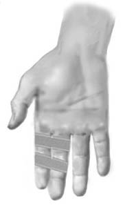
Objective: Stabilise finger joints.

Application: Two anchor strips are applied, one on each side of the affected joint on the finger. Strips are similarly applied to the neighbouring finger. The two fingers are subsequently taped together with strips on the anchors. The healthy finger can in this manner stabilise the injured finger, whilst both fingers can still be bent and stretched.

Treatment(3)

SportNetDoc

Early active mobilisation of volar plate avulsion fractures.

Gaine WJ, Beardsmore J, Fahmy N. Injury 1998 Oct;29(8):589-91.

This is a prospective follow up of 190 consecutive cases of volar plate avulsion fractures. A standard management regimen of immediate, active movement was followed in all cases and physiotherapy was rarely required. Of the 190 patients, 162 were followed up for at least one year. An excellent or good outcome was achieved in 98 per cent. Patients presenting more than three weeks from injury had a worse outcome. The size and displacement of the avulsed fragment did not affect the outcome. For the stable joint, early active mobilisation with minimal or no splintage provides a good result.

Treatment(2)

SportNetDoc

Role of MR imaging in the management of “skier’s thumb” injuries.

Plancher KD, Ho CP, Cofield SS, Viola R, Hawkins RJ. Magn Reson Imaging Clin N Am 1999 Feb;7(1):73-84, viii.

“Skier’s thumb” is an acute rupture of the ulnar collateral ligament (UCL) of the metacarpophalangeal (MCP) joint of the thumb. As the method of choice in evaluating soft tissue injuries, MR imaging is useful in evaluating UCL injuries. This article reviews current concepts regarding the rupture of the UCL, including a study of 34 UCL injuries in which MR imaging was used as the main diagnostic tool. When correlated with surgical findings, MR imaging resulted in identifying UCL tears with 96% sensitivity and 95% specificity.