Kategoriarkiv: Golf elbow

KONDITION

step4

Training ladder for:
GOLF ELBOW
(EPICONDYLITIS MEDIALIS)

STEP 4

KONDITION
Unlimited: Cycling. Swimming. Running.

UDSPÆNDING
(20 min)

Sit at a table with the injured arm hanging over the edge with the palm facing downwards. Use the other arm to apply pressure on the back of the injured hand so that the injured lower arm becomes increasingly stretched on the upper side. Repeat the exercise where the injured hand is alternately pressed from side to side. The injured arm should finally be turned over and the exercise repeated by pressing the underside of the hand so that the injured lower arm becomes increasingly stretched on the under side. The stretching positions should be held for 20 seconds followed by 20 seconds of rest before repeating.

Bend your wrist up as far as possible and press with the opposite hand. Hold the position for 20 seconds. Repeat the exercise pressing the wrist downwards. Hold the position for 20 seconds.

Press your hands together and lift your elbows while holding your arms in front of your chest. Hold the position for 20 seconds and relax for 20 seconds before repeating.

Sit on a table holding your arms in to your body. Turn your hands so that the fingers face backwards and thumbs to the side. Your arms should be outstretched. Slowly lean your upper body backwards so that your forearms become increasingly stretched. Hold the position for 20 seconds and relax for 20 seconds before repeating.

STYRKE
(40 min)

Using an elastic band around the back of the injured hand, move the wrist upwards while keeping the elastic taut.

Sit at a table with the side of the injured hand resting on the table edge. Place the elastic around the thumb, draw it downwards over the back of the hand and hold with the good hand. Slowly rotate the lower arm from side to side to stretch the elastic.

Let the injured hand hang over the edge of a table with the palm facing downwards. Move the hand slowly up and down while holding a weight. Support the injured arm with the good arm.

Lie on your back with support at the elbow. Slowly bend and stretch the elbow while holding a weight.

Squeeze a soft ball.

Put an elastic band around your fingers. Spread your fingers so that the elastic is stretched.

Let the injured hand hang over the edge of a table with the back of the hand facing downwards. Holding a weight, slowly move your hand up and down while supporting the arm with the good hand.

Stretching is carried out in the following way: stretch the muscle group for 3-5 seconds. Relax for 3-5 seconds. The muscle group should subsequently be stretched for 20 seconds. The muscle is allowed to be tender, but must not hurt. Relax for 20 seconds, after which the procedure can be repeated.The time consumed for stretching, coordination and strength training can be altered depending on the training opportunities available and individual requirements.

KONDITION

step3

Training ladder for:
GOLF ELBOW
(EPICONDYLITIS MEDIALIS)

STEP 3

KONDITION
Unlimited: Cycling. Swimming. Running.

UDSPÆNDING
(20 min)

Sit at a table with the injured arm hanging over the edge with the palm facing downwards. Use the other arm to apply pressure on the back of the injured hand so that the injured lower arm becomes increasingly stretched on the upper side. Repeat the exercise where the injured hand is alternately pressed from side to side. The injured arm should finally be turned over and the exercise repeated by pressing the underside of the hand so that the injured lower arm becomes increasingly stretched on the under side. The stretching positions should be held for 20 seconds followed by 20 seconds of rest before repeating.

Bend your wrist up as far as possible and press with the opposite hand. Hold the position for 20 seconds. Repeat the exercise pressing the wrist downwards. Hold the position for 20 seconds.

Press your hands together and lift your elbows while holding your arms in front of your chest. Hold the position for 20 seconds and relax for 20 seconds before repeating.

Sit on a table holding your arms in to your body. Turn your hands so that the fingers face backwards and thumbs to the side. Your arms should be outstretched. Slowly lean your upper body backwards so that your forearms become increasingly stretched. Hold the position for 20 seconds and relax for 20 seconds before repeating.

STYRKE
(40 min)

Using an elastic band around the back of the injured hand, move the wrist upwards while keeping the elastic taut.

Sit at a table with the side of the injured hand resting on the table edge. Place the elastic around the thumb, draw it downwards over the back of the hand and hold with the good hand. Slowly rotate the lower arm from side to side to stretch the elastic.

Let the injured hand hang over the edge of a table with the palm facing downwards. Move the hand slowly up and down while holding a weight. Support the injured arm with the good arm.

Lie on your back with support at the elbow. Slowly bend and stretch the elbow while holding a weight.

Squeeze a soft ball.

Put an elastic band around your fingers. Spread your fingers so that the elastic is stretched.

Stretching is carried out in the following way: stretch the muscle group for 3-5 seconds. Relax for 3-5 seconds. The muscle group should subsequently be stretched for 20 seconds. The muscle is allowed to be tender, but must not hurt. Relax for 20 seconds, after which the procedure can be repeated.The time consumed for stretching, coordination and strength training can be altered depending on the training opportunities available and individual requirements.

KONDITION

step2

Training ladder for:
GOLF ELBOW
(EPICONDYLITIS MEDIALIS)

STEP 2

KONDITION
Unlimited: Cycling. Running.

UDSPÆNDING
(20 min)

Sit at a table with the injured arm hanging over the edge with the palm facing downwards. Use the other arm to apply pressure on the back of the injured hand so that the injured lower arm becomes increasingly stretched on the upper side. Repeat the exercise where the injured hand is alternately pressed from side to side. The injured arm should finally be turned over and the exercise repeated by pressing the underside of the hand so that the injured lower arm becomes increasingly stretched on the under side. The stretching positions should be held for 20 seconds followed by 20 seconds of rest before repeating.

Bend your wrist up as far as possible and press with the opposite hand. Hold the position for 20 seconds. Repeat the exercise pressing the wrist downwards. Hold the position for 20 seconds.

Press your hands together and lift your elbows while holding your arms in front of your chest. Hold the position for 20 seconds and relax for 20 seconds before repeating.

Sit on a table holding your arms in to your body. Turn your hands so that the fingers face backwards and thumbs to the side. Your arms should be outstretched. Slowly lean your upper body backwards so that your forearms become increasingly stretched. Hold the position for 20 seconds and relax for 20 seconds before repeating.

STYRKE
(40 min)

Sit at a table with the injured hand over the edge and the palm facing upwards. Use the good hand to place slight pressure on the injured hand and hold the position for 10 seconds. Rest for 10 seconds before repeating.

Squeeze a soft ball.

Put an elastic band around your fingers. Spread your fingers so that the elastic is stretched.

Curl a tea towel with outstretched arms.

Stretching is carried out in the following way: stretch the muscle group for 3-5 seconds. Relax for 3-5 seconds. The muscle group should subsequently be stretched for 20 seconds. The muscle is allowed to be tender, but must not hurt. Relax for 20 seconds, after which the procedure can be repeated.The time consumed for stretching, coordination and strength training can be altered depending on the training opportunities available and individual requirements.

KONDITION

step1

Training ladder for:
GOLF ELBOW
(EPICONDYLITIS MEDIALIS)

STEP 1

The indications of time after stretching, coordination training and strength training show the division of time for the respective type of training when training for a period of one hour. The time indications are therefore not a definition of the daily training needs, as the daily training is determined on an individual basis.

KONDITION
Unlimited: Cycling. Running.

UDSPÆNDING
(20 min)

Sit at a table with the injured arm hanging over the edge with the palm facing downwards. Use the other arm to apply pressure on the back of the injured hand so that the injured lower arm becomes increasingly stretched on the upper side. Repeat the exercise where the injured hand is alternately pressed from side to side. The injured arm should finally be turned over and the exercise repeated by pressing the underside of the hand so that the injured lower arm becomes increasingly stretched on the under side. The stretching positions should be held for 20 seconds followed by 20 seconds of rest before repeating.

Bend your wrist up as far as possible and press with the opposite hand. Hold the position for 20 seconds. Repeat the exercise pressing the wrist downwards. Hold the position for 20 seconds.

Press your hands together and lift your elbows while holding your arms in front of your chest. Hold the position for 20 seconds and relax for 20 seconds before repeating.

Sit on a table holding your arms in to your body. Turn your hands so that the fingers face backwards and thumbs to the side. Your arms should be outstretched. Slowly lean your upper body backwards so that your forearms become increasingly stretched. Hold the position for 20 seconds and relax for 20 seconds before repeating.

STYRKE
(40 min)

Sit at a table with the injured hand on a ball and roll slowly from side to side.

Sit at a table with the injured hand over the edge with the palm facing upwards. Bend and stretch the wrist.

Sit at a table with the injured hand over the edge with the palm facing downwards. Bend and stretch the wrist.

Sit at a table with the injured hand on a ball and roll slowly backwards and forwards.

Sit at a table with the injured hand over the edge and the palm facing upwards. Use the good hand to place slight pressure on the injured hand and hold the position for 10 seconds. Rest for 10 seconds before repeating.

Stretching is carried out in the following way: stretch the muscle group for 3-5 seconds. Relax for 3-5 seconds. The muscle group should subsequently be stretched for 20 seconds. The muscle is allowed to be tender, but must not hurt. Relax for 20 seconds, after which the procedure can be repeated.The time consumed for stretching, coordination and strength training can be altered depending on the training opportunities available and individual requirements.

tape-instruction

Tapening


Type: TENNIS ELBOW & GOLF ELBOW:

Objective: The load on the tendon fastening can be adjusted with use of tape, so that the maximum load is moved to another part of the tendon fastening allowing the injured part to be relieved.

Application: One or two tape “anchors” are applied around the forearm directly under the inner and outer elbow projection (medial and lateral epicondyle). The tape must not be fixed so tightly so as to cause any discomfort from the forearm or hand (cold sensation, sleeping sensation).

treatment-article2

SportNetDoc

Pragmatic randomised controlled trial of local corticosteroid injection and naproxen for treatment of lateral epicondylitis of elbow in primary care.

Hay EM, Paterson SM, Lewis M, Hosie G, Croft P. BMJ 1999 Oct 9;319(7215):964-8.

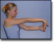
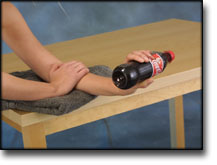
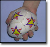
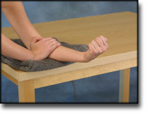
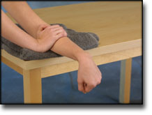
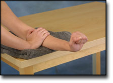
OBJECTIVE: To compare the clinical effectiveness of local corticosteroid injection, standard non-steroidal anti-inflammatory drugs, and simple analgesics for the early treatment of lateral epicondylitis in primary care. DESIGN: Multicentre pragmatic randomised controlled trial. SETTING: 23 general practices in North Staffordshire and South Cheshire. PARTICIPANTS: 164 patients aged 18-70 years presenting with a new episode of lateral epicondylitis. Interventions: Local injection of 20 mg methylprednisolone plus lignocaine, naproxen 500 mg twice daily for two weeks, or placebo tablets. All participants received a standard advice sheet and co-codamol as required. MAIN OUTCOME MEASURES: Participants’ global assessment of improvement (five point scale) at four weeks. Pain, function, and “main complaint” measured on 10 point Likert scales at 4 weeks, 6 months, and 12 months. RESULTS: Over 2 years, 53 subjects were randomised to injection, 53 to naproxen, and 58 to placebo. Prognostic variables were similar between groups at baseline. At 4 weeks, 48 patients (92%) in the injection group were completely better or improved compared with 30 (57%) in the naproxen group (P<0.001) and 28 (50%) in the placebo group (P<0.001). At 12 months, 43 patients (84%) in the injection group had pain scores 0.05). CONCLUSIONS: Early local corticosteroid injection is effective for lateral epicondylitis. Outcome at one year was good in all groups, and effective early treatment does not seem to influence this.

treatment-article1

SportNetDoc

Lateral and Medial Epicondylitis of the Elbow.

Jobe FW, Ciccotti MG. J Am Acad Orthop Surg 1994 Jan;2(1):1-8.

Epicondylitis of the elbow involves pathologic alteration in the musculotendinous origins at the lateral or medial epicondyle. Although commonly referred to as “tennis elbow” when it occurs laterally and “golfer’s elbow” when it occurs medially, the condition may in fact be caused by a variety of sports and occupational activities. The accurate diagnosis of these entities requires a thorough understanding of the anatomic, epidemiologic, and pathophysiologic factors. Nonoperative treatment should be tried first in all patients, beginning with an initial phase of rest, ice, nonsteroidal anti-inflammatory agents, and possibly corticosteroid injection. A second phase includes coordinated rehabilitation, consisting of range-of-motion and strengthening exercises and counterforce bracing, as well as technique enhancement and equipment modification if a sport or occupation is causative. Nonoperative treatment has been deemed highly successful, yet the few prospective reports available suggest that symptoms frequently persist or recur. Operative treatment is indicated for debilitating pain that is diagnosed after the exclusion of other pathologic causes for pain and that persists in spite of a well-managed nonoperative regimen spanning a minimum of 6 months. The surgical technique involves excision of the pathologic portion of the tendon, repair of the resulting defect, and reattachment of the origin to the lateral or medial epicondyle. Surgical treatment results in a high degree of subjective relief, although objective strength deficits may persist.

examination-article

SportNetDoc

Novel use of laser doppler imaging for investigating epicondylitis.

Ferrell WR, Balint PV, Sturrock RD. Rheumatology (Oxford) 2000 Nov;39(11):1214-7.

OBJECTIVE: This investigation evaluated a novel form of tissue perfusion measurement, laser Doppler imaging (LDI), in a case of lateral epicondylitis to establish if it might have applications in assessing soft tissue lesions. LDI was used in conjunction with ultrasonography to provide information about tissue oedema as well as the power Doppler signal as an alternative method of assessing blood flow. METHODS: A laser Doppler imager with a near-infrared (NIR) laser source was used to improve tissue penetration and yield measurements of perfusion (flux) from structures under the skin. Skin temperature over the lateral epicondylar region was also measured. Ultrasonography was used in both grey-scale and power Doppler modes. LDI, temperature measurements and ultrasonographic data were obtained before treatment and serially after local injection of methylprednisolone. RESULTS: Before treatment there was increased perfusion and skin temperature and the presence of a power Doppler sign associated with the right lateral epicondyle as well as oedema at the extensor origin. None of these was present at the asymptomatic contralateral epicondylar region. Twenty-four hours after methylprednisolone administration, both perfusion and skin temperature had increased, and they declined over the subsequent 48 h. Although skin temperature had declined to normal (referenced to the contralateral epicondyle) by the third day after injection, it took until the eleventh day after injection for perfusion to normalize. CONCLUSIONS: LDI using an NIR laser source appears to be an effective non-invasive method for the examination of inflammatory responses in soft tissue, with greater sensitivity than thermally based methods. In addition, LDI was found to correlate with power Doppler ultrasonography.