Kategoriarkiv: Thigh

rehabilitation-article

SportNetDoc

Femoral shaft stress fractures in athletes.

Hershman EB, Lombardo J, Bergfeld JA. Clin Sports Med 1990 Jan;9(1):111-9.

Stress fractures of the femoral shaft in athletes occur most commonly in the proximal third of the femur. They can, however, also be found in the mid- or distal third. Conservative treatment is highly successful in healing these fractures without complications. Athletes can usually return to activity in 8 to 14 weeks. Recognition of the symptoms characteristic of these fractures (vague thigh pain, diffuse tenderness, no trauma) will assist early diagnosis. Early definitive diagnosis can be made by radionuclide scanning or later, by plain radiography, if symptoms have been present for a sufficient period. Diagnosis is not limited to novice runners since runners with significant mileage, or baseball or basketball players, can develop femoral shaft stress fractures.

treatment-article

SportNetDoc

Interventions for preventing and treating stress fractures and stress reactions of bone of the lower limbs in young adults.

Gillespie WJ, Grant I. Cochrane Database Syst Rev 2000;(2):CD000450.

BACKGROUND.
Stress reaction in bone, which may proceed to a fracture, is a significant problem in military recruits and in athletes, particularly long distance runners.

OBJECTIVES.
To evaluate the evidence from controlled trials of treatments and programmes for prevention or management of lower limb stress fractures and stress reactions of bone in active young adults.

SEARCH STRATEGY.
We searched the Cochrane Musculoskeletal Injuries Group Trials Register, The Cochrane Library, MEDLINE, EMBASE, Current Contents, Dissertation Abstracts, Index to UK Theses and the bibliographies of identified articles. Date of last search: December 1997.

SELECTION CRITERIA.
Any randomised or quasi-randomised trial evaluating a programme or treatment to prevent or treat lower limb stress reactions of bone or stress fractures in active young adults.

DATA COLLECTION AND ANALYSIS.
Searching, a decision on inclusion or exclusion, methodological assessment, and data extraction were carried out according to a predetermined protocol included in the body of the review. Analysis using Review Manager software allowed pooling of data and calculation of Peto odds ratios and absolute risk reductions, each with 95% confidence intervals.

MAIN RESULTS.
The use of “shock absorbing” insoles, evaluated in four trials, appears to reduce the incidence of stress fractures and stress reactions of bone (Peto odds ratio 0.47, 95% confidence interval 0. 30 to 0.76). Incomplete data from one trial indicated that reduction of running and jumping intensity may also be effective. The use of pneumatic braces in the rehabilitation of tibial stress fractures significantly reduces the time to recommencing training (weighted mean difference -42.6 days, 95% confidence interval -55.8 to -29.4 days).

REVIEWER’S CONCLUSIONS.
The use of shock absorbing insoles in footwear reduces the incidence of stress fractures in athletes and military personnel. Rehabilitation after tibial stress fracture is aided by the use of pneumatic bracing.

examination-article2

SportNetDoc

Femoral stress fractures.

Boden BP, Speer KP. Clin Sports Med 1997 Apr;16(2):307-17.

Stress fractures are common overuse injuries attributed to the repetitive trauma associated with vigorous weightbearing activities. A high index of suspicion is necessary to diagnose stress fractures of the femur because the symptoms may be vague. The precipitating factors, whether related to training errors or medical conditions, should be thoroughly evaluated. Early diagnosis of distraction femoral neck stress fractures is critical to avoid serious complications. Femoral shaft stress fractures have excellent healing potential when diagnosed early and treated non-operatively. Stress fractures of the femoral condyles are uncommon, but should be included in the differential of knee pain.

examination-article1

SportNetDoc

Stress fractures of the femoral shaft in athletes–more common than expected. A new clinical test.

Johnson AW, Weiss CB Jr, Wheeler DL. Am J Sports Med 1994 Mar-Apr;22(2):248-56.

Athletes from 20 Division I AA collegiate varsity sports and 1 club sport were followed carefully for the development of stress fractures during the 1990 to 1991 and the 1991 to 1992 academic years. During this period, among 914 athletes, 34 stress fractures were sustained. Seven of these, or 20.6%, were of the femoral shaft. This represents a much higher incidence than previously observed in athletes. A new clinical test is described that significantly aids in the early diagnosis and follow-up treatment of femoral shaft stress fractures.

cause-article1

SportNetDoc

Risk factors for stress fractures.

Bennell K, Matheson G, Meeuwisse W, Brukner P. Sports Med 1999 Aug;28(2):91-122.

Preventing stress fractures requires knowledge of the risk factors that predispose to this injury. The aetiology of stress fractures is multifactorial, but methodological limitations and expediency often lead to research study designs that evaluate individual risk factors. Intrinsic risk factors include mechanical factors such as bone density, skeletal alignment and body size and composition, physiological factors such as bone turnover rate, flexibility, and muscular strength and endurance, as well as hormonal and nutritional factors. Extrinsic risk factors include mechanical factors such as surface, footwear and external loading as well as physical training parameters. Psychological traits may also play a role in increasing stress fracture risk. Equally important to these types of analyses of individual risk factors is the integration of information to produce a composite picture of risk. The purpose of this paper is to critically appraise the existing literature by evaluating study design and quality, in order to provide a current synopsis of the known scientific information related to stress fracture risk factors. The literature is not fully complete with well conducted studies on this topic, but a great deal of information has accumulated over the past 20 years. Although stress fractures result from repeated loading, the exact contribution of training factors (volume, intensity, surface) has not been clearly established. From what we do know, menstrual disturbances, caloric restriction, lower bone density, muscle weakness and leg length differences are risk factors for stress fracture. Other time-honoured risk factors such as lower extremity alignment have not been shown to be causative even though anecdotal evidence indicates they are likely to play an important role in stress fracture pathogenesis.

complications-article2

SportNetDoc

Clinical measurement of longitudinal femoral overgrowth following fracture in children.

Nordin S, Ros MD, Faisham WI. Singapore Med J 2001 Dec;42(12):563-5

We have studied residual limb length inequality following femoral shaft fractures in 62 children. From 61.2% of the children who had shortening of more than 1 cm at union, 34.21% still maintained the shortening at the completion of study. The longitudinal femoral overgrowth occurred significantly during the first 18 months of the fracture in 77.4% of the children, with an average of 1.17 cm. Children with proximal-third fractures and those who sustained the fractures before eight years of age have higher capability to correct the limb length disparity.

complications-article1

SportNetDoc

Complications of rigid intramedullary rodding of femoral shaft fractures in children.

Letts M, Jarvis J, Lawton L, Davidson D. J Trauma 2002 Mar;52(3):504-16

BACKGROUND: Intramedullary rodding of femoral shaft fractures has been frequently performed in adults, but until recently rarely in children. It was the purpose of this study to investigate the experience with this treatment method at a pediatric trauma center. METHODS: From 1987 to 1998, 54 children were treated for traumatic femoral fractures with intramedullary rods at a major pediatric trauma center. The average age was 15 years 3 months, ranging between 11 years 4 months and 17 years 11 months. The average follow-up was 5 years 3 months, ranging from 20 months to 10 years 1 month. RESULTS: All of the fractures occurred secondary to trauma and the most common anatomic fracture site was the femoral midshaft. Complications encountered included 8 instances of minor limb length discrepancy, 11 instances of discomfort because of rod prominence, 1 case of avascular necrosis of the femoral head, 2 instances of heterotopic ossification over the rod tip, 1 broken rod, and 3 cases that demonstrated decreased external rotation of the affected limb. One child developed osteomyelitis after intramedullary rodding for a fracture previously treated with external fixation. There were no cases of surgically induced nonunion or malunion and only one delayed union secondary to infection. CONCLUSION: Results of this series demonstrate intramedullary rodding to be an effective treatment modality for femoral fractures in skeletally mature children. In children with open femoral physes, rigid rodding should be avoided because of the small but serious occurrence of avascular necrosis of the femoral head. Intramedullary rodding is not recommended in children initially treated with external fixation because of the increased risk of infection.

treatment-article2

SportNetDoc

Plating of femoral shaft fractures. A review of 15 cases.

Seligson D, Mulier T, Keirsbilck S, Been J.

The objective of this study was to define the role, indications and outcome of plating in femur shaft fractures. All femoral shaft fractures admitted and treated by the authors during a 2-year period were analysed. The authors personally treated a total of 135 femur fractures. Of these 135 fractures, 15 (11%) were treated with primary plating. The femoral fractures were classified as grade I (n = 4), grade III (n = 3), grade IV (n = 4), grade V (n = 3), and grade VII (n = 1) (OTA classification). Three patients sustained open fractures (one grade I and two grade II, Gustilo and Anderson classification). Pelvic (6) or ipsilateral lower extremity injuries (4) occurred in 10 of the 15 patients. A total of 23 body areas were injured, most commonly the chest (n = 10), abdomen (n = 5), head (n = 6) and blood vessels (n = 3). There were no infections reported. Two implant failures were noted. Femur plating is a useful technique in polytrauma patients for specific indications where intramedullary nailing (IMN) may be contra-indicated or technically not feasible. Although the postoperative morbidity (ARDS, death) in our study seems to be lower after plating than after intramedullary nailing, the rate of complications of fracture healing (30%) is significantly greater with femur plating than with intramedullary nailing (12%).

treatment-article1

SportNetDoc

Immediate hip spica casting for femur fractures in pediatric patients. A review of 175 patients.

Infante AF Jr, Albert MC, Jennings WB, Lehner JT. Clin Orthop 2000 Jul;(376):106-12

Immediate closed reduction and application of a well-molded hip spica cast is a safe and effective treatment option for closed, isolated femur fractures in children who weigh between 10 and 100 pounds. Between 1988 and 1996, 190 immediate hip spica casts were placed on children with isolated femoral shaft fractures who weight between 10 and 100 pounds. Fifteen patients were lost to followup leaving 175 children who were evaluated and followed up for at least 2 years after the hip spica cast was removed (range 2-10 years). The femur fractures were reduced closed and placed in a 1 1/2 hip spica cast in the emergency room with the patient under conscious sedation or in the operating room with the patient under general anesthesia. All of the children returned home within 24 hours of the procedure. All 175 femur fractures united within 8 weeks. The only complication was a refracture in a 25 pound child who fell 1 week after the cast was removed. No significant residual angular deformities were present in any of the children at last followup. None of the children required external shoe lifts, epiphysiodesis, antibiotics, irrigation and debridements, or limb lengthening procedures for leg length inequalities. The authors think that immediate closed reduction and placement of a well-molded hip spica cast is a safe and reliable treatment option for isolated, closed femur fractures in children from birth to 10 years of age who weigh less than 80 pounds.

thigh stress fracture

STRESS FRACTURE

Diagnosis: STRESS FRACTURE
(Stress fracture)


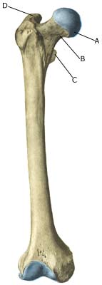
Anatomy:
The femur is the only bone in the thigh. Innumerable muscles are attached to the bone.

  1. Caput femoris
  2. Collum femoris
  3. Trochanter minor
  4. Trochanter major

THIGH BONE FROM THE FRONT

Cause: Repeated loads, particularly when walking or running, can cause such great stress that cracks (stress fractures) appear in the shaft of the femur (article).

Symptoms: Pain and tired sensation in the thigh. The pain is aggravated upon applying pressure (direct and indirect tenderness) and applying load (walking, running).

Examination: X-ray. Since many stress fractures are not visible early in the course, x-ray examination can be repeated after a few weeks, if stress fractures are still suspected. Scintigraphy, CT, MRI and ultrasound scans can often diagnose stress fractures far earlier than x-rays (Ultrasonic image). The frequency of stress fractures in the femur is probably more often than presumed (article). It is crucial for the result of the treatment that the diagnosis is made as early as possible (article).

Treatment: The treatment primarily comprises relief. Only in special cases is surgery necessary (article).

Rehabilitation: The rehabilitation is completely dependant on the type of fracture and the treatment (relief or surgical). A rehabilitation period of 2-4 months must be expected before maximum participation in sports activity can be resumed (article).

Complications: If progress is not smooth, you should be medically re-evaluated to ensure that the fracture is healing according to plan. In some cases a false joint is formed (pseudoarthrosis), which requires surgical treatment.