|
||
|
||
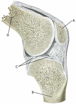
| Cause: An inflammation of the synovial membrane (synovialis) can occur following a twist in the knee joint, which subsequently thickens and produces fluid causing the joint to swell. The injury can occur when, for example, a soccer player strikes the ball with the outermost toe, and thereby twists in the foot and knee.
Symptoms: Swelling of the joint. Pain upon movement of the knee joint. Trouble flexing the knee completely. Acute treatment: Click here. Examination: Swelling of joints always requires medical examination. The diagnosis is usually made following a normal medical examination, where it is not possible to show damage to other structures (ligaments, meniscus). Smaller fluid accumulations in the knee can only be seen with ultrasound (article). Treatment: Relief. If the swelling does not decrease despite relief, the treatment can be supplemented with medicinal treatment in the form of rheumatic medicine (NSAID) or the injection of corticosteroid in the joint, possibly preceded by drainage and evaluation of the joint fluid which can advantageously be performed under ultrasound guidance (article). Complications: If smooth progress is not achieved, it should be considered whether the diagnosis is correct or whether complications have arisen. Amongst others the following should be considered: |