|
||
|
||
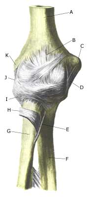
| Cause: With a pull on the arm, the articular head of the one of the forearm bones (caput radii) can become partially dislocated. The condition is most often seen up to the age of about 5 years (article)
Symptoms: Sudden insetting pain around the elbow and pain induced restriction of mobility of the arm (article). Examination: Sudden, strong pain in the arm with restriction of movement should always lead to a medical examination. A General Practitioner will in the vast majority of cases be able to make the diagnosis and treat the condition. X-rays or ultrasound scans are very seldom necessary (article). Treatment: In uncomplicated cases the dislocation can normally be put into place by use of simple manipulation (article). In some circles, a couple of days of relief is subsequently recommended (article). Rehabilitation of children and adolescents: There is not usually a need for specific rehabilitation when the dislocation has been put into place. |