|
||
|
||
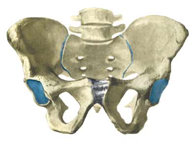
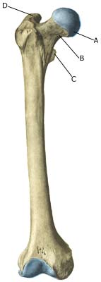
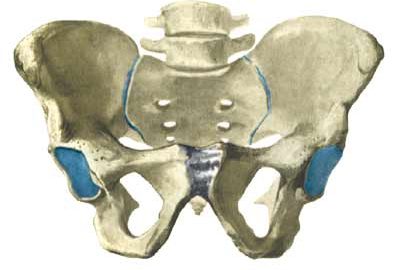
| Cause: Repeated loads, especially when walking or running can cause cracks (stress fractures) in the femoral neck (collum femoris) (article-1) (article-2).
Symptoms: Pain in the hip when applying pressure (direct and indirect tenderness) and when under load (walking, running). Examination: X-ray. Since many stress fractures cannot be seen early in the course of events, X-ray examination can be repeated after a few weeks. Scintigraphy, CT- and MRI and ultrasound scan can often diagnose stress fractures far earlier than X-rays (Ultrasonic image). Treatment: Relief. In some cases surgery is necessary (article). Rehabilitation: The rehabilitation is completely dependent on the type of fracture and treatment (conservative or operative). Complications: If progress is not smooth, you should be re-examined to ensure that the fracture heals according to plan. In some cases a false joint can be formed (pseudoarthrosis), which requires surgical treatment. Special: Shock absorbing shoes or inlays will reduce the load. |