Alle indlæg af urtehave_com

examination-article

SportNetDoc

The staging of osteochondritis dissecans in the knee and ankle joints with MR tomography. A comparison with conventional radiology and arthroscopy.

Bachmann G, Jurgensen I, Siaplaouras J. Rofo Fortschr Geb Rontgenstr Neuen Bildgeb Verfahr 1995 Jul;163(1):38-44

PURPOSE: A prospective study was performed on 50 patients suffering from osteochondritis dissecans of the knee and ankle to define criteria for stability and fixation of osteochondral lesions. METHODS: Morphological parameters in MRI (size, fragmentation, cartilage, interface) and conventional radiology (separation, fragmentation) were registered and compared with arthroscopic staging. MRI staging based on different types of interfaces was demonstrated on T1- and T2-weighted images. RESULTS: MRI could correctly predict a Grade 1 lesion in 50%, a Grade 2 lesion in 90%, a Grade 3 lesion in 0%, and a Grade 4 lesion in 79%. Stable lesions were differentiated from unstable lesions in 90%. Radiographic findings corresponded with arthroscopic staging in only 56% of the cases because fibrotic connection may guarantee stability in cases of bony separation. CONCLUSION: MRI should be performed before therapy to select those patients who do not need surgical therapy or arthroscopy.

treatment-article1

SportNetDoc

Treatment strategies for acute fractures and nonunions of the proximal fifth metatarsal.

Rosenberg GA, Sferra JJ. J Am Acad Orthop Surg 2000 Sep-Oct;8(5):332-8.

There are at least three distinct fracture patterns that occur in the proximal fifth metatarsal: tuberosity avulsion fractures, acute Jones fractures, and diaphyseal stress fractures. Each of these fracture patterns has its own mechanism of injury, location, treatment options, and prognosis regarding delayed union and nonunion. Tuberosity avulsion fractures are the most common in this region of the foot. The majority heal with symptomatic care in a hard-soled shoe. The true Jones fracture is an acute injury involving the fourth-fifth intermetatarsal facet. These injuries are best treated with non-weight-bearing cast immobilization for 6 to 8 weeks. The rate of successful union with this treatment has been reported to be between 72% and 93%. For the high-performance athlete with an acute Jones fracture, early intramedullary-screw fixation is an accepted treatment option. Nonacute diaphyseal stress fractures of the proximal fifth metatarsal and Jones fractures that develop into delayed unions and nonunions can both be managed with operative fixation with either closed axial intramedullary-screw fixation or autogenous corticocancellous grafting. Early results with the use of electrical stimulation are promising; however, prospective studies are needed to better define the role of this modality in managing these injuries.

examination-article2

SportNetDoc

Sensitivity of a clinical examination to predict need for radiography in children with ankle injuries: a prospective study.

Boutis K, Komar L, Jaramillo D, Babyn P, Alman B, Snyder B, Mandl KD, Schuh S. Lancet 2001 Dec 22-29;358(9299):2118-21

BACKGROUND: Radiographs are ordered routinely for children with ankle trauma. We assessed the predictive value of a clinical examination to identify a predefined group of low-risk injuries, management of which would not be affected by absence of a radiograph. We aimed to show that no more than 1% of children with low-risk examinations (signs restricted to the distal fibula) would have high-risk fractures (all fractures except avulsion, buckle, and non-displaced Salter-Harris I and II fractures of the distal fibula), and to compare the potential reduction in radiography in children with low-risk examinations with that obtained by application of the Ottawa ankle rules (OAR). METHODS: Standard clinical examinations and subsequent radiographs were prospectively and independently evaluated in two tertiary-care paediatric emergency departments in North America. Eligible participants were healthy children aged 3-16 years with acute ankle injuries. Sample size, negative and positive predictive values, sensitivity, and specificity were calculated. McNemar’s test was used to compare differences in the potential reduction in radiographs between the low-risk examination and the OAR. FINDINGS: 607 children were enrolled; 581 (95.7%) received follow-up. None of the 381 children with low-risk examinations had a high-risk fracture (negative predictive value 100% [95% CI 99.2-100]; sensitivity 100% [93.3-100]). Radiographs could be omitted in 62.8% of children with low-risk examinations, compared with only 12.0% reduction obtained by application of the OAR (p<0.0001). INTERPRETATION: A low-risk clinical examination in children with ankle injuries identifies 100% of high-risk diagnoses and may result in greater reduction of radiographic referrals than the OAR.

examination-article1

SportNetDoc

Acute paediatric ankle trauma: MRI versus plain radiography.

Lohman M, Kivisaari A, Kallio P, Puntila J, Vehmas T, Kivisaari L. Skeletal Radiol 2001 Sep;30(9):504-11

OBJECTIVE: To evaluate the diagnosis of acute physeal ankle fractures on plain radiographs using MRI as the gold standard. METHODS: Sixty consecutive children, 29 with a clinical diagnosis of lateral ligament injury and 31 with physeal ankle fractures, were examined using both radiographs and MRI in the acute period. The imaging data were reviewed by three “masked” radiologists. The fracture diagnosis and Slater-Harris classification of radiographs were compared with findings on MRI. RESULTS: Plain radiography produced five of 28 (18%) false negative and 12 of 92 (13%) false positive fracture diagnoses compared with MRI. Six of the 12 false positive fractures were due to a misclassification of lateral ligament disruption as SH1 fractures, Altogether a difference was found in 21% of cases in either the diagnosis or the classification of the fractures according to Salter-Harris. All bone bruises in the distal tibia and fibula and 64% of bone bruises in the talus were seen in association with lateral ligament injuries. Talar bone bruises in association with fractures occurred on the same side as the malleolar fracture; talar bone bruises in association with lateral ligament disruption were seen in different locations. The errors identified on radiographs by MRI did not affect the management of the injury. CONCLUSIONS: The incidence of false negative ankle fractures in plain radiographs was small and no complex ankle fractures were missed on radiographs. The total extent of complex fractures was, however, not always obvious on radiographs. In an unselected series of relatively mild ankle injuries, we were unable to show a single case where the treatment or prognosis based on plain radiography should have been significantly altered after having done a routine MRI examination. Plain radiography is still the diagnostic cornerstone of paediatric ankle injuries.

tape-description


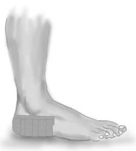
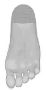
Type: HEEL PAD:

Objective: Gather the heel pad under the heel to improve shock absorption.

Application: Apply a semicircle around the heel (A) – “anchor”. The heel pad is gathered under the heel with several strips of tape (B). No bare skin should be visible. Finish with a further strip on top of the “anchor” A.

Symptoms

SportNetDoc

Black heel a minor hazard of sport.

Wilkinson DS. Cutis 1977 Sep;20(3):393-6.

“Black heel” (calcaneal petechiae) is a traumatic lesion affecting the back or posterolateral aspect of the heel. It is seen almost exclusively in adolescentes or young adults engaged in active sports, notably basketball, but also football, lacrosse, tennis, and so forth. The lesion is disposed horizontally at the upper dege of the calcaneal fat-pad and consists of grouped punctate hemorrhages, the nature of which is revealed by repeated paring of the lesion. The nature of the pigment is shown by specialized stains. “Black heel” is probably more common than is realized. It is likely to be cuased by a shearing or pinching stress from abrupt contact of th foot with a floor or hard ground. As it si usually symptomless, it may be disregarded or only observed by chance. However, it has been confused clinically with a melaonoa, and as it is such a trivial self-healing process, it is important that it be recognized for what it is.

Treatment

SportNetDoc

Metacarpal and phalangeal fractures in athletes.

Capo JT, Hastings H 2nd. Clin Sports Med 1998 Jul;17(3):491-511.

The high demands placed on the upper extremity in sporting activities subject the competitive athlete to common injuries of the hand. Treatment options are based on the fracture configuration, associated extremity injuries, and status of the surrounding soft tissue. Metacarpal and phalangeal fractures may usually be treated by closed, nonoperative methods, and most athletes may quickly return to play with a protective orthosis. Supplemental methods of fixation, such as percutaneous pins and tension-band wires, may be used for unstable fractures. When required, open reduction and internal fixation can provide optimum stability to the fracture, which allows immediate range-of-motion and early return to play.

Treatment

SportNetDoc

Complications of plate fixation in fresh displaced midclavicular fractures.

Bostman O, Manninen M, Pihlajamaki H. J Trauma 1997 Nov;43(5):778-83.

BACKGROUND.
The role of plate fixation in the management of fresh displaced midclavicular fractures is unsettled. The objective of this study was to evaluate the drawbacks and pitfalls of this treatment method.

METHODS.
We analyzed the complications encountered in 103 consecutive adult patients with severely displaced fresh fractures of the middle third of the clavicle who were treated by open reduction and internal fixation using AO/ASIF plates. These 103 patients accounted for 9.5% of the 1,081 patients with fresh midclavicular fractures seen between 1989 and 1995. The mean age of the 103 patients was 33.4 years (range, 19-62 years).

RESULTS.
Seventy-nine patients had an uneventful recovery, whereas 24 (23%) suffered one or several complications. The major complications included deep infection, plate breakage, nonunion, and refracture after plate removal. The most common of the minor complications was plate loosening resulting in malunion. The infection rate was 7.8%. A total of 14 reoperations were performed because of the complications. Permanent nonunion ensued in two patients. A severely comminuted fracture (relative risk of failure, 5.15) as well as a state of alcohol intoxication on admission (relative risk of failure, 3.12) were identified as markers of increased complication risk.

CONCLUSIONS.
Patient noncompliance with the postoperative regimen could be suspected to have been a major cause of the failures. The high complication rate supports a reserved attitude toward plate fixation of fresh midclavicular fractures. The method should be reserved for patients who have trustworthy personal motives for quick pain relief and functional recovery.

Treatment(3)

SportNetDoc

Displaced supracondylar fractures of the humerus in children. Audit changes practice. 

O’Hara LJ, Barlow JW, Clarke NM. J Bone Joint Surg Br 2000 Mar;82(2):204-10

We performed an audit of 71 children with consecutive displaced, extension-type supracondylar fractures of the humerus over a period of 30 months. The fractures were classified according to the Wilkins modification of the Gartland system. There were 29 type IIA, 22 type IIB and 20 type III. We assessed the effectiveness of guidelines proposed after a previous four-year review of 83 supracondylar fractures. These recommended that: 1) an experienced surgeon should be responsible for the initial management; 2) closed or open reduction of type-IIB and type-III fractures must be supplemented by stabilisation with Kirschner (K-) wires; and 3) K-wires of adequate thickness (1.6 mm) must be used in a crossed configuration. The guidelines were followed in 52 of the 71 cases. When they were observed there were no reoperations and no malunion. In 19 children in whom they had not been observed more than one-third required further operation and six had a varus deformity. Failure to institute treatment according to the guidelines led to an unsatisfactory result in 11 patients. When they were followed the result of treatment was much better. We have devised a protocol for the management of these difficult injuries.