Kategoriarkiv: Knee

Jumpers’s knee

JUMPERS KNEE

Diagnosis: JUMPER’S KNEE


Anatomy:
The large anterior thigh muscle (musculus quadriceps femoris) consists of four muscles (m vastus lateralis, m vastus medialis, m vastus intermedius & m rectus femoris). All the muscles fasten on the upper edge of the knee cap. The knee cap ligament (ligamentum patellae) connects the lower edge of the knee cap with the front part of the shin bone (tuberositas tibiae). The function of the knee cap ligament is therefore, when the knee is stretched, to transfer the power the large thigh muscle produces.


  1. M. rectus femoris
  2. M. vastus medialis
  3. Retinaculum patellae mediale
  4. Retinaculum patellae mediale
  5. Tuberositas tibiae
  6. Lig. Patellae
  7. Retinaculum patellae laterale
  8. M. vastus lateralis

KNEE FROM THE FRONT

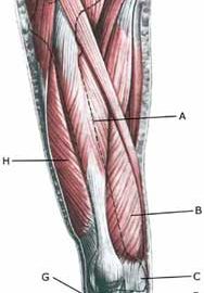
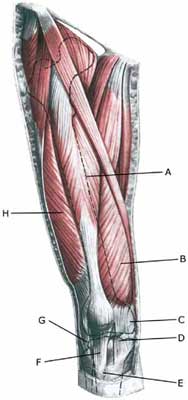
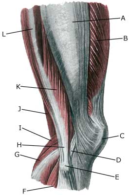
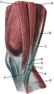
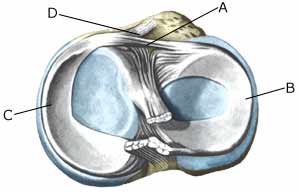
Cause: Repeated uniform loads on the knee cap ligament (jumping, kicking) causes microscopic ruptures at the knee cap ligament fastening on the lower edge of the knee cap. As the load often continues despite tenderness, which in the early stages diminishes after warm-up, a chronic inflammation gradually occurs in the ligament. In some cases, the symptoms will arise at the ligament fastening from the anterior thigh muscle on the upper edge of the knee cap (Photo)

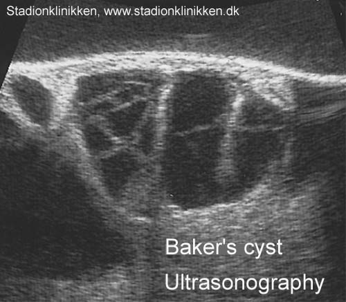
Symptoms: Jumping and similar activity will initially cause tenderness at the lower edge of the knee cap. The pain diminishes during the first weeks/months after warm-up. If the sport activity continues the pain will increase, resulting in the activity eventually becoming impossible.

Acute treatment: Click here.

Examination: Correct treatment from the outset is essential for a good result. A medical examination is consequently recommended as soon as possible after the initial onset of pain under the knee cap. The diagnosis is often made following a normal medical examination. At deterioration or lack of progress, an ultrasound scan should be performed which will be able to reveal a thickening of the ligament, granuloma, scar tissue formation, partial rupture, calcification in the ligament, bursitis and inflammation of the tissue surrounding the ligament (peridenitis) (Ultrasonic image) (article).

Treatment: The treatment consists of relief from the pain inducing activity (jumping, kicking) as soon as possible after the onset of symptoms. The injury can in some cases heal within a few weeks if the treatment is instigated quickly. A rehabilitation period of several months must be anticipated if the pain has been in evidence for some months, and especially if an ultrasound scan has found thickening or changes of the ligament. Emphasis is put on stretching and strength training of the anterior thigh muscle, by activating the muscle simultaneously with stretching (eccentric training). Ice treatment can be repeated every time tenderness is provoked in the knee cap during the rehabilitation period. If there is a lack of progress following relief and strict rehabilitation, medicinal treatment in the form of rheumatic medicine (NSAID) can be considered, or injection of corticosteroid in the area surrounding the thickened part of the ligament. Research has shown that ultrasound guided injection of corticosteroid is extremely effective in reducing the extent of the thickened ligament, allowing more active rehabilitation to begin (Ultrasonic image) (article-1), (article-2). As the injection of corticosteroid is always an element in the long term rehabilitation of a very serious, chronic injury, it is vital that the rehabilitation period lasts over several months in order to reduce the risk of a relapse or rupture. The ligament is naturally unable to accommodate maximum strain or load after a prolonged injury period following only a short rehabilitation period. If the diagnosis is made by use of ultrasound scanning, the injections are performed under guidance of ultrasound, and the rehabilitation is progressed in accordance with the guidelines mentioned, then the treatment involving corticosteroid injections has very few risks connected (article). It is not unusual for a rehabilitation period of six months before maximum strain or load in the form of jumping is permitted. During recent years, different types of experimental treatment have been seen such as sclerosis injection (where injections are performed around the tendon with a drug to destroy the small blood vessels (and nerves) that infiltrate the sick tendons), and shock-wave (ultrasound treatment). However, there is no sure or clear documentation for the effect of these kinds of treatment. If satisfactory progress is not made in the rehabilitation and conservative treatment, surgical intervention can be considered. Long-term results of operations are often disappointing (article).

Bandage: Some patients have the opinion that the application of tape or bandage around the shin bone directly under the knee cap can relieve the discomfort (tape-instruction).

Complications: In case of lack of progress it should be considered whether the diagnosis is correct or whether complications have arisen. Special consideration should be given to:

Special: Shock absorbing shoes or inlays will reduce the load.

Inflammation of the bursa

INFLAMMATION OF THE BURSA

Diagnosis: INFLAMMATION OF THE BURSA
(Bursitis)


Anatomy:
There are numerous bursas around the knee for the purpose of reducing the pressure on the muscles, tendons and ligaments which lie close to bone projections. Bursas can be present at all muscle fastenings around the knee, externally (i.e. bursa pes anserinus, bursa subtendinea m bicipitis), internally, to the front (i.e. bursae prepatellaris, bursa infrapatellaris profunda) and to the rear of the knee (i.e. bursae m semimembranosus, bursa subtendinea m gastrocnemii medialis & lateralis, Baker cyst).

  1. M. gastrocnemius
  2. M. plantaris
  3. M. soleus
  4. Tendo m. gastrocnemii
  5. Tendo calcaneus (Achillis)
  6. M. popliteus
  7. Bursa m. semimembranosi
  8. M. semimembranosus
  9. Bursa subtendinea m. gastrocnemii medialis

KNEE FROM THE REAR

Cause: The bursas can become inflamed, produce fluid, swell and become painful with repeated over-load or due to blows. Although the condition is termed inflammation of the bursa, there is not often an infection in the bursa.

Symptoms: Pain when applying pressure to the bursa, which sometimes, but far from always, can give the impression of being swollen. The pain is aggravated when the muscle above the bursa is activated.

Acute treatment: Click here.

Examination: Medical examination is usually not required in light cases with only minimal tenderness. With more pronounced pain, or lack of improvement, medical examination should always be performed to confirm the diagnosis and commencement of treatment if required. The diagnosis is usually made from a normal medical examination, however, if any doubts arise an ultrasound scan can be performed which is most well suited to confirm the diagnosis.

Treatment: Treatment is primarily concentrated on providing rest. Treatment can be supplemented with rheumatic medicine (NSAID) or injection of corticosteroid in the bursa preceded by draining, which can be best performed under ultrasound guidance (article).

Rehabilitation: Treatment is completely dependent upon which bursa is inflamed, but the sports activity can usually be cautiously resumed when the pain has diminished, especially if the provoking factor has been identified and removed.
Also read rehabilitation, general.

Complications: If there is not a steady improvement in the condition consideration must be given as to whether the diagnosis is correct, or if complications have arisen:

In rare cases, the bursa can be infected with bacteria. This is a serious condition if the bursa becomes red, warm and increasingly swollen and tender. This condition requires immediate examination and treatment. If relief and medicinal treatment (including ultrasound guided injection of corticosteroid) does not produce any progress, a surgical removal of the bursa can be attempted.

Special: Shock absorbing shoes or inlays will reduce the load. If there is a lack of progress or a relapse after successful rehabilitation, consideration must be given to performing a running style analysis to establish whether a correction of the running style should be recommended.

Synovial fluid in the popliteal space

SYNOVIAL FLUID IN THE POPLITEAL SPACE


Diagnosis: SYNOVIAL FLUID IN THE POPLITEAL SPACE
(Baker’s cyst)


Anatomy:
If an excess of fluid is formed in the knee joint, the synovial fluid will be pressed through the weakest point of the rear of the joint-capsule, and accumulate in an outpouching in the hollow (popliteal space) of the knee (Baker’s cyst).

Cause: Injuries in the knee that bring about inflammation of the synovial membrane (synovitis), causing formation of an excess of synovial fluid in the knee. The fluid is pressed out through the joint-capsule and accumulates in the popliteal space (Baker cyst). The Baker cyst is consequently a symptom of something not right in the knee. The connection from the joint to the Baker cyst can in some cases become strangulated, meaning that the Baker cyst can still be present even thought he injury in the knee has healed.

Symptoms: A sensation of the popliteal space being filled up, and difficulty in flexing the knee completely. There is often also discomfort from the changes in the knee that provoked the Baker cyst.

Examination: As the presence of a Baker cyst is usually a symptom of an injury in the knee, anyone with a Baker cyst or discomfort in the knee should undergo a medical examination. It will often prove difficult to diagnose even a large Baker cyst from a normal examination, and the diagnosis is made easiest and swiftest from an ultrasound scan (Ultrasonic image).

Treatment: Treatment is naturally dependant upon the injury in the knee joint that has provoked the Baker cyst. The accumulation in the knee and the Baker cyst can be treated with rheumatic medicine (NSAID) or more effectively by injection of corticosteroid in the knee (or the Baker cyst), preceded by draining of the fluid which can advantageously be performed under ultrasound guidance. Synovial fluid can be drained from the Baker cyst if there is a connection between the knee joint and the Baker cyst, and ultrasound scanning will show the injected corticosteroid spread through the knee joint as well as the Baker cyst (article). The content of the Baker cyst is often quite thick (gelatinous) if the Baker cyst does not communicate with the knee joint.

Rehabilitation: Treatment is completely dependant upon the provoking cause of the Baker cyst.
Also read rehabilitation, general.

Complications: If smooth progress is not achieved, it should be considered whether the diagnosis is correct. Amongst others, the following should be considered:

The bursa can become infected with bacteria in rare cases. This is a serious condition where the bursa becomes red, warm and increasingly swollen and tender. This condition requires immediate medical examination and treatment. If relief and medicinal treatment (including ultrasound guided injection of corticosteroid) does not produce any progress, a surgical removal of the bursa can be attempted.


Fluid accumulation in the joint

FLUID ACCUMULATION IN THE JOINT

Diagnosis: FLUID ACCUMULATION IN THE JOINT
(Traumatic arthritis/synovitis)


Anatomy:
The bones in the knee joint comprise the thigh bone (femur), shin bone (tibia) and the knee cap (patella). The joint cavity is coated with a very thin synovial membrane.

  1. Patella
  2. Tibiae
  3. Meniscus lateralis
  4. Femur

KNEE JOINT

Cause: An inflammation of the synovial membrane (synovialis) can occur following a twist in the knee joint, which subsequently thickens and produces fluid causing the joint to swell. The injury can occur when, for example, a soccer player strikes the ball with the outermost toe, and thereby twists in the foot and knee.

Symptoms: Swelling of the joint. Pain upon movement of the knee joint. Trouble flexing the knee completely.

Acute treatment: Click here.

Examination: Swelling of joints always requires medical examination. The diagnosis is usually made following a normal medical examination, where it is not possible to show damage to other structures (ligaments, meniscus). Smaller fluid accumulations in the knee can only be seen with ultrasound (article).

Treatment: Relief. If the swelling does not decrease despite relief, the treatment can be supplemented with medicinal treatment in the form of rheumatic medicine (NSAID) or the injection of corticosteroid in the joint, possibly preceded by drainage and evaluation of the joint fluid which can advantageously be performed under ultrasound guidance (article).

Complications: If smooth progress is not achieved, it should be considered whether the diagnosis is correct or whether complications have arisen. Amongst others the following should be considered:

Tendinitis

TENDINITIS

Diagnosis: TENDINITIS


Anatomy:
The tendons from the thigh to the shin bone pass the knee joint on the inner and outer sides, as well as to the front and rear of the knee.

  1. Tractus iliotibialis
  2. M. vastus lateralis
  3. Patella
  4. Ligamentum collaterale laterale/fibulare
  5. Caput fibulae
  6. M. soleus
  7. M. gastrocnemius
  8. Tendo m. biciptis femoris
  9. M. plantaris
  10. M. semimembranosus
  11. M. biceps femoris (caput breve)
  12. M. biceps femoris (caput longum)

OUTER KNEE

  1. M. gracilis
  2. M. semimembranosus
  3. M. semitendinosus
  4. Tendo m. semimembranosi
  5. M. gastrocnemius
  6. M. sartorius

MUSCLES AND TENDONS IN THE KNEE REGION
FROM THE MEDIAL POSITION

Cause: Tendinitis occurs as a result of repeated uniform loads causing microscopic ruptures in the tendon, and especially at the tendon fastening, which causes an inflammation. Tendinitis is a warning that the exercise performed is too strenuous for the particular muscle tendons, and if the load is not reduced a rupture can occur (“pulled muscle”) or a chronic inflammation with a substantially longer rehabilitation period as a consequence.

Symptoms: Pain upon applying pressure along the tendon, aggravated when stretching or activation of the muscle tendon.

Acute treatment: Click here.

Examination: Medical examination is not necessarily required in slight cases. Severe cases or cases that are not improved by relief should be medically evaluated to ensure a precise diagnosis. The diagnosis is usually made on the basis of a normal medical examination, however, if any doubts surround the diagnosis an ultrasound scan can be performed as this is the best type of examination to ensure the diagnosis.

Treatment: Relief, stretching and slowly increasing load within the pain threshold. If there is not sufficient progress with relief and regular rehabilitation, medical treatment in the form of rheumatic medicine (NSAID) or the injection of corticosteroid along the inflamed tendon can be considered. As the injection of corticosteroid is always a part of a long term rehabilitation of a chronic injury, it is often necessary for the rehabilitation period to stretch over several weeks to reduce the risk of relapse and ruptures. The tendon is naturally unable to accommodate maximum strain or load after a prolonged injury period after only a short rehabilitation period. If the diagnosis is made by use of ultrasound scanning and the injections are performed under ultrasound guidance, and the rehabilitation is progressed in accordance with the guidelines mentioned, then the treatment involving corticosteroid injections has very few risks connected. In cases of lack of progress with rehabilitation and medicinal treatment, surgery can be considered. However, this is very rarely necessary after a regular and strict rehabilitation period.

The rehabilitation is dependant upon which tendons are involved, but it is usually the tendons in the popliteal space of the knee (tendons contributing to the flexing of the knee).

Complications: If smooth progress is not achieved, it should be considered whether the diagnosis is correct or whether complications have arisen. Amongst others the following should be considered:

Special: Shock absorbing shoes or inlays will reduce the load. In case of lack of progress or recurrence after successful rehabilitation, a running style analysis can be considered to evaluate whether correction of the running style is indicated.

Inflammation of the mucous fold

INFLAMMATION OF THE MUCOUS FOLD

Diagnosis: INFLAMMATION OF THE MUCOUS FOLD
(Plica synovialis)


Anatomy:
The joint cavity in the knee joint is covered with a thin synovial membrane (synovialis). Four mucous folds (plica synovialis) can develop in the membrane: one fold in the inner joint chamber (medial plica), outer joint chamber (lateral plica), upper (suprapatellar) and front (anterior plica). A mucous fold is found in the inner joint chamber (medial plica) in almost 50% of all people (article).

Cause: The mucous fold will become inflamed if an entrapment of the fold occurs, or if the fold suffers internal bleeding. This will result in thickening and subsequently cause pain.

Symptoms: The mucous fold in the inner joint chamber (medial plica) will most often give symptoms which are difficult to distinguish from the symptoms of a meniscus lesion. The pain is usually localised on the inner side of the knee cap, in front of the inner joint line. The pain often occurs quite suddenly following certain movements, and can be accompanied by swelling in the knee. The knee can lock if a flap of the mucous fold becomes entrapped (article).

Examination: A medical examination is always necessary to ensure the diagnosis if there is any suspicion of an inflamed mucous fold in the knee. A tender string inside the knee cap can occasionally give a slipping sensation, but often a normal clinical examination is not sufficient. It is often necessary to perform an arthroscopic examination (telescopic examination of the joint) or MR-scan to make the diagnosis (article).

Treatment: Treatment comprises relief and careful rehabilitation of the knee. If the discomfort does not slowly diminish, the treatment can be supplemented with rheumatic medicine (NSAID) or injection of corticosteroidi in the mucous fold. Medial synovial shelf plica syndrome. Treatment by intraplical steroid injection. If this does not give the desired results, the mucous fold can be removed by arthroscopy (telescopic examination of the knee).

Exerting load on the knee can commence as soon as the pain and swelling in the knee has diminished. In the best cases, full activity is possible after a period of a few weeks. In uncomplicated cases, it should be possible to resume a full level of sports activity during the course of a month. Rehabilitation must not be allowed to cause increased swelling (or pain) in the knee.

Complications: If insufficient progress is made prior to an operation it must be considered if the diagnosis is correct. Supplementary examinations will often be required (X-ray, ultrasound or MR scan). In particular the following should be considered:

Following an arthroscopic examination fluid accumulation in the joint (traumatic arthritis/synovitis), should be considered, as well as infection in the scar or knee joint, which will always require medical attention as soon as possible.

Meniscus lesion

MENISCUS LESION

Diagnosis: MENISCUS LESION


Anatomy:
The bones in the knee joint comprise the thigh bone (femur), shin bone (tibia) and the knee cap (patella). The articular surfaces on the thigh bone, shin bone and the knee cap are covered with a few mm thick cartilage coating which serves to reduce the strain on the articular surfaces. There is a ring-formed cartilage disk (meniscus) internally and externally in the joint. The knee joint is strengthened by a joint capsule which is in turn strengthened on the sides by an outer and an inner collateral ligament (ligamentum collaterale laterale (LCL) and ligamentum collaterale mediale (MCL)). The internal meniscus is attached to the inner collateral ligament, whereas the external meniscus is not attached to the outer collateral ligament.

  1. Ligamentum cruciatum posterius
  2. Ligamentum collaterale mediale/tibiale
  3. Meniscus medialis
  4. Insertio anterior menisci medialis
  5. Ligamentum transversum genus
  6. Tibiae
  7. Fibulae
  8. Ligamentum cruciatum anterius
  9. Ligamentum collaterale laterale/fibulare
  10. Meniscus lateralis
  11. Femur

KNEE JOINT FROM THE FRONT


  1. Ligamentum transversum genus
  2. Meniscus lateralis
  3. Meniscus medialis
  4. Insertio anterior menisci

KNEE JOINT FROM ABOVE

Cause: Meniscus lesions often occur when the knee is stretched at the same time as rotating. The meniscus will consequently be squeezed between the thigh bone and the shin bone. The inner meniscus is more often damaged than the external. Meniscus injuries in children is very rare (article).

Symptoms: Pain along the joint line when applying pressure and when rotating the knee. The pain often occurs suddenly when performing certain movements, and can be accompanied by swelling of the knee. In some instances the knee can lock due to a splinter from the meniscus becoming jammed. In other cases, a small and tender outpouching of fluid can be sensed at the joint line (meniscus cyst). Prolonged discomfort will often cause the thigh muscle to diminish.

Acute treatment: Click here.

Examination: A medical examination is always necessary if there is any suspicion of injury within the knee joint. A normal clinical examination is often not sufficient. The doctor can perform various knee tests (meniscus test), however, there is typically tenderness along the joint line which is aggravated when rotating the knee joint. It is often necessary to perform an arthroscopy (telescopic examination of the joint), MR-scan or ultrasound scan to make the diagnosis (article) (Ultrasonic image).

Treatment: In uncertain cases the treatment comprises relief and rehabilitation of the knee. If the discomfort does not slowly diminish, if there are instances of “locking” or if it is clear that the meniscus lesion is large (e.g. assessed from an ultrasound scan), surgery should be considered. Under an operation, it can be attempted to sew the torn piece of meniscus back (which will require a considerably longer rehabilitation period, but will probably in the long term reduce the risk of degenerative arthritis in the knee) (article). The torn piece of meniscus is usually removed, but in rare cases the whole meniscus is taken out.

Following an operation, exerting load on the knee can commence as soon as the pain and swelling in the knee has diminished. In the best cases, full activity is possible after a period of a few weeks. In uncomplicated cases, it should be possible to resume a full level of sports activity during the course of a month. Rehabilitation must not be allowed to cause increased swelling (or pain) in the knee.

Complications: If insufficient progress is made prior to an operation it must be considered if the diagnosis is correct. Supplementary examinations will often be required (X-ray, ultrasound or MR scan). In particular the following should be considered:

Following an arthroscopy, fluid accumulation in the joint (traumatic arthritis/synovitis) should be considered, and infection in the scar or knee joint, which will always require medical attention as soon as possible. Special: Since there is a risk that the injury can cause permanent disability, the injury should be reported to your insurance company.

Rupture of the posterior cruciate ligament

RUPTURE OF THE POSTERIOR CRUCIATE LIGAMENT

Diagnosis: RUPTURE OF THE POSTERIOR CRUCIATE LIGAMENT
(Ruptura ligamentum cruciatum posterius)


Anatomy:
The bones in the knee joint comprise the thigh bone (femur), shin bone (tibia) and the knee cap (patella). There is furthermore a small joint between the shin bone and the calf bone (fibula). The knee joint is strengthened by a joint capsule which is in turn strengthened on the sides by an outer and an inner collateral ligament (ligamentum collaterale laterale (LCL) and ligamentum collaterale mediale (MCL)). Inside the knee there are two ligaments, the anterior and posterior cruciate ligaments (ligamentum cruciatum anterius and ligamentum cruciatum posterius).

  1. Ligamentum cruciatum posterius
  2. Ligamentum collaterale mediale/tibiale
  3. Meniscus medialis
  4. Insertio anterior
    menisci medialis
  5. Ligamentum transversum genus
  6. Tibiae
  7. Fibulae
  8. Ligamentum cruciatum anterius
  9. Ligamentum collaterale laterale/fibulare
  10. Meniscus lateralis
  11. Femur

KNEE JOINT FROM THE FRONT

Cause: Rupture of the posterior cruciate ligament usually occurs following a blow or kick direct on the front of the shin bone just below the knee (Photo).

Symptoms: Usually a snap can be heard or felt and continued sports activity must be aborted. The knee can swell within the first few hours, after which the knee can not bend completely. You can subsequently often sense that the leg gives way (knee failure).

Acute treatment: Click here.

Examination: If a partial or complete rupture of the cruciate ligament is suspected, you should seek medical attention (casualty ward) immediately, to obtain a diagnosis. The doctor can perform various tests on the knee (rear drawer looseness) to examine the stability of the knee. It should be noted that the looseness in the knee can often only be demonstrated after two weeks. It is often necessary to supplement the examination with a MR-scan, ultrasound scan (Ultrasonic image) (article), or arthroscopy to make the diagnosis with certainty.

Treatment: Treatment of a rupture of the posterior cruciate ligament usually comprises relief and rehabilitation. It is only in cases of pronounced looseness, or if the rupture is combined with other ligament ruptures, that surgery is recommended (article).

An intensive rehabilitation period of at least six months is to be expected.

Bandage: Hinge bandages (Don-Joy) can be utilised the first few weeks. Tape treatment of cruciate ligament ruptures in the knee has no sure effect.

Complications: If smooth progress is not achieved, it should be considered whether the diagnosis is correct or whether complications have arisen. Amongst others the following should be considered:

Special: Since there is a risk that the injury can cause permanent disability, the injury should be reported to your insurance company.

Rupture of the anterior cruciate ligament

RUPTURE OF THE ANTERIOR CRUCIATE LIGAMENT

Diagnosis: RUPTURE OF THE ANTERIOR CRUCIATE LIGAMENT
(Ruptura ligamentum cruciatum anterius)


Anatomy:
The bones in the knee joint comprise the thigh bone (femur), shin bone (tibia) and the knee cap (patella). There is furthermore a small joint between the shin bone and the calf bone (fibula). The knee joint is strengthened by a joint capsule which is in turn strengthened on the sides by an outer and an inner collateral ligament (ligamentum collaterale laterale (LCL) and ligamentum collaterale mediale (MCL)). Inside the knee there are two ligaments, the anterior and posterior cruciate ligaments (ligamentum cruciatum anterius and ligamentum cruciatum posterius).

  1. Ligamentum cruciatum posterius
  2. Ligamentum collaterale mediale/tibiale
  3. Meniscus medialis
  4. Insertio anterior menisci medialis
  5. Ligamentum transversum genus
  6. Tibiae
  7. Fibulae
  8. Ligamentum cruciatum anterius
  9. Ligamentum collaterale laterale/fibulare
  10. Meniscus lateralis
  11. Femur

KNEE JOINT FROM THE FRONT

Cause: Rupture of the anterior cruciate ligament usually occurs during running with fast changes of direction or as the result of a fall while skiing. Most injuries occur without contact with an opponent.

Symptoms: Usually a snap can be heard or felt and continued sports activity must be aborted. The knee swells within the first few hours, after which the knee can not bend completely. You can subsequently often sense that the leg gives way (knee failure).

Acute treatment: Click here.

Examination: If a partial or complete rupture of the cruciate ligament is suspected, you should seek medical attention (casualty ward) immediately, to obtain a diagnosis. The doctor can perform various tests on the knee (front drawer looseness, Lachmann, Pivot shift) to examine the stability of the knee. It should be noted that the looseness in the knee can often only be demonstrated after two weeks. The fluid in the knee can be drained. Blood in the knee provides a very strong suspicion of a rupture of the anterior cruciate ligament. The bleeding that occurs after a rupture of the anterior cruciate ligament can usually be seen in an ultrasound scan (Ultrasonic image) (article) or MR-scan. Arthroscopy (a telescopic examination of the knee) is the best suited examination if there is any doubt in the diagnosis.

Treatment: Young athletes or others with physically demanding work will usually be advised surgery with the insertion of a new cruciate ligament (article).

An intensive rehabilitation period of at least six months is to be expected. It is important that the knee is stretched completely at least twice a day. The surgeon should be consulted if problems occur with stretching the knee completely.

Bandage: Hinge bandages (Don-Joy) can be utilised the first few weeks. Tape treatment of cruciate ligament ruptures in the knee has no sure effect.

Complications: In case of lack of progress you should consider various complications to the surgery:

In cases where the knee cap tendon is used as a new cruciate ligament an inflammation, is often seen where the knee cap tendon has been removed. With prolonged discomfort which does not diminish following relief, medical treatment in form of rheumatic medicine (NSAID) or injection of corticosteroid along the inflamed part of the knee cap tendon can be attempted (article).
Less than half the athletes suffering a rupture of the anterior cruciate ligament are able to resume sports activity on the same level as prior to the injury (article).

Special: Since there is a risk that the injury can cause permanent disability, the injury should be reported to your insurance company.

Outer collateral ligament rupture

OUTER COLLATERAL LIGAMENT RUPTURE

Diagnosis: OUTER COLLATERAL LIGAMENT RUPTURE
(Ruptura ligamentum collaterale laterale/fibulare)


Anatomy:
The bones in the knee joint comprise the thigh bone (femur), shin bone (tibia) and the kneecap (patella). There is furthermore a small joint between the shin bone and the calf bone (fibula). The knee joint is strengthened by a joint capsule which is in turn strengthened on the sides by an outer and an inner collateral ligament (ligamentum collaterale laterale (LCL) and ligamentum collaterale mediale (MCL)). Inside the knee there are two ligaments, the anterior and posterior cruciate ligaments (ligamentum cruciatum anterius and ligamentum cruciatum posterius).


  1. Ligamentum cruciatum posterius
  2. Ligamentum collaterale mediale/tibiale
  3. Meniscus medialis
  4. Insertio anterior menisci medialis
  5. Ligamentum transversum genus
  6. Tibiae
  7. Fibulae
  8. Ligamentum cruciatum anterius
  9. Ligamentum collaterale laterale/fibulare
  10. Meniscus lateralis
  11. Femur

KNEE FROM THE FRONT


  1. M. gastrocnemius
  2. Caput fibulae
  3. B. subtendinea m. bicipitis femoris inferior
  4. M. biceps femoris
  5. Ligamentum collaterale laterale/fibulare
  6. M. plantaris

OUTER LOWER LEG

Cause: When the shin bone is pressed inwards in relation to the thigh bone the outer side-ligament is stretched. If the load becomes sufficiently powerful (as is the case when the load is sudden, so that the thigh muscles do not have time to tighten and stabilize the joint) the ligament can rupture.

Symptoms: Sudden pain on the outside of the knee. Occasionally a snap is felt when the ligament ruptures. In severe cases the athlete complains about a sense of looseness in the knee.

Acute treatment: Click here.

Examination: If a total or partial rupture of the ligaments in the knee is suspected, you should seek medical attention for a diagnosis. The doctor can perform various knee tests to examine the stability of the knee (external side-instability-test) . If the knee is stable the injury is called a “sprain” of the outer side-ligament. If the knee is loose the injury is called a “rupture” of the ligament. The diagnosis is usually made from a normal medical examination. If there are any doubts surrounding the diagnosis, an ultrasound scan can be performed which will reveal the rupture and the bleeding along the ligaments (Ultrasonic image).

Treatment: The treatment of a partial or total rupture of the outer side-ligament involves relief and rehabilitation. If the knee is markedly loose, you can, for a short period, use a support splint (Don Joy). With pronounced sideways looseness surgery can be considered (article). (However if the rupture of the outer side-ligament is combined with other ligament ruptures, as is often the case, surgery is recommended).

Bandage: Hinge bandages can be used initially (Don Joy). Tape treatment of ligament ruptures in the knee have no sure effect.

Complications: If smooth progress is not achieved, it should be considered whether the diagnosis is correct or whether complications have arisen. Supplementary examinations (X-ray, ultrasound, MR scan or arthroscopy) will often be required. The following should be considered:

An inflammation by the ligament fastening is sometimes seen later in the course of the injury, and in some cases bursitis at the ligament fastening is seen. The inflammation and bursitis can possibly be treated with medication (rheumatic medicine (NSAID) or the injection of corticosteroid), if further relief has no effect (article).

Special: Since there is a risk that the injury can cause permanent disability, the injury should be reported to your insurance company.