Kategoriarkiv: Adults injuries

Inflammation of the bursa at the attachment of the Achilles to the heel bone

INFLAMMATION OF THE BURSA AT THE ATTACHMENT OF THE ACHILLES TO THE HEEL BONE

Diagnosis: INFLAMMATION OF THE BURSA AT THE ATTACHMENT OF THE ACHILLES
TO THE HEEL BONE

(BURSITIS ACHILLES)


Anatomy:
The calf muscle (M Gastrocnemicus) is comprised of two muscle heads which gather in a wide tendinous ligament and continue in to the Achilles tendon. Another of the larger calf muscles (M Soleus) is attached to the front side of the Achilles tendon and thus forms a part of the Achilles tendon. The Achilles is attached to the heel bone (calcaneus). At the heel bone there is a bursa in front of the Achilles anchor point (bursae tendinis Achilles), as well as behind (bursae subcutanea calcanei). The bursa reduce the pressure against the heel bone.

Cause: Inflammation of the bursa in front of and behind the Achilles occurs with continued overload, where the bursa is squeezed against the heel bone (for example an ill-fitting heel cap on the shoe).

Symptoms: Pain when activating the Achilles tendon (running and jumping) and when applying pressure at the point of attachment of the tendon on the heel bone. Contrary to the tenderness occurring with inflammation of the Achilles tendon, the tenderness is localised to the point of attachment to the heel bone.

Acute treatment: Click here.

Examination: Medical examination is not necessarily required in light cases where the tenderness is minimal. In all cases where smooth improvement is not experienced, medical attention should be sought as soon as possible to exclude a (partial) rupture of the Achilles tendon or rupture of the soleus muscle. This situation is best determined by use of ultrasound scanning, as a number of injuries requiring treatment can easily be overlooked during a clinical examination (Ultrasonic image). Ultrasound scanning enables an evaluation of the extent of the change in the tendon; inflammation of the tendon (tendinitis), development of cicatricial tissue (tendinosis), calcification, inflammation of the tissue surrounding the tendon (peritendinitis), inflammation of the bursa (bursitis), as well as (partial) rupture (article).

Treatment: Treatment is primarily comprised of relief from the painful activity (running). It is important that shoes do not pinch the heel. If satisfactory progress is not made during the rehabilitation, medical treatment can be considered in the form of rheumatic medicine (NSAID) or injection of corticosteroid in the bursa. Injections should be performed under ultrasound guidance to ensure optimal effect and reduce the risk of injecting into the Achilles itself. If progress is not made neither through rehabilitation nor medicinal treatment, surgical treatment can be attempted (article-1)(article-2).

Bandage: In some cases a ring of felt (for example) can be taped around the tender bursa which will reduce the pressure from shoes. It is naturally important that the hole in the ring is positioned directly above the bursa.

Complications: If there is not a steady improvement in the condition an ultrasound scan should be performed to exclude:

Rupture of the Achilles tendon

Diagnosis: RUPTURE OF THE ACHILLES TENDON
(Ruptura traumatica tendinis achilles)


Anatomy:
The calf muscle (M Gastrocnemicus) is comprised of two muscle heads which gather in a wide tendinous ligament and continue in to the Achilles tendon. Another of the larger calf muscles (M Soleus) is attached to the front side of the Achilles tendon and thus forms a part of the Achilles tendon. The Achilles is attached to the heel bone (calcaneus). The weakest area of the Achilles tendon is located approx. 3 cm. above the point of attachment to the heel bone.


  1. M. soleus
  2. Tuber calcanei
  3. Tendo calcaneus (Achillis)
  4. M. gastrocnemius

LOWER LEG FROM THE REAR

Cause: Full or partial rupture of the Achilles tendon most usually occurs when the Achilles is stretched simultaneously with the calf muscle contracting (eccentric contraction), which happens for example when a badminton player lands after a smash and at the same time starts out to reach the net quickly. Full or partial rupture of the Achilles always occurs with the background of degeneration in the tendon. Under half the cases have experienced symptoms prior to the rupture, however, as good as all the tendons show signs of degeneration during the subsequent examination.

Symptoms: Sudden pain in the Achilles tendon, where there is often a sensation of feeling and hearing a “crack”. Many believe that they have been kicked from behind. The pain is aggravated when activating the Achilles (walking), pressure on the tendon and when stretching the tendon. It is often possible to feel a defect in the tendon, and it is usually impossible to walk on the toes.

Acute treatment: Click here.

Examination: In all cases when there is a sense of a “crack”, or sudden shooting pains in the Achilles tendon, medical attention should be sought as soon as possible. Ultrasound scanning is used to advantage when making the diagnosis, as even full ruptures can easily be overlooked without the aid of ultrasound scanning. Ultrasound scanning enables an evaluation of the extent of the change in the tendon; full or partial rupture, inflammation of the tendon (tendinitis), development of cicatricial tissue (tendinosis), clacification, inflammation of the tissue surrounding the tendon (peritendinitis) and inflammation of the bursa (bursitis) (article), (Ultrasonic image).

Treatment: Ruptures can be treated with bandaging or surgical intervention. Operation is usually recommended for athletes and others with physical work. A period of 9-12 months must in all cases be expected to elapse before the sports activity can be resumed on the same level. Shoes with a slight heel elevation will relieve the pressure on the Achilles tendon. Treatment with ice for a period of at least 20 minutes after straining the tendon is recommended as long as the tendon remains sore (article-1), (article-2), (article-3).

Bandage: Taping to relieve problems with the Achilles tendon is of questionable significance, but can be attempted as the tape will not invoke further injury if applied in the correct manner (tape-instruction).

Complications: If there is not a steady improvement in the condition an ultrasound scan should be performed to exclude a renewed rupture of the Achilles tendon or:

It is unfortunately often the case that it is not possible to return to the same level of sports activity despite correctly administered treatment and training.

Special: As there is a risk that the injury can be permanent, all cases should be reported to your insurance company. It is important that running shoes fit well (tight heel cap, slight heel elevation, shock absorbing soles).

Inflammation of Achilles tendon

INFLAMMATION OF THE ACHILLES TENDON

Diagnosis: INFLAMMATION OF THE ACHILLES TENDON
(TENDINITIS ACHILLES)


Anatomy:
The calf muscle (M Gastrocnemicus) is comprised of two muscle heads which gather in a wide tendinous ligament and continue in to the Achilles tendon. Another of the larger calf muscles (M Soleus) is attached to the front side of the Achilles tendon and thus forms a part of the Achilles tendon. The Achilles is attached to the heel bone (calcaneus). The weakest area of the Achilles tendon is found approx. 3 cm. above the point of attachment on the heel bone.


  1. M. soleus
  2. Tuber calcanei
  3. Tendo calcaneus (Achillis)
  4. M. gastrocnemius

LOWER LEG FROM THE REAR

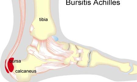
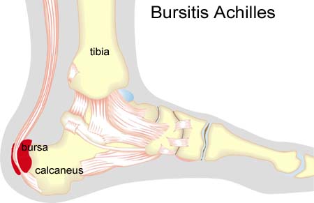
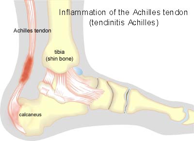
Cause: Inflammation occurs with continued overload in the form of running and jumping. The risk of over-load injuries of the Achilles tendon increases with age.

Symptoms: Pain when activating the Achilles tendon (running and jumping), when applying pressure and with stretching of the tendon. The tendon often feels thickened.

Acute treatment: Click here.

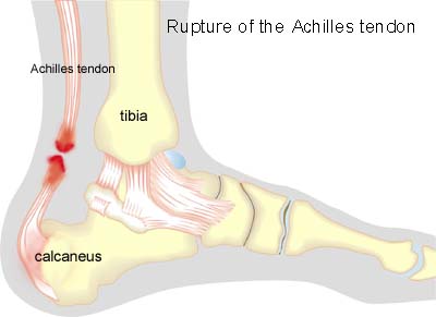
Examination: Medical examination is not necessarily required in slight, early cases where the tenderness is slowly increasing without sudden worsening. In cases when there is a sense of a “crack”, or sudden shooting pains in the Achilles tendon, medical attention should be sought as soon as possible to exclude a (partial) rupture of the Achilles tendon or rupture of the soleus muscle. This situation is best determined by use of ultrasound scanning, as a number of injuries requiring treatment can easily be overlooked during a normal clinical examination. In all cases where satisfactory progress is not in evidence, an ultrasound examination should be performed as early as possible. Ultrasound scanning enables an evaluation of the extent of the change in the tendon; inflammation of the tendon (tendinitis), development of cicatricial tissue (tendinosis), clacification, inflammation of the tissue surrounding the tendon (peritendinitis), inflammation of the bursa (bursitis), as well as (partial) rupture (article) (Ultrasonic image).

Treatment: Treatment is primarily comprised of relief from the painful activity (running). If the treatment is commenced early, the injury can in some cases heal within a few weeks. If the pain has been experienced for several months, and if ultrasound scanning has revealed thickening of, and changes in the tendon, a rehabilitation period of several months must be anticipated. Special emphasis is put on fitness training where the tendon is activated simultaneously with stretching (eccentric training) (article). It is naturally crucial that footwear is in good condition (good running shoes with shock absorbing heel and close fitting heel cap). Pressure on the Achilles tendon can be relieved by using shoes with an elevated heel, whilst a heel cushion in the shoe is of less significance since the heightening achieved by this method is greatly limited. If experiencing tenderness in the Achilles tendon during the rehabilitation period, treatment with ice for a period of at least 20 minutes is recommended. If the rehabilitation does not progress satisfactorily, medicinal treatment in the form of rheumatic medicine (NSAID) can be considered, or corticosteroid injection in the area surrounding the thickened part of the tendon. It is vital for safety that injections in the treatment of chronic Achilles tendon conditions are performed under ultrasound guidance. Studies have shown that ultrasound -guided injections of corticosteroid are extremely effective in reducing the extent of the thickened tendons, to enable more active rehabilitation to take place (article). As the injection of corticosteroid is always an element in the long term rehabilitation of a very serious, chronic injury, it is vital that the rehabilitation period lasts over several months in order to reduce the risk of a relapse or (further) rupture. The tendon is naturally unable to accommodate maximum strain or load after a prolonged injury period after only a short rehabilitation period. If the diagnosis is made by use of ultrasound scanning, the injections are performed under guidance of ultrasound, and the rehabilitation is progressed in accordance with the guidelines mentioned, then the treatment involving corticosteroid injections has very few risks connected (article). It is not unusual for a rehabilitation period of six months before maximum strain or load in the form of jumping is permitted. During recent years, different types of experimental treatment have been seen such as sclerosis injection (where injections are performed around the tendon with a drug to destroy the small blood vessels (and nerves) that infiltrate the sick tendons), and shock-wave (ultrasound treatment). However, there is no sure or clear documentation for the effect of these kinds of treatment. If satisfactory progress is not made in the rehabilitation and medicinal treatment, surgical intervention can be considered. Long-term results of operations are often disappointing (article-1), (article-2).

Bandage: Taping to relieve problems with the Achilles tendon is of questionable significance, but can be attempted as the tape will not invoke further injury if applied in the correct manner (tape-instruction).

Complications: If there is not a steady improvement in the condition an ultrasound scan should be performed to exclude:

Few sports injuries carry as large a risk of chronic, permanent sporting disability as inflammation of the Achilles tendon. In the severe, chronic cases, all treatment and rehabilitation attempts will often result in permanent inability to continue the sports activity.

Special: As inflammation of the Achilles tendon is extremely difficult to treat, it is important to prevent the injury from arising or recurring. The principles in (rehabilitation, general) should be followed to ensure that quickly increasing training loads at the season start, or after an injury period, are avoided. It is important that running shoes fit well (tight heel cap, shock absorbing soles). Ultrasound scanning of symptom-free athletes has shown changes in the tendon which increases the risk of developing symptoms during the next year. Preventive fitness and agility training (“Rehabilitation, specific”) can commence prior to the injury giving symptoms. It is therefore recommended that elite athletes over the age of 25 with high-load activities (running, jumping) have an ultrasound examination of the Achilles tendon once a year (article). In the event of unsatisfactory progress, or relapse after successful rehabilitation, consideration must be given to performing an analysis of the patient’s running style to establish whether a correction of the running style should be recommended.

Disclocation of the elbow

DISCLOCATION OF THE ELBOW

Diagnosis: DISCLOCATION OF THE ELBOW
(LUXATIO ARTICULI CIBITI)


Anatomy:
The elbow joint is comprised of the upper arm bone (humerus), and one of the two forearm bones (ulna). The other forearm bone (radius) forms a joint with ulna (art. Radioulnaris proximalis). A joint capsule and several strengthening ligaments surround the elbow joint.

  1. Humerus
  2. Capsula articularis
  3. Epicondylus medialis
  4. Lig. collaterale ulnare
  5. Chorda obliqua
  6. Ulna
  7. Radius
  8. Tendo m. bicipitis brachii
  9. Lig. anulare radii
  10. Lig. collaterale radiale
  11. Epicondylus lateralis

Elbow joint

Cause: A dislocation of the elbow can occur following a direct fall on an outstretched arm. The dislocation can in some cases be complicated by a bone fracture, vascular damage or nerve damage.

Symptoms: Sudden insetting pain around the elbow, with pain-conditional restriction of mobility of the arm following a sudden, violent load (fall).

Acute treatment:
Click here.

Examination: Sudden, strong pain in the arm with restriction of movement following a fall should always lead to acute medical examination. Acute medical assistance should be sought due to the risk of damage to blood vessels and nerves. An X-ray examination will usually reveal the dislocation and rule out bone fracture.

Treatment: The dislocation can usually, in uncomplicated cases, be put in place without the need of surgery. Some recommend a short time where bandaging is used after the dislocation has been put into place. Surgery is often necessary in cases where complications arise from the dislocation in the form of bone fracture, vascular damage or nerve damage. Rehabilitation with exercises involving movement should be commenced as soon as possible, (article-1)(article-2).

Rehabilitation can commence shortly after the dislocation is put into place (and possible bandaging has been removed) in uncomplicated cases without bone fracture, vascular damage or nerve damage. Recommendations from your doctor must be taken into consideration in the rehabilitation program if the dislocation has involved complications and has possibly required surgery.
Also read rehabilitation, general.

Complications: Tears or ruptures around the elbow will in the vast majority of cases heal without complication. Some cases will experience persistent stiffness in the elbow, looseness of the elbow, calcification in the muscles surrounding the elbow and vascular or nerve damege. Dislocation of the elbow can in some cases be complicated by ligament injuries in the wrist, (article).

Fracture of the humeral shaft at the elbow

FRACTURE OF THE HUMERAL SHAFT AT THE ELBOW

Diagnosis: FRACTURE OF THE HUMERAL SHAFT AT THE ELBOW
(fractura supracondylaris humeri)


Anatomy:
The upper arm (humerus) consists of the capitulum, the neck (collum), the long tubular bone (corpus) and the articulated part in the elbow (epicondylus).

  1. Caput humeri
  2. Collum chirurgicum
  3. Epicondylus medialis
  4. Epicondylus lateralis
  5. Tuberculum minus
  6. Sulcus intertubercularis
  7. Tuberculum majus
  8. Collum anatomicum

UPPER ARM FROM THE FRONT

Cause: A fracture of the humerus can occur in cases of a direct fall on the outstretched arm. The fracture may occur anywhere on the humerus, but a fracture through the neck of the humerus (collum) and the middle of the long tubular bone (corpus) are the most common locations. However, in children especially a fracture of the upper arm just above the elbow is often seen (fractura supracondylaris humeri), which in rare cases can cause entrapment of blood vessels and nerves.

Symptoms: Sudden pain in the elbow region and pain induced constriction of movement of the arm after a fall.

Acute treatment: Click here.

Examination: Sudden, powerful pains in the arm with constriction of movement after a fall, should always lead to acute medical examination due to the risk of damage to the blood vessels and nerves. The fracture is usually visible on x-rays, and on the basis of the type of fracture, the correct treatment can be determined.

Treatment: The fracture can normally be set in place under an aenesthetic, followed by bandaging for a few weeks. Surgical fixation may be required for certain types of fractures.

Rehabilitation: When pain has decreased and the bandage has been removed fitness training in the form of cycling and running may be started along with retraining as specified under  rehabilitation, general. Children will often be able to resume sports activity within 2-3 months.

Complications: In the vast majority of cases the fracture heals without complications although in some instances a poor healing occurs affecting the blood and nerve supply to the arm, or development of muscle acute compartment syndrome (article).

Inflammation of the bursa

INFLAMMATION OF THE BURSA

Diagnosis: INFLAMMATION OF THE BURSA
(BURSITIS)


Anatomy:
There are numerous bursas surrounding the elbow, with the purpose of reducing the pressure on muscles and tendons, where these lie close to the bone.

Cause: In cases of repeated loads or blows the bursas can become inflamed, produce fluid, swell and become painful. One of the most frequent bursitis forms in the elbow region is inflammation of the bursa located between the biceps tendon and the fastening on the radius (tuberositas radii), (article).

Symptoms: Pain upon applying pressure on the bursa, which sometimes (but far from always) may feel swollen. Aggravated upon activation of the muscle located immediately above the bursa.

Acute treatment: Click here.

Examination: In light cases with only minimal tenderness, medical examination is not necessarily required. In cases of more pronounced pain or lack of progress, a medical examination should be carried out to ensure a correct diagnosis and treatment. The diagnosis is made most easily and quickly with an ultrasound scan.

Treatment: The treatment primarily consists of relief with removal of the provoking cause if known. The treatment can be supplemented with rheumatic medicine (NSAID) or the injection of corticosteroid in the bursa preceded by draining of the bursa, which can advantageously be performed under ultrasound guidance.

Rehabilitation: The treatment is completely dependant on which bursa is inflamed, but sports activity can usually be cautiously resumed once pain has decreased, particularly if it has been possible to remove the provoking cause.
See also rehabilitation, general.

Complications: If progress is not smooth, it should be considered if the diagnosis is correct or whether complications have arisen: In rare cases the bursa can become infected with bacteria. This is a serious condition where the bursa becomes red, warm and increasingly swollen and tender, and requires immediate medical examination and treatment. 
If there is no progress with relief, medical treatment rheumatic medicine (NSAID) and the ultrasound guided injection of corticosteroid
surgical removal of the bursa may be attempted.

Inflammation of the bursa at the elbow

INFLAMMATION OF THE BURSA AT THE ELBOW

Diagnosis: INFLAMMATION OF THE BURSA AT THE ELBOW
(BURSITIS OLECRANEI)


Anatomy:
There is a large bursa on the point of the elbow (olecranon) which protects the elbow bone against blows and pressure.

Cause: A blow or fall on the point of the elbow (football goal-keeper) can cause the bursa to become inflamed, swell and become tender.

Symptoms: Tenderness and swelling on the point of the elbow with pain conditioned constriction of movement of the arm following a strenuous load (fall). The pain is aggravated when the elbow is supported on a table top (the injury is also called “student’s elbow”).

Acute treatment: Click here.

Examination: The diagnosis is usually made on the basis of a normal medical examination, however, if any doubt surrounds the diagnosis an ultrasound scan can be performed which will clearly show the bursa (Ultrasonic image).

Treatment: Relief and protection from further blows. If no change for the better is experienced, the treatment can be supplemented with rheumatic medicine (NSAID) or injection of corticosteroid in the bursa preceded by draining of the bursa fluid (which can be sent for bacteriological examination) (article).

Rehabilitation: Normal training can generally be resumed taking care to avoid further blows to the elbow.
See also: rehabilitation, general.

Complications: If the bursa does not diminish following the treatment outlined, the fluid should be drained once again to rule out a bacterial infection in the bursa. If it proves impossible to make the bursa diminish despite repeated treatment, the bursa can be surgically removed, however this will often result in discomfort when resting the elbow on a table top or receiving blows of a similar nature.

Special: The injury can be partially guarded against by use of elbow protection (volley ball players, football goal-keepers).

Golf elbow

Diagnosis: GOLF ELBOW
(EPICONDYLITIS MEDIALIS)


Anatomy:
A large number of the muscles in the forearm that are designed to flex the wrist and fingers fasten on the inner bone projection on the elbow (epicondylus medialis).


  1. M. biceps brachii
  2. Epikondylus mediale
  3. Aponeurosis m. bicipitis brachii
  4. M. pronator teres
  5. M. flexor carpi radialis
  6. M. palmaris longus
  7. M. flexor digitorum superficialis
  8. M. flexor carpi ulnaris

FLEXORS OF THE FOREARM

Cause: The strength of the muscle fastening will be exceeded if subjected to repeated uniform (over)loads, causing microscopic ruptures in the tendon, and especially at the tendon fastening, resulting in an inflammation. This tendinitis is a warning that the training performed is too strenuous for the muscle tendons in question, and if the load is not reduced a chronic inflammation can arise which is problematic to treat. The condition is also called “golf elbow”, and is often a consequence of incorrect stroke technique but can be the result of a number of other causes.

Symptoms: Tenderness and pain in the area of the inner bone projection elbow (epicondylus medialis) on the elbow which is aggravated when activating the muscle group which fastens there (flexing of the wrist (flexion) against resistance and when stretching).

Acute treatment: Click here.

Examination: The diagnosis is usually made based on a normal medical examination, however, if there are any doubts surrounding the diagnosis an ultrasound scan can be performed which will often reveal the inflammatory changes at the muscle fastening (article). Novel use of laser doppler imaging for investigating epicondylitis. With prolonged discomfort a fraying of the bone membrane (“entesopati”) (Ultrasonic image) can be observed, as well as calcification of the soft parts which in places can have the characteristics of a calcaneal spur.

Treatment: Correction of stroke technique and adjustment of equipment are naturally vital elements for a successful rehabilitation. Treatment primarily comprises relief, stretching and strength training of the forearm muscles (article). If the discomfort does not abate, the treatment can be supplemented with medicinal treatment in the form of rheumatic medicine (NSAID) or injection of corticosteroid (article). Surgical treatment can be considered if there is no change for the better, however, the results are far from convincing.

Bandage: Some patients experience an improvement in the symptoms by applying tape (or a bandage) around the forearm just below the elbow (tape-instruction)

Complications: If satisfactory progress is not achieved it should be considered whether the diagnosis is correct or whether complications have arisen, which can amongst others be:

Tennis elbow

TENNIS ELBOW

Diagnosis: TENNIS ELBOW
(EPICONDYLITIS LATERALIS)


Anatomy:
A large number of the muscles in the forearm that are designed to flex the wrist backwards and stretch the fingers, fasten on the outer bone projection on the elbow.


  1. M. extensor carpi radialis brevis
  2. M. extensor digitorum
  3. M. extensor carpi ulnaris
  4. M. anconeus
  5. Olecranon
  6. Epicondylus lateralis

EXTENSORS OF THE FOREARM

Cause: The strength of the muscle fastening will be exceeded if subjected to repeated uniform (over)loads, causing microscopic ruptures in the tendon, and especially at the tendon fastening, resulting in an inflammation. This tendinitis is a warning that the training performed is too strenuous for the muscle tendons in question, and if the load is not reduced a chronic inflammation can arise which is problematic to treat. The condition is also called “tennis elbow”, and is often a consequence of incorrect stroke technique or unsuitable equipment (racket).

Symptoms: Tenderness and pain in the area of the outer bone projection on the elbow (epicondylus lateralis) which is aggravated when activating the muscle group which fastens there (backwards flexing of the wrist (extension) against resistance and when stretching).

Acute treatment: Click here.

Examination: The diagnosis is usually made based on a normal medical examination, however, if there are any doubts surrounding the diagnosis an ultrasound scan can be performed which will often reveal the inflammatory changes at the muscle fastening (article). With prolonged discomfort a fraying of the bone membrane (entesopatia) can be observed, as well as calcification of the soft parts which in places can have the characteristics of a calcaneal spur (Ultrasonic image).

Treatment: Correction of stroke technique and adjustment of equipment are naturally vital elements for a successful rehabilitation. Treatment primarily comprises relief, stretching and strength training of the forearm muscles (article). If the discomfort does not abate, the treatment can be supplemented with medicinal treatment in the form of rheumatic medicine (NSAID) or injection of corticosteroid (article). During recent years, different types of experimental treatment have been seen such as sclerosis injection (where injections are performed around the tendon with a drug to destroy the small blood vessels (and nerves) that infiltrate the sick tendons), and shock-wave (ultrasound treatment). However, there is no sure or clear documentation for the effect of these kinds of treatment. Surgical treatment can be considered if there is no change for the better, however, the results are far from convincing.

Bandage: Some patients experience an improvement in the symptoms by applying tape (or a bandage) around the forearm just below the elbow (tape-instruction).

Complications: If satisfactory progress is not achieved it should be considered whether the diagnosis is correct or whether complications have arisen, which can amongst others be:

Rib fracture

RIB FRACTURE

Diagnosis: RIB FRACTURE
(Fractura costae)


Anatomy:
The cavity of the chest with the lungs and heart is protected by 12 sets of ribs which are attached to the thoracic vertebrae to the rear, and the breastbone at the front. Muscles are fastened on the ribs which assist breathing amongst other things.


  1. Costa (Rib)
  2. Sternum (Breastbone)
  3. Vertebra lumbalis (processus costalis)
  4. L II (2. lumbar vertebrae)

SKELETON OF THE CHEST

Cause: Blows or repeated forceful loads (i.e. golf) can cause a fracture of the ribs (article).

Symptoms: Pain when applying pressure (direct or indirect tenderness), and when breathing deeply.

Examination: The diagnosis can usually be made by normal medical examination. The fracture can be seen by ultrasonography (Ultrasonic image), but rare by X-rays (article). X-ray examination is rarely indicated.

Treatment: Treatment primarily comprises rest. Paracetamol possibly combined with weak morphine type drugs, can be used as painkillers.

Rehabilitation: Rehabilitation can begin as soon as the pain can permit. Elapse of a couple of months must be expected before contact sports can be resumed.
Also read rehabilitation, general.

Tape: Compression bandage has only limited pain relieving effect on rib fractures.

Complications: If there is not a steady improvement in the condition a medical examination should be performed once more to ensure that the diagnosis is correct. If sudden shortness of breath occurs, this can be due to the sharp end of the rib fracture having punctured the lung. If this is suspected, medical attention should be sought immediately for examination and possible treatment.