Kategoriarkiv: Shoulder

Rupture of the deltoid muscle

RUPTURE OF THE DELTOID MUSCLE

Diagnosis: RUPTURE OF THE DELTOID MUSCLE
(Ruptura M deltoideus)


Anatomy:
The large Deltoid muscle (M Deltoideus) is a thick triangular muscle, emanating around the shoulder, covering the shoulder joint and forming the rounding of the shoulder. The muscle is fastened on the humerus. The various parts of the deltoid muscle take part in nearly all movements of the shoulder joint.

  1. Clavicula
  2. M. biceps brachii
  3. Tuberculum majus
  4. M. deltoideus
  5. Acromion

SHOULDER AND UPPER ARM MUSCLES FROM THE FRONT

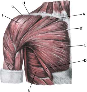
Cause: When a muscle is subjected to a load beyond the strength of the muscle (butterfly swimming, weight training, weight lifting) a rupture occurs. Most ruptures in athletes are partial ruptures. The rupture can be located both in the front and the back as well as at the attachment to the upper arm (humerus).

Symptoms: In light cases a localised tenderness can be felt following the load (“muscle strain”, “imminent pulled muscle”). In more severe cases sudden shooting pains in the muscle can be felt (“partial muscle rupture”) and in the worst cases a sudden snap is felt, rendering the muscle unusable (“total muscle rupture”) this is very rare, and is almost only seen in conjunction with other damage in the shoulder. With muscle injuries the following three symptoms are characteristic: pain upon pressure, stretching and activating against resistance.

Acute treatment: Click here.

Examination: Light cases with only minimal tenderness and no discomfort when using the arm do not necessarily require medical examination. The extent of the tenderness is, however, not always a mark of the degree of the injury. In case of more pronounced tenderness medical examination is advised with the aim of securing a correct diagnosis and treatment. Pain will be present when pressure is applied to the damaged muscle, which will be aggravated when the muscle is activated against resistance and when the muscle is stretched. Ultrasound is well suited to ensure the diagnosis.

Treatment: The treatment primarily involves relief, discontinuance of the injury inducing activity, stretching and increasing fitness training.

Complications: If satisfactory progress is not made, a physician should be consulted to ensure that the diagnosis is correct and that no complications have arisen. Amongst others the following should be considered:

Inflammation of the biceps tendon

INFLAMMATION OF THE BICEPS TENDON

Diagnosis: INFLAMMATION OF THE BICEPS TENDON
(Tendinitis M biceps brachii)


Anatomy:
The biceps muscle (M biceps brachii) has two tendons, fastened on the shoulder blade. One tendon (caput longum) passes through the shoulder joint. The two tendons join the biceps muscle on the upper arm and are anchored just below the elbow on the forearm. The function of the biceps muscle is to bend the elbow and supination (rotation) of the forearm. The long head of the biceps tendon has a tendon sheath that communicates with the shoulder joint.


  1. Sulcus bicipitalis medialis
  2. M. biceps brachii
  3. Epicondylus medialis
  4. Aponeurosis
    m. bicipitis brachii
  5. Sulcus bicipitalis lateralis
  6. Caput longum
    (m. bicipitis brachii)
  7. Caput breve
    (m. bicipitis brachii)
  8. M. coracobrachialis
  9. Processus coracoideus

SHOULDER AND UPPER ARM MUSCLES FROM THE FRONT

Cause: In case of repeated (over)loading (swimming, throwing) the long head of the biceps muscle (caput longum biceps bracii) becomes inflamed. Inflammation of the biceps tendon is also called a “swimming shoulder”. In some cases fluid is formed in the tendon sheath on the front of the upper arm (tenosynovitis). Inflammation of the biceps tendon is very often seen in conjunction with other damage in the shoulder, such as: impingement syndrome, inflammation of the upper shoulder blade muscle, rupture of the upper shoulder blade muscle, inflammation of the bursa (bursitis subacromialis), meniscus lesion in the shoulder (laesio labrum glenoidale).
With age and repeated loads the biceps muscle becomes marked by wear and tear, which increases the chance of ruptures. Ruptures occur most often when the muscle is contracting while being stretched (eccentric contraction). Long-term inflammation of the tendon sheath (tenosynovitis) increases the chance of a rupture of the tendon. It is rare for healthy tendons to rupture.

Symptoms: Slowly insetting localises tenderness exterior and to the front of the shoulder after load. Sometimes radiating down the upper arm. Pain deteriorates, when external pressure is applied to the biceps tendon, the front of the shoulder, when the muscle is activated (attempting to bend the arm against resistance) and when the muscle is taut (stretching and at the same time rotating inwards in the elbow (pronating)).

Acute treatment: Click here.

Examination: In light cases with only minimal tenderness medical examination is not necessarily required. In case of more pronounced pain and in lieu of progress a medical examination should be carried out to ensure the correctness of the diagnosis and to commence the correct treatment. Ordinary medical examination is often sufficient to form the diagnosis, but if uncertainty exists, it should be supplanted by ultrasound, which is the most suited examination for shoulder injuries (article) (Ultrasonic image).

Treatment: The treatment consists of relief and slow rehabilitation of the biceps muscle and the other muscles around the shoulder. Only in cases of total rupture of the biceps muscle should surgery be considered, the vast majority are treated with rehabilitation. If no progress is made with relief of the tendinitis, medical treatment may be considered in the form of rheumatic medicine (NSAID) or draining of the fluid in the tendon sheath and injection of corticosteroid in the tendon sheath, which must be performed with ultrasound guidance. Since the injection of corticosteroid is part of a long-term rehabilitation of a long-term injury, it is often necessary that the rehabilitation period stretches over several weeks, to reduce the risk of recurrences and ruptures. Naturally the tendon can not sustain maximum load after only a short rehabilitation period.

Complications: If smooth progress is not achieved the correctness of the diagnosis, or whether complications have arisen should be considered. Amongst others the following should be considered:

and ailment inside the shoulder (the fluid in the biceps tendon may be from the shoulder joint).

Rupture of the biceps tendon

RUPTURE OF THE BICEPS TENDON

Diagnosis: RUPTURE OF THE BICEPS TENDON
(Ruptura biceps brachii)


Anatomy:
The biceps muscle (M biceps brachii) has two tendons, fastened on the shoulder blade. One tendon (caput longum) passes through the shoulder joint. The two tendons join the biceps muscle on the upper arm and are anchored just below the elbow on the forearm. The function of the biceps muscle is to bend the elbow and supination (rotation) of the forearm. The long head of the biceps tendon has a tendon sheath that communicates with the shoulder joint.


  1. Sulcus bicipitalis medialis
  2. M. biceps brachii
  3. Epicondylus medialis
  4. Aponeurosis
    m. bicipitis brachii
  5. Sulcus bicipitalis lateralis
  6. Caput longum
    (m. bicipitis brachii)
  7. Caput breve
    (m. bicipitis brachii)
  8. M. coracobrachialis
  9. Processus coracoideus

SHOULDER AND UPPER ARM MUSCLES FROM THE FRONT

Cause: A rupture of the biceps muscle (caput longum) often occurs due to changes in the tendon due to wear and tear following many years’ repetitive load or attributable to a single violent load (for example weight lifting). Ruptures occur most often when the muscle is contracting while being stretched (eccentric contraction). Long-term inflammation of the tendon sheath (tenosynovitis) increases the chance of a rupture of the tendon. It is rare for healthy tendons to rupture.

Symptoms: Sudden insetting localised tenderness exterior and to the front of the shoulder after load. Sometimes radiating down the upper arm. Pain is aggravated when external pressure is applied to the biceps tendon, the front of the shoulder, when the muscle is activated (attempting to bend the arm against resistance) and when the muscle is taut (stretching and at the same time rotating inwards in the elbow (pronating)). A swelling can often be observed (the contracted biceps muscular belly) above the bend of the arm (cubital fossa).

Acute treatment: Click here.

Examination: In case of sudden insetting pain and swelling above the elbow a medical examination should be carried out to ensure the correctness of the diagnosis and to commence the correct treatment. Normal medical examination is often sufficient to form the diagnosis, but if any doubts exist the examination should be supplemented by an ultrasound scan, which is the most suited examination for shoulder injuries (article) (Ultrasonic image).

Treatment: The treatment consists of relief and slow rehabilitation of the biceps muscle and the other muscles around the shoulder. Only in cases of total rupture of the biceps muscle should surgery be considered, as the vast majority of ruptures are treated with rehabilitation.

Complications: If smooth progress is not achieved the correctness of the diagnosis should be considered, or whether complications have arisen. Amongst others the following should be considered:

and pain inside the shoulder (the fluid in the biceps tendon may emanate from the shoulder joint).

Rupture of the large chest nuscle

BRISTNING AF DEN STORE BRYSTMUSKEL

Diagnosis: RUPTURE OF THE LARGE CHEST MUSCLE
(Ruptura M pectoralis major)


Anatomy:
The large chest muscle (M pectoralis major) is fan shaped covers the front surface of the chest wall and fastens on humerus (crista tuberculi majoris). The function of the chest muscle is, amongst others, to bring the arm along the body and to bend and rotate inwards in the shoulder (like when throwing). The large chest muscle forms the forward fold in the armpit.

  1. M. pectoralis majo
    (pars clavicularis)
  2. Sternum (Breastbone)
  3. M. pectoralis major
    (pars sternocostalis)
  4. M. pectoralis major
    (pars abdominalis)
  5. M. serratus anterior
  6. M. deltoideus
  7. Acromion
  8. Clavicula (Collar bone)

SHOULDER MUSCLES FROM THE FRONT

Cause: When a muscle is subjected to a load beyond the strength of the muscle (fitness training, weight lifting, wrestling), a rupture occurs. The vast majority of ruptures in athletes are total. The rupture is usually located at the anchor on the humerus.

Symptoms: In light cases a local tenderness is felt after the load (“muscle strain”, “imminent pulled muscle”). In severe cases a sudden jolting pain is felt in the muscle (partial “muscle rupture” or “pulled muscle”) and in the worst case a violent snap, rendering the muscle unusable (“total muscle rupture”). In muscle injuries the following three symptoms are characteristic: pain upon pressure, stretching and activation against resistance. In total ruptures a defect can often be seen and felt at the forward fold in the armpit.

Acute treatment: Click here.

Examination: In light cases with only minimal tenderness and no inconveniences with normal usage of the arm, medical examination is not necessarily required. Though the severity of the tenderness is not always a measure of the severity of the injury. In case of more pronounced tenderness or pain medical examination is required to ensure the diagnosis and treatment. A normal medical examination is usually sufficient in order to make the diagnosis. Pain will be present when pressure is applied to the damaged and deteriorated muscle, when the arm is squeezed to the body against resistance (adduction) and when the muscle is stretched. If there is uncertainty regarding the diagnosis, the medical examination should be supplemented with an ultrasound or MRI scan, which is the most suitable examinations to ensure the diagnosis (article).

Treatment: In athletes with total ruptures located at the point of attachment on the humerus, most advise surgery (article). If the rupture is not total or if the rupture is in the muscle belly, relief and careful rehabilitation is recommended. In total ruptures a rehabilitation period stretching over several months must be expected before maximal loading can be resumed.

Rehabilitation naturally depends on the degree of rupture and the treatment (conservative / operative). It is advised to avoid fitness training the first couple of months, and hereafter resume it with a very low load so as not to rupture the muscle again.

Complications: If progress is not smooth, it should be considered whether the diagnosis is correct or whether complications have arisen. Amongst others the following should be considered:

Rupture of the lower shoulder blade muscle

RUPTURE OF THE LOWER SHOULDER BLADE MUSCLE

Diagnosis: RUPTURE OF THE LOWER SHOULDER BLADE MUSCLE
(Tendinitis M infraspinatus)


Anatomy:
Four muscles are part of the rotator cuff surrounding the shoulder joint, and controls, coordinates and assists movement of the shoulder: M infraspinatus (the lower shoulder blade muscle), M supraspinatus, M subscapularis and M teres minor. The infraspinatus muscle rotates the arm outwards.


  1. Acromion
  2. Tuberculum majus humeri
  3. M. infraspinatus
  4. Spina scapulare
  5. M. supraspinatus

SHOULDER BLADE MUSCLES FROM THE REAR

Cause: When a muscle is suddenly subjected to a load in excess of the strength of the muscle, a rupture will occur in the muscle. Rupture of the infraspinatus (the lower shoulder blade muscle) is a common throwing injury (handball). Inflammation of the tendon (tendinitis) often precede larger ruptures and tendinitis is not rare combined with inflammation of the bursa (bursitis subacromialis).

Symptoms: In light cases a local tenderness is felt after the load (“muscle strain”, “imminent pulled muscle”, “inflammation of the tendon”), which can precede larger ruptures if the exercise intensity is not altered. In severe cases a sudden shooting pains are felt in the muscle (“partial muscle rupture”, “pulled muscle”) and in the worst case a violent snap is felt, rendering the muscle unusable (“total muscle rupture”). In case of muscle damage the following three symptoms are characteristic: pain upon pressure (externally, on the back of the shoulder), stretching and activation against resistance (rotation outwards against resistance).

Acute treatment: Click here.

Examination: In light cases with only minimal tenderness medical examination is not necessarily required. In case of more pronounced pain and in lack of progress a medical examination should be carried out to ensure the correctness of the diagnosis and to commence the correct treatment (article-1) (article-2). A normal medical examination is usually sufficient in order to make the diagnosis, however, if there is any doubt concerning the diagnosis the medical examination should be supplemented with an ultrasound scan, which is the most suitable examination for shoulder damage (Ultrasonic image) (article).

Treatment: The treatment primarily involves relief from the pain inducing activity, stretching and rehabilitation of the muscles around the shoulder. In light cases with slowly insetting tenderness after load, without the sensation of a snap and good function of the muscle (“muscle strain”, “imminent pulled muscle”, “inflammation of the tendon”), medical treatment in the form of rheumatic medicine (NSAID) or injection of corticosteroid (usually in the subacromiale bursa over the infraspinatus muscle). Since the injection of corticosteroider is part of a long-term rehabilitation of a long-term injury, it is often necessary for rehabilitation to stretch over several weeks to months to reduce the risk of recurrences and ruptures. The tendon can of course not sustain maximum load after a prolonged injury-period and only a short-term rehabilitation period. Ultrasound guided injection gives the maximal effect with minimal risk, since “blind” injections hit the wrong structures in more than half the cases (article). In case of lack of progress in rehabilitation and medical treatment, an operative treatment can be attempted.

Complications: If progress is not smooth, it should be considered whether the diagnosis is correct or whether complications have arisen. Amongst others the following should be considered:

Shoulder

Inflammation of the tendon sheath of the upper shoulder blade muscle

INFLAMMATION OF THE TENDON SHEATH OF THE UPPER SHOULDER BLADE MUSCLE

Diagnosis:

INFLAMMATION OF THE TENDON SHEATH OF THE UPPER SHOULDER BLADE MUSCLE
(Tendinitis M supraspinatus)

Anatomy: Four muscles are part of the rotator cuff surrounding the shoulder joint, and controls, coordinates and assists movement of the shoulder: M supraspinatus (the upper shoulder blade muscle), M infraspinatus, M subscapularis and M teres minor. When the arm is moved away from the body and above the head (abducated) the supraspinatus muscle slips under the upper bone projection of the shoulder blade (acromion).

  1. Acromion
  2. Tuberc. majus
    (humeri)
  3. M. infraspinatus
  4. Spina scapulae
  5. M. supraspinatus

SHOULDER BLADE MUSCLES FROM THE REAR
Shoulder

Cause: In case of repeated loads with the arm above the head (tennis, swimming) the upper shoulder blade muscle tendon (M supraspinatus) become inflamed swells and may become squeezed between the head of the upper arm (caput humeri) and the upper bone projection of the shoulder blade (acromion). Additionally this causes fraying and weakening of the tendon with risk of ruptures. It is not uncommon for tendinitis to be seen in conjunction with inflammation of the bursa (bursitis subacromialis).

Symptoms: Slowly insetting local tenderness after load, exterior and to the front of the shoulder. Sometimes radiating down the upper arm. The pain deteriorates when external pressure is applied to the supraspinatus muscle, on the front of the shoulder, when the muscle is activated (the arm is lifted to the side) and when the muscle is stretched (hand is brought to the loin). Due to the inflammation and wear and tear changes, calcification can occur in the shoulder muscle.

Acute treatment: Click here.

Examination:
In light cases with only minimal tenderness medical examination is not necessarily required. In case of more pronounced pain or trouble lifting the arm away from the body and in case of lack of progress, a medical examination should be performed to ensure the correctness of the diagnosis and treatment. Ordinary clinical examination is often sufficient to make the diagnosis (article-1) (article-2). If there is uncertainty surrounding the diagnosis, it should be supplemented with an ultrasound scan, which is the most suitable examination for shoulder injuries (article).

Treatment: The treatment primarily involves relief from the pain inducing activities, stretching and rehabilitation of the muscles around the shoulder. If lack of progress in rehabilitation a medical treatment in the form of rheumatic medicine (NSAID) or injection of corticosteroid (usually in the bursa above the supraspinatus muscle) may be considered. Since the injection of corticosteroid is part of a long-term rehabilitation of a long-term injury, it is often necessary that the rehabilitation period stretches over several weeks to months, to reduce the risk of recurrences and ruptures. Naturally the tendon can not sustain maximum load after only a short rehabilitation period. If calcification is present in the shoulder muscle, an attempt may be made to extract this at the same time corticosteroid is injected around the calcification (article). The optimal effect at minimal risk can be accomplished by performing the injections guided by ultrasound. The wrong structures are hit in more than half the cases where the injection is done blindly (article). In lack of progress with rehabilitation and medical treatment a surgical treatment can be attempted.

Complications: If smooth progress is not achieved, it should be considered whether the diagnosis is correct or whether complications have arisen. Amongst others the following should be considered:

Rupture of the upper shoulder blade muscle

BRISTNING AF ØVRE SKULDERBLADSMUSKEL

Diagnosis: RUPTURE OF THE UPPER SHOULDER BLADE MUSCLE
(Ruptura M supraspinatus)


Anatomy:
Four muscles are part of the rotator cuff surrounding the shoulder joint, and controls, coordinates and assists movement of the shoulder: M supraspinatus (the upper shoulder blade muscle), M infraspinatus, M subscapularis and M teres minor. When the arm is moved away from the body and above the head (abducated) the supraspinatus slips under the upper bone projection of the shoulder blade (acromion).


  1. Acromion
  2. Tuberculum majus humeri
  3. M. infraspinatus
  4. Spina scapulare
  5. M. supraspinatus

SHOULDER BLADE MUSCLES FROM THE REAR

Cause: With age and repeated loads with the arm above the head (tennis, swimming) the upper shoulder blade muscle tendon (M supraspinatus) becomes stiffer and less flexible, and is more easily squeezed between the head of the upper arm (caput humeri) and the acromion, causing the tendon to become frayed and weakened with risk of ruptures. When a muscle is suddenly subjected to a load in excess of the strength of the muscle, a rupture occurs (fall on an outstretched arm, lifting or throwing of heavy objects). The vast majority of ruptures are partial muscle ruptures. The vast majority of total ruptures are seen in older people with wear and tear changes in the muscle tendon (in very old people total ruptures are very frequent). It is very rare for healthy tendons to rupture.

Symptoms: In light cases a local tenderness is felt after the load (“muscle strain”, “imminent pulled muscle”). In severe cases a sudden shooting pain in the muscle is felt (partial “muscle rupture” or “pulled muscle”) and in the worst case a sudden snap is felt, rendering the muscle unusable (“total muscle rupture”). With muscle damage the following three symptoms are characteristic: pain upon pressure, stretching (hand to the loin) and activation against resistance (lifting the arm to the side or above the head). The pain is localized on top and external to the shoulder. After a longer period, loss of muscle can be observed on the back of the shoulder blade. With many older people the rupture has not produced symptoms.

Acute treatment: Click here.

Examination: Light cases with only minimal tenderness do not necessarily require medical examination. In case of more pronounced pain or trouble lifting the arm away from the body and in case of lack of progress, a medical examination should be carried out to ensure the correct diagnosis and treatment. Ordinary medical examination is often sufficient to make the diagnosis (article-1) (article-2). Returning to the bedside: using the history and physical examination to identify rotator cuff tears, 2) The ‘dropping’ and ‘hornblower’s’ signs in evaluation of rotator cuff tears. If there is uncertainty about the diagnosis it should be supplemented with ultrasound, which is the most suitable examination for shoulder injuries (article) (Ultrasonic image). If there is doubt as to whether it is a total or partial rupture, the doctor can inject a local anaesthetic around the rupture. If, after this, the arm is able to move freely, it is not a total rupture (diagnostic blockade).

Treatment: Only in cases of near total rupture of the supraspinatus muscle can surgery be considered. Renewed ruptures occur relatively frequently after surgery (article). In partial ruptures and total ruptures in the elderly the treatment is short-term relief and careful rehabilitation of the part of the supraspinatus muscle, which has not ruptured.

Complications: If progress is not smooth, it should be considered whether the diagnosis is correct or whether complications have arisen. Amongst others the following should be considered:

Shoulder

Inflammation of the bursa

INFLAMMATION OF THE BURSA

Diagnosis: INFLAMMATION OF THE BURSA
(Bursitis)

Anatomy: Around the shoulder joint are numerous bursas, reducing the pressure on muscles, tendons and ligaments, where these lie close to bone projections. Between the upper bone-projection (acromion) and the upper shoulder blade muscle (M Supraspinatus) lies the largest bursa in the shoulder (bursae subacromiale), which often communicates with the shoulder joint (Photo).


  1. Acromion
  2. Tuberc. majus
    (humeri)
  3. M. infraspinatus
  4. Spina scapulae
  5. M. supraspinatus

SHOULDER BLADE MUSCLES FROM THE REAR
Shoulder

Cause: Upon repeated loads or blows, the bursa can become inflamed, increasing the production of fluids, swelling and becoming painful. Inflammation of the subacromiale bursae is often caused by working with the arm above the level of the head. When the arm is moved away from the body and above the level of the head (abducted) the supraspinatus muscle slips under the upper bone projection of the shoulder blade (acromion). With age, the supraspinatus tendon becomes stiff and unremitting, and is more easily squeezed between the head of the upper arm (caput humeri) and the acromion, causing the bursa to be squeezed and swell.

Symptoms: Pain upon pressure on the bursa, which occasionally (but far from always) may feel swollen. Pain in the subacromiale bursae is worsened when the arm is at a right angle to the body. Inflammation of the bursa often causes nightly pain, and pain when lying on the side of the inflamed bursa.

Acute treatment: Click here.

Examination:
In light cases with only minimal tenderness medical examination is not necessarily required. In case of pain that is more pronounced or lack of progress, a medical examination should be carried out to ensure the correct diagnosis and treatment. The doctor may carry out various clinical examinations, which not always allows a certain diagnosis (article). The diagnosis is most rapidly made with ultrasound (allowing simultaneous treatment) (article), (Ultrasonic image).

Treatment: The treatment primarily involves relief. Possible removal of the provoking factor, if known. The treatment may be supplemented with rheumatic medicine (NSAID) or injection of corticosteroid in the bursa, possibly preceded by draining of the bursa. This procedure can advantageously be guided by ultrasound, (article). In the absence of any effect, an operative solution with removal of the bursa may be attempted (and in cases of bursitis subacromiale removal of a part of the shoulder’s upper bone projection (acromion). Thereby avoiding that the supraspinatus muscle and the bursa is squeezed between the head of the upper arm and the acromion, when the arm is raised).

Rehabilitation, specific: The treatment is dependent on which bursa is inflamed, but sports activity can be carefully resumed once pain has diminished, especially if the triggering cause has been determined, and subsequently removed. Generally careful training of the shoulder muscles is recommended, primarily with the arm below the level of the head.

Complications: If smooth progress is not achieved the correctness of the diagnosis, or whether complications have arisen should be considered. Amongst others the following should be considered: