Kategoriarkiv: Inflammation of the growth zone of the heel bone

Rupture of the Soleus muscle

Diagnosis: RUPTURE OF THE SOLEUS MUSCLE
(Ruptura M soleus)


Anatomy:
The calf muscle (M Gastrocnemicus) is comprised of two muscle heads which gather in a wide tendinous ligament and continue in to the Achilles tendon. Another of the larger calf muscles (M Soleus) is attached to the front side of the Achilles tendon and thus forms a part of the Achilles tendon. The Achilles is attached to the heel bone (calcaneus).

  1. Tendo calcaneus (Achillis)
  2. M. soleus
  3. M. gastrocnemius

LOWER LEG

  1. M. gastrocnemius
  2. M. plantaris
  3. M. soleus
  4. Tendo m. gastrocnemii
  5. Tendo calcaneus (Achillis)
  6. M. popliteus
  7. Bursa m. semimembranosi
  8. M. semimembranosus
  9. Bursa subtendinea m. gastrocnemii medialis

KNEE FROM THE REAR

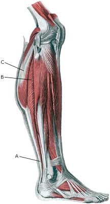
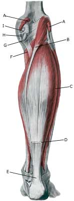
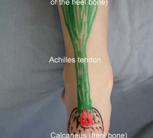
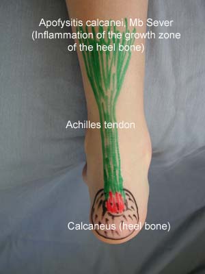
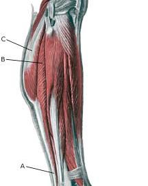
Cause: Full or partial rupture of the soleus muscle usually occurs when the calf muscle becomes stretched while it is contracting (eccentric contraction). Partial ruptures represent the majority of the ruptures. The rupture occurs in many instances at the point of attachment of the soleus muscle to the Achilles tendon, which will often trigger an inflammation of the Achilles tendon as a result of the soleus rupture.

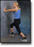
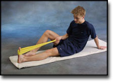
Symptoms: Pain when activating the calf muscle (running and jumping), when applying pressure on the Achilles tendon approx. 4 cm. above the anchor point on the heel bone or higher up in the calf muscle, and when stretching the tendon. Walking on tip-toe will aggravate the pain.

Acute treatment: Click here.

Examination: In all cases when there is a sense of a “crack”, or sudden shooting pains in the Achilles tendon, medical attention should be sought as soon as possible. Ultrasound scanning or MRI examination is used to advantage when making the diagnosis, as even full ruptures can easily be overlooked by normal clinical examination.

Treatment: Treatment of the rupture can comprise rest, stretching and training.

Complications: If there is not a steady improvement in the condition an ultrasound scan should be performed to exclude:

inflammation of the bursa

Diagnosis: INFLAMMATION OF THE BURSA
(Bursitis)


Anatomy:
There are numerous bursas around the foot for the purpose of reducing the pressure on the muscles, tendons and ligaments which lie close to bone projections. The bursas at the achilles tendon are those which most often give rise to symptoms.

Cause: The bursas can become inflamed, produce fluid, swell and become painful with repeated over-load or due to blows.

Symptoms: Pain when applying pressure to the bursa, which sometimes, but far from always, can give the impression of being swollen.

Acute treatment: Click here.

Examination: Medical examination is usually not required in light cases with only minimal tenderness. With more pronounced pain, or lack of improvement, medical examination should always be performed for confirm the diagnosis and commencement of treatment if required. The diagnosis is best made using ultrasound examination.

Treatment: Treatment is primarily concentrated on providing rest. If the provoking factor is known (i.e. tight shoes), this should naturally be corrected. Treatment can be supplemented with rheumatic medicine (NSAID) or injection of corticosteroid in the bursa preceded by draining, which can be best performed if ultrasound-guided.

Rehabilitation: Treatment is completely dependent upon which bursa is inflamed, but the sports activity can be cautiously resumed when the pain has diminished, especially if the provoking factor has been identified and removed.
Also read rehabilitation, general.

Bandage: In some cases a ring of felt (for example) can be taped around the tender bursa which will reduce the pressure from shoes. It is naturally important that the hole in the ring is positioned directly above the bursa.

Complications: If there is not a steady improvement in the condition consideration must be given as to whether the diagnosis is correct, or if complications have arisen:

In rare cases, the bursa can be infected with bacteria. This is a serious condition if the bursa becomes red, warm and increasingly swollen and tender. This condition requires immediate examination and treatment.

KONDITION

STEP4

TRAINING LADDER FOR CHILDREN AND ADOLESCENTS:
FOR INFLAMMATION OF THE GROWTH ZONE OF THE HEEL BONE
(APOPHYSITIS CALCANEI, MB SEVER)

STEP 4

The majority of young patients can manage with relief until the pain subsides, following which the sports activity can be slowly resumed. The following rehabilitation program will cover the needs for the vast majority of children with inflammation of the growth zone of the heel bone. Older teenagers involved in sports at a high level can advantageously use the rehabilitation program for adults.
KONDITION
Unlimited: Cycling. Swimming. Running with increasing distance on a soft surface.

UDSPÆNDING
(5 min)

Stand with the injured leg stretched backwards with the toes facing front. Slowly bend the knee so that the calf muscles become increasingly stretched. Hold the position for 20 seconds and relax for 20 seconds before repeating.

Stand with the injured leg outstretched with the ankle joint bent up against the wall. Press your abdomen against the wall so that the calf muscles become increasingly stretched. Hold the position for 20 seconds and relax for 20 seconds before repeating.

KOORDINATION
(5 min)

Seesaw. Balance on two legs, possibly using a hand as support against the wall, balancing subsequently on one leg without support. Look straight ahead and keep knees bent.

STYRKE
(10 min)

Sit on the floor. Tip the foot up and down with elastic under the forefoot so that the elastic becomes taut.

Sit on the floor. Tip the foot up and down with elastic on top of the forefoot so that the elastic becomes taut.

Sit on the floor. Tip the foot from side to side with elastic on the outer side of the foot, without moving the knee.

Sit on the floor. Tip the foot from side to side with elastic on the inner side of the foot, without moving the knee.

Stand with both forefeet on a stool or doorstep with the heel out over the edge. Slowly rise up on to your toes with your weight on the healthy leg. Go slowly down on the injured leg as far as you can go. Use the healthy leg to rise up on to your toes again. The exercise should be performed with stretched, as well as bent knee. Wearing a rucksack and gradually increasing the ballast in the rucksack can increase the load.

Stretching is carried out in the following way: stretch the muscle group for 3-5 seconds. Relax for 3-5 seconds. The muscle group should subsequently be stretched for 20 seconds. The muscle is allowed to be tender, but must not hurt. Relax for 20 seconds, after which the procedure can be repeated.

The time consumed for stretching, coordination and strength training can be altered depending on the training opportunities available and individual requirements.

KONDITION

STEP3

TRAINING LADDER FOR CHILDREN AND ADOLESCENTS:
FOR INFLAMMATION OF THE GROWTH ZONE OF THE HEEL BONE
(APOPHYSITIS CALCANEI, MB SEVER)

STEP 3

The majority of young patients can manage with relief until the pain subsides, following which the sports activity can be slowly resumed. The following rehabilitation program will cover the needs for the vast majority of children with inflammation of the growth zone of the heel bone. Older teenagers involved in sports at a high level can advantageously use the rehabilitation program for adults.
KONDITION
Unlimited: Cycling. Swimming. Light jogging (few minutes) on a smooth surface.

UDSPÆNDING
(5 min)

Stand with the injured leg stretched backwards with the toes facing front. Slowly bend the knee so that the calf muscles become increasingly stretched. Hold the position for 20 seconds and relax for 20 seconds before repeating.

Stand with the injured leg outstretched with the ankle joint bent up against the wall. Press your abdomen against the wall so that the calf muscles become increasingly stretched. Hold the position for 20 seconds and relax for 20 seconds before repeating.


STYRKE
(10 min)


Sit on the floor. Tip the foot up and down with elastic under the forefoot so that the elastic becomes taut.

Sit on the floor. Tip the foot up and down with elastic on top of the forefoot so that the elastic becomes taut.

Sit on the floor. Tip the foot from side to side with elastic on the outer side of the foot, without moving the knee.

Sit on the floor. Tip the foot from side to side with elastic on the inner side of the foot, without moving the knee.

Stretching is carried out in the following way: stretch the muscle group for 3-5 seconds. Relax for 3-5 seconds. The muscle group should subsequently be stretched for 20 seconds. The muscle is allowed to be tender, but must not hurt. Relax for 20 seconds, after which the procedure can be repeated.

The time consumed for stretching, coordination and strength training can be altered depending on the training opportunities available and individual requirements.

KONDITION

STEP2

TRAINING LADDER FOR CHILDREN AND ADOLESCENTS:
FOR INFLAMMATION OF THE GROWTH ZONE OF THE HEEL BONE
(APOPHYSITIS CALCANEI, MB SEVER)

STEP 2

The majority of young patients can manage with relief until the pain subsides, following which the sports activity can be slowly resumed. The following rehabilitation program will cover the needs for the vast majority of children with inflammation of the growth zone of the heel bone. Older teenagers involved in sports at a high level can advantageously use the rehabilitation program for adults.
KONDITION
Unlimited: Cycling. Swimming.

UDSPÆNDING
(5 min)

Stand with the injured leg stretched backwards with the toes facing front. Slowly bend the knee so that the calf muscles become increasingly stretched. Hold the position for 20 seconds and relax for 20 seconds before repeating.

Stand with the injured leg outstretched with the ankle joint bent up against the wall. Press your abdomen against the wall so that the calf muscles become increasingly stretched. Hold the position for 20 seconds and relax for 20 seconds before repeating.

STYRKE
(5 min)

Sit on the floor. Tip the foot up and down with elastic on top of the forefoot so that the elastic becomes taut.

Sit on the floor. Tip the foot from side to side with elastic on the outer side of the foot, without moving the knee.

Sit on the floor. Tip the foot from side to side with elastic on the inner side of the foot, without moving the knee.

Stretching is carried out in the following way: stretch the muscle group for 3-5 seconds. Relax for 3-5 seconds. The muscle group should subsequently be stretched for 20 seconds. The muscle is allowed to be tender, but must not hurt. Relax for 20 seconds, after which the procedure can be repeated.

The time consumed for stretching, coordination and strength training can be altered depending on the training opportunities available and individual requirements.

KONDITION

STEP1

GENOPTRÆNING

TRAINING LADDER FOR CHILDREN AND ADOLESCENTS:
FOR INFLAMMATION OF THE GROWTH ZONE OF THE HEEL BONE
(APOPHYSITIS CALCANEI, MB SEVER)

STEP 1

The majority of young patients can manage with relief until the pain subsides, following which the sports activity can be slowly resumed. The following rehabilitation program will cover the needs for the vast majority of children with inflammation of the growth zone of the heel bone. Older teenagers involved in sports at a high level can advantageously use the rehabilitation program for adults.
KONDITION
Unlimited: Cycling. Swimming.

UDSPÆNDING
(5 min)

Stand with the injured leg stretched backwards with the toes facing front. Slowly bend the knee so that the calf muscles become increasingly stretched. Hold the position for 20 seconds and relax for 20 seconds before repeating.

Stand with the injured leg outstretched with the ankle joint bent up against the wall. Press your abdomen against the wall so that the calf muscles become increasingly stretched. Hold the position for 20 seconds and relax for 20 seconds before repeating.

Stretching is carried out in the following way: stretch the muscle group for 3-5 seconds. Relax for 3-5 seconds. The muscle group should subsequently be stretched for 20 seconds. The muscle is allowed to be tender, but must not hurt. Relax for 20 seconds, after which the procedure can be repeated.

The time consumed for stretching, coordination and strength training can be altered depending on the training opportunities available and individual requirements.

Treatment(2)

SportNetDoc

Local corticosteroid injection in sport: review of literature and guidelines for treatment.

Fredberg U. Scand J Med Sci Sports 1997 Jun;7(3):131-9.

The risks and benefits of local injection therapy of overuse sports injuries with corticosteroids are reviewed here. Injection of corticosteroid inside the tendon has a deleterious effect on the tendon tissue and should be unanimously condemned. No reliable proof exists of the deleterious effects of peritendinous injections. Too many conclusions in the literature are based on poor scientific evidence and it is just the reiteration of a dogma if all steroid injections are abandoned. The corticosteroids represent an adjuvant treatment in the overall management of sports injuries: basic treatment is ‘active’ rest and graduated rehabilitation within the limits of pain. With proper indications there are only few and trivial complications that may occur with corticosteroid injections. Guidelines for proper local injection therapy with corticosteroids are given.

Treatment(1)

Prevention and management of calcaneal apophysitis in children: an overuse syndrome.

Micheli LJ, Ireland ML. J Pediatr Orthop 1987 Jan-Feb;7(1):34-8.

Calcaneal apophysitis (Sever disease) is a common cause of heel pain, particularly in the athletically active child. Eighty-five children (137 heels) with calcaneal apophysitis were reviewed. Both heels were affected in 52 (61%) patients. The most common associated foot condition was pronation, occurring in 16 patients. Sixty-eight patients complained that pain was made worse by a specific sport, with soccer leading the list. All patients were treated with a physical therapy program of lower extremity stretching, especially of the heel cords, and ankle dorsiflexion strengthening. Soft Plastizote orthotics or heel cups were used in 98% of patients. Proper athletic shoewear was advised. All patients improved and were able to return to their sport of choice 2 months after the diagnosis. There were two recurrences.

Examination

SportNetDoc

Ultrasonography in the differential diagnosis of Achilles tendon injuries and related disorders. A comparison between pre-operative ultrasonography and surgical findings.

Paavola M, Paakkala T, Kannus P, Jarvinen M. Acta Radiol 1998 Nov;39(6):612-9.

PURPOSE.
To assess the value of US in the diagnosis of various Achilles tendon disorders.

MATERIAL AND METHODS.
Pre-operative US was compared with surgical findings in 79 patients with an Achilles tendon complaint.

RESULTS.
US was highly reliable for verifying a complete Achilles tendon rupture, only one false-negative US examination was found in the 26 surgically verified cases. For diagnosing retrocalcanear bursitis, US was accurate: 6 out of the 8 cases of bursitis were found and there were no false-positive cases. There were also no false-positive US findings in patients with peritendinitis/tendinitis, but 7 false-negative US cases among the 40 surgically verified peritendinitis/tendinitis patients indicated that a negative US finding in a clinically suspected case of peritendinitis/tendinitis is unreliable. US also seemed to be inadequate for differentiating partial tendon rupture from a focal tendon degeneration. Nevertheless, the occurrence and location of such a lesion could be adequately determined by US.

CONCLUSION.
US can reliably be used for locating the Achilles tendon abnormality, estimating its severity, and determining most of the conditions requiring surgical intervention. However, US is not completely reliable for diagnosing peritendinitis and tendinitis, and it cannot be used to differentiate partial tendon ruptures from focal degenerative lesions.