Kategoriarkiv: Tendinitis at the ischiatic bone

Tendonitis at the ischiatic bone

Diagnosis: TENDINITIS AT THE ISCHIATIC BONE
(ENTESOPATIA TUBER ISCHIADICUM


Anatomy:
The large posterior thigh muscles (hamstring muscles) have a common muscle tendon fastening on the ischiatic bone (tuber ischiadicum). The posterior thigh muscles flex the knee and stretch the hip.

  1. Bursa trochanterica m. glutei maximi
  2. M. gluteus maximus
  3. M. biceps femoris (caput longum)
  4. M. semitendinosus
  5. M. semimembranosus
  6. M. adductor magnus
  7. M. gracilis
  8. M. quadratus femoris
  9. Bursa ischiadica m. glutei maximi

(Photo)

RIGHT GLUTEAL MUSCLES
FROM THE REAR

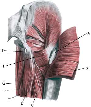
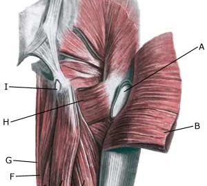
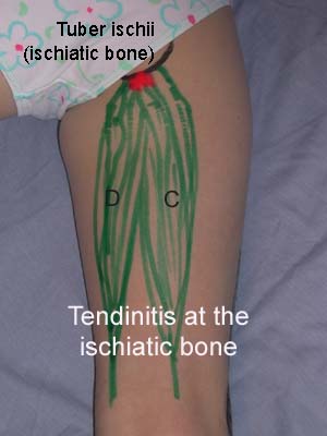
Cause: Inflammation of the tendon fastenings (tendinitis) at the ischiatic bone (tuber ischiadicum) occurs following repeated uniform (over)loads (e.g. running, sprinting) causing microscopic ruptures in the tendon, and especially at the tendon fastening. Tendinitis is a warning that the training performed is too strenuous for the muscles in question, and if the load is not reduced a rupture of the posterior thigh muscle fastening on the ischiatic bone (“pulled muscle”) may occur. This will result in a considerably prolonged rehabilitation period.

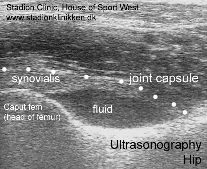
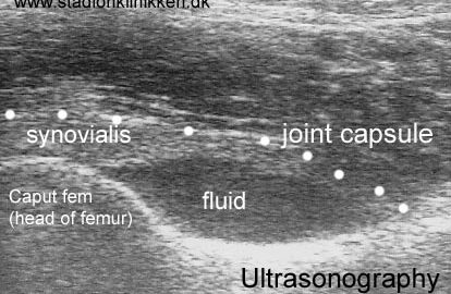
Symptoms: Pain in the ischiatic bone can occasionally radiate down into the rear of the thigh. The pain is aggravated when applying pressure on the bone (e.g. sitting position), stretching and activating the posterior thigh muscles (flexing the knee against resistance).

Acute treatment: Click here.

Examination: In slight cases with only minimal tenderness and no discomfort with walking, medical examination is not necessarily required. The extent of the tenderness is, however, not always a mark of the degree of the injury. In cases of more pronounced pain or tenderness, medical examination is required to ensure the correct diagnosis and treatment. The diagnosis is usually made on the basis of a normal medical examination, however, if there is any doubt concerning the diagnosis, this can be confirmed by ultrasound scanning or MR scanning (article).

Treatment: The treatment usually comprises relief, stretching and rehabilitation (article). If the rehabilitation does not progress satisfactorily, medicinal treatment in the form of rheumatic medicine (NSAID) can be considered, or corticosteroid injection in the area surrounding the inflamed tendon fastening on the ischiatic bone. As the injection of corticosteroid is always an element in the long term rehabilitation of a very serious, chronic injury, it is vital that the rehabilitation period lasts over several months in order to reduce the risk of a relapse or ruptures. The tendon can naturally not sustain maximal load after a long-term injury period and only a short-term rehabilitation period. If the diagnosis is made by use of ultrasound scanning, the injections are performed under guidance of ultrasound, and the rehabilitation is progressed in accordance with the guidelines mentioned, then the treatment involving corticosteroid injections has very few risks connected. If satisfactory progress is not made following the rehabilitation and medicinal treatment, surgical intervention can be considered. Long-term results of operations are often disappointing, despite publication of a minor series with good results (article).

Complications: If the treatment does not progress according to plan, it should be considered if the diagnosis is correct or whether complications have arisen. The following should in particular be considered:

Scheuermann disease

Diagnosis: SCHEUERMANN’S DISEASE


Anatomy:
The back is constructed of box-like vertebrae which are held in place partly with the help of the shape of the bones, partly by ligaments and partly by the large and small back and stomach muscles.


  1. Vertebra prominens
  2. Vertebra coccygea I
  3. Promontorium
  4. L I
  5. Th I
  6. Axis

THE BACK SEEN FROM THE SIDE

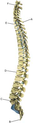
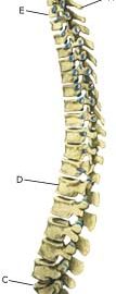
Cause: Scheuermann’s disease occurring in approximately 4% of the population (article). A curvature of the back occurs (bending over forwards) due to the vertebrae becoming wedge shaped. There are also characteristic x-ray finds. The cause of the condition is unknown, but evidence tends to suggest that the condition is hereditary (article).

Symptoms: Back curvature localised high in the back (thoracal Scheuermann) gives often only few, if any, symptoms. Back curvature localised in the lower back (thoracolumbal or lumbal Scheuermann) does entail back pain for the majority (article).

Examination: The diagnosis is usually made following a medical examination supplemented with an x-ray (at least 3 adjacent vertebrae with at least 5 degrees wedge form, Schmorlske impressions, flattening of discs, irregular end plates) (article). The crooked back is often mistaken in the beginning for “bad posture”. In some cases, CT or MRI scanning is recommended.

Treatment: The vast majority of cases can be treated with training, attempting to maintain the mobility of the back, counteract the curvature tendency and strengthen the stomach and back muscles. A corset can in some cases be used until the young person is fully grown. An operation can be performed only in very rare cases. The condition has a good prognosis (article), and even after an operation it is still possible to take part in many different forms of sport (article).

Rehabilitation of children and adolescents: INSTRUCTION

Complications: In some cases a crooked back can have other causes (infection, nerve disease, inborn bone change, rheumatic illness, bone disease, metabolic disorder).

Cause-article4

SportNetDoc

Apophyseal injuries in the young athlete.

Peck DM. Am Fam Physician 1995 Jun;51(8):1891-5, 1897-8

Apophyseal injuries, which are unique in the adolescent athlete, cause inflammation at the site of a major tendinous insertion onto a growing bony prominence. These injuries typically occur in active adolescents between the ages of eight and 15 years and usually present as periarticular pain associated with growth, skeletal immaturity, repetitive microtrauma and muscle-tendon imbalance. Common apophyseal injuries, and their sites, include Sever’s disease (posterior calcaneus), Osgood-Schlatter disease (tibial tuberosity), Sindig-Larsen-Johansson syndrome (inferior patella), medial epicondylitis (humeral medial epicondyle) and apophysitis of the hip (iliac crest, ischial tuberosity). Conservative therapy, including rest, ice, compression, elevation, nonsteroidal anti-inflammatory agents, modification of the athlete’s activity level and exercises for increased flexibility and strengthening, is usually effective.

Cause-article3

SportNetDoc

Hamstring strains in athletes: diagnosis and treatment.

Clanton TO, Coupe KJ. J Am Acad Orthop Surg 1998 Jul-Aug;6(4):237-48.

Hamstring strains are among the most common injuries (and reinjuries) in athletes. Studies combining electromyography with gait analysis have elucidated the timing of activity of the three muscles of the hamstring group; they function during the early-stance phase for knee support, during the late-stance phase for propulsion, and during midswing to control the momentum of the leg. Muscle injury, whether partial or complete, occurs at the myotendinous junction, where force is concentrated. The healing response begins with inflammation, associated edema, and localized hemorrhage. After an initial period of reduced tension, the healing muscle regains strength rapidly as long as reinjury does not occur. Although the use of anti-inflammatory medication is a keystone of treatment, a certain degree of inflammation is necessary for removing necrotic muscle fibers and rescaffolding to allow optimal recovery. The protocol of rest, ice, compression, and elevation is still the preferred first-aid approach. After a brief period of immobilization (usually less than 1 week for even the most severe strain), mobilization is begun to properly align the regenerating muscle fibers and limit the extent of connective tissue fibrosis. Concurrent pain-free stretching and strengthening exercises (beginning with isometrics and progressing to isotonics and isokinetics) are essential to regain flexibility and prevent further injury and inflammation. Readiness for return to competition can be assessed by isokinetic testing to confirm that muscle-strength imbalances have been corrected, the hamstring-quadriceps ratio is 50% to 60%, and the strength of the injured leg has been restored to within 10% of that of the unaffected leg. The only indication for surgery is a complete rupture at or near the origin from the ischial tuberosity or distally at its insertion (either soft-tissue avulsion with a large defect or bone avulsion with displacement by 2 cm).

Cause-article2

SportNetDoc

Hamstring injuries. Current trends in treatment and prevention.

Kujala UM, Orava S, Jarvinen M. Sports Med 1997 Jun;23(6):397-404.

Pre-exercise stretching and adequate warm-up are important in the prevention of hamstring injuries. A previous mild injury or fatigue may increase the risk of injury. Hamstring muscle tear is typically partial and takes place during eccentric exercise when the muscle develops tension while lengthening, but variation in injury mechanisms is possible. Diagnosis of typical hamstring muscle injury is usually based on typical injury mechanism and clinical findings of local pain and loss of function. Diagnosis of avulsion in the ischial tuberosity, with the need for longer immobilisation, and a complete rupture of the hamstring origin, in which immediate operative treatment is necessary, poses a challenge to the treating physician. X-rays, ultrasonography or magnetic resonance imaging (MRI) may be helpful in differential diagnostics. After first aid with rest, compression, cold and elevation, the treatment of hamstring muscle injury must be tailored to the grade of injury. Conservative treatment is based on a knowledge of the biological background of the healing process of the muscle. Experimental studies have shown that a short period of immobilisation is needed to accelerate formation of the granulation tissue matrix following injury. The length of the immobilisation is, however, dependent on the grade of injury and should be optimised so that the scar can bear the pulling forces operating on it without re-rupture. Mobilisation, on the other hand, is required in order to regain the original strength of the muscle and to achieve good final results in resorption of the connective tissue scar and re-capillarisation of the damaged area. Another important aim of mobilisation–especially in sports medical practice–is to avoid muscle atrophy and loss of strength and extensibility, which rapidly result from prolonged immobilisation. Complete ruptures with loss of function should be operated on, as should cases resistant to conservative therapy in which, in the late phase of repair, the scar and adhesions prevent the normal function of the hamstring muscle.

Cause-article1

SportNetDoc

Ischial tuberosity apophysitis and avulsion among athletes.

Kujala UM, Orava S, Karpakka J, Leppavuori J, Mattila K. Int J Sports Med 1997 Feb;18(2):149-55

Ischial tuberosity pain in athletes may be caused by several clinical entities, which include acute and old bony or periosteal avulsions and apophysitis. We studied the natural course of these injuries based on our clinical case series of fourteen patients with apophysitis and twenty-one with avulsion of the ischial tuberosity. Only patients with the diagnosis confirmed by X-ray finding were included. The clinical diagnostic criteria by ischial apophysitis consisted of gradually increasing functional and palpatory pain at the ischial tuberosity without any major trauma at the beginning of the symptoms. Typically there was asymmetry on plain radiographs of the ischial tuberosities in apophysitis; the involved apophyseal area became sclerotic, wider than the non-symptomatic apophysis, osteoporotic patches developed and the lower margin of the ischial tuberosity became irregular. The patients with avulsion reported an acute trauma at the beginning of the symptoms and an avulsion fragment was immediately after injury or later seen in plain radiographs. The mean age of the patients with apophysitis (14.1 yrs) was lower than that of the subjects with avulsions (18.9 yrs). Apophysitis of the ischial tuberosity usually healed well without complications. Avulsions often caused more prolonged pain with referral pain to the posterior parts of the thigh which often required operative interventions. A small bony or periosteal avulsion sometimes grew to a pseudotumor calcification. We recommend conservative treatment as the primary treatment modality for both ischial tuberosity apophysitis and avulsion fractures.