INFLAMMATION OF THE BURSA
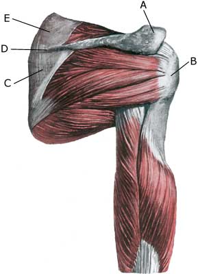
Anatomy: Around the shoulder joint are numerous bursas, reducing the pressure on muscles, tendons and ligaments, where these lie close to bone projections. Between the upper bone-projection (acromion) and the upper shoulder blade muscle (M Supraspinatus) lies the largest bursa in the shoulder (bursae subacromiale), which often communicates with the shoulder joint (Photo). |
||
|
||
| Cause: Upon repeated loads or blows, the bursa can become inflamed, increasing the production of fluids, swelling and becoming painful. Inflammation of the subacromiale bursae is often caused by working with the arm above the level of the head. When the arm is moved away from the body and above the level of the head (abducted) the supraspinatus muscle slips under the upper bone projection of the shoulder blade (acromion). With age, the supraspinatus tendon becomes stiff and unremitting, and is more easily squeezed between the head of the upper arm (caput humeri) and the acromion, causing the bursa to be squeezed and swell. Symptoms: Pain upon pressure on the bursa, which occasionally (but far from always) may feel swollen. Pain in the subacromiale bursae is worsened when the arm is at a right angle to the body. Inflammation of the bursa often causes nightly pain, and pain when lying on the side of the inflamed bursa. Acute treatment: Click here. Examination: In light cases with only minimal tenderness medical examination is not necessarily required. In case of pain that is more pronounced or lack of progress, a medical examination should be carried out to ensure the correct diagnosis and treatment. The doctor may carry out various clinical examinations, which not always allows a certain diagnosis (article). The diagnosis is most rapidly made with ultrasound (allowing simultaneous treatment) (article), (Ultrasonic image). Treatment: The treatment primarily involves relief. Possible removal of the provoking factor, if known. The treatment may be supplemented with rheumatic medicine (NSAID) or injection of corticosteroid in the bursa, possibly preceded by draining of the bursa. This procedure can advantageously be guided by ultrasound, (article). In the absence of any effect, an operative solution with removal of the bursa may be attempted (and in cases of bursitis subacromiale removal of a part of the shoulder’s upper bone projection (acromion). Thereby avoiding that the supraspinatus muscle and the bursa is squeezed between the head of the upper arm and the acromion, when the arm is raised). Rehabilitation, specific: The treatment is dependent on which bursa is inflamed, but sports activity can be carefully resumed once pain has diminished, especially if the triggering cause has been determined, and subsequently removed. Generally careful training of the shoulder muscles is recommended, primarily with the arm below the level of the head. Complications: If smooth progress is not achieved the correctness of the diagnosis, or whether complications have arisen should be considered. Amongst others the following should be considered:
|