Alle indlæg af urtehave_com

Fluid accumulation in the hip joint

Untitled Document

Diagnosis: FLUID ACCUMULATION IN THE HIP JOINT
(SYNOVITIS/COXITIS)


Anatomy:
The hip joint consists of the hip socket (acetabulum) and the femoral head (caput femoris). The articular surfaces are coated with a cartilage layer a few mm thick, which reduces the load on the articular surfaces.

Cause: Many repeated loads or one violent load can cause an inflammation of the synovial membrane (synovitis), fluid formation, swelling, restriction of movement and pain in the hip joint. The condition is relatively often seen in children(Drawing).

Symptoms: Smerter i leddet ved bevægelse med belastning. Ofte vil der være bevægelsesindskrænkning ved rotation i hofteleddet.

Examination: It will often be necessary to supplement the ordinary clinical examination with an ultrasound scan, where the fluid in the hip joint can easily be seen (Ultrasonic image).

Treatment: The treatment primarily comprises relief from the pain inducing activity until the swelling has abated. Rehabilitation, within the pain threshold, can subsequently be commenced. In cases of lack of progress with relief the treatment can be supplemented with a medical treatment in the form of rheumatic medicine (NSAID) or by drainage of the joint fluid which should be done with ultrasound guidance.

Rehabilitation of children and adolescents: When the pain has diminished, walking and gradually thereafter running, can be cautiously resumed within the pain threshold, in accordance with rehabilitation of children and adolescents in general.

Complications: lack of progress it should be considered if the diagnosis is correct (article). In particular you should consider a bacterial infection in the joint (pyarthron), which is determined by ultrasound guided draining of the joint fluid, the child hip diseases epifysiolysis capitis femoris (article), Calvé-Legg-Perthes disease (article) and inguinal hernia.

Inguinal hernia

Diagnosis: INGUINAL HERNIA
(HERNIA INGUINALIS)


Anatomy:
The various muscles of the abdominal wall are penetrated by the inguinal canal, which contains nerves (N ilioinguinalis and the genital branch of N genitofemoralis) and in men the spermatic cord (funilicus spermaticus). In women the inguinal canal instead contains a small fibrous ligament. Where the inguinal canal penetrates the abdominal wall weak spots arise (anulus inguinalis superficialis and anulus inguinalis profundus).

  1. M. recti abdominis
  2. Funiculus spermaticus
  3. Ligamentum inguinale
  4. Spina iliaca anterior superior
  5. M. obliquus externus abdominis

Inguinal canal

  1. Anulus inguinalis superficialis
  2. Crus mediale
  3. Funiculus spermaticus et m. cremaster
  4. V. femoralis
  5. Hiatus saphenus
  6. Lig. lacunare
  7. Anulus femoralis
  8. Margo falciformis (cornu superius)
  9. Lig inguinale
  10. Fibrae intercrurales
  11. M. obliquus externus abdominis

Right inguinal canal

Cause: If the abdominal wall becomes too weak, the intestines can be pressed through the weak spots in the abdominal wall causing a hernia.

Symptoms: Pain in the groin, aggravated when coughing. Sometimes a swelling in the groin is visible. Usually the swelling (containing intestine) can be pushed into place. If the swelling is painful and cannot be pressed into place the hernia may be strangulated (which requires acute medical assistance).

Examination: In obvious cases with visible swelling in the groin the diagnosis is easy to make. With lack of swelling in the groin the diagnosis is naturally difficult (article 1). A normal medical examination is usually sufficient in order to make the diagnosis, however, if there is any doubt concerning the diagnosis a MRI-scan or a dynamic ultrasound scan, where weaknesses in the abdominal wall can be detected while increasing the pressure in the abdominal cavity by applying pressure can be performed (article 2), (article 3).

Treatment: With modest discomfort, you can primarily attempt training of the abdominal muscles (article) . If discomfort continues, surgery is advised. If strangulated hernia is suspected you should seek acute medical assistance to evaluate the indication for acute surgery, as there is a risk of lasting damage to the intestine. With uncomplicated surgery, a relief period of approx. two months should be expected before maximal sports activity can be resumed (very dependent on the sport in question).

Rehabilitation of children and adolescents: In normal cases, sports activity will be able to be resumed after a few weeks without special rehabilitation. See: Rehabilitation of children and adolescents in general

Slipping of the femoral head in the growth zone

Diagnosis: SLIPPING OF THE FEMORAL HEADIN THE GROWTH ZONE
(epifysiolyse caput femoris)


Anatomy:
The hip joint consists of the hip socket (acetabulum) and the femoral head (caput femoris). The articular surface is covered with a few mm thick cartilage layer, which reduces the load on the articular surfaces.

  1. Caput femoris
  2. Collum femoris
  3. Trochanter minor
  4. Trochanter major

Femoral head

Cause: For unknown reasons, a slipping of the femoral head in the growth zone can occur in some cases. The condition is mainly seen in boys in the 11-16 years age group (article).

Symptoms: There will only be slight pain initially, with tiredness in the hip joint upon movement and being subjected to load, as well as limping and constriction of movement of the joint. Constriction of movement when rotating the hip joint will often be seen. Increased pain will be felt later on, with more pronounced limping. The pain can occasionally be felt in the knee instead of the hip.

Examination: It is important to undergo a medical examination as soon as possible so that the diagnosis can be made, as this is vital to achieve a good result from the treatment. It will often be necessary to supplement the examination with an x-ray (X-ray) (inclusive of Lauensteins projection), MRI scan or an ultrasound scan (article).

Treatment: Treatment should be commenced as soon as ever possible, and comprises surgery where the articular head is put back into place and fixed if necessary, as well as relief (article).

Rehabilitation of children and adolescents: The type of rehabilitation and load which can be permitted is completely dependant upon the severity of the condition. The rehabilitation should therefore be performed in close cooperation with the doctors controlling the treatment.

Complications: The condition can cause the risk of a lasting injury to the articular head (caput necrose) (article), and degenerative arthritis in the hip joint, as well as shortening of the leg and reduced mobility of the hip joint.

Calve Legg Perthe

Diagnosis: CALVÉ-LEGG-PERTHES DISEASE


Anatomy:
The hip joint consists of the hip socket (acetabulum) and the femoral head (caput femoris). The articular surface is covered with a few mm thick cartilage layer, which reduces the load on the articular surfaces.

  1. Caput femoris
  2. Collum femoris
  3. Trochanter minor
  4. Trochanter major

Femoral head

Cause: For unknown reasons, a slow collapse of the femoral head (aseptic necrosis of the bone) can occur, resulting in a disintegration and flattening of the femoral head and consequently irritation in the hip joint. However, secondhand smoke is recognised as playing a role  (article). The condition is mainly seen in children in the 3-11 age group, with boys being affected three times more often than girls. 10% of cases are on both sides of the hip.

Symptoms: There will only be slight pain initially, with tiredness in the hip joint upon movement and being subjected to load, as well as limping and constriction of movement of the joint. Constriction of movement when rotating the hip joint will often be observed. Increased pain will be felt later on, with more pronounced limping due to the shortening of the leg. The pain can occasionally be felt in the knee instead of the hip.

Examination: It is important to undergo a medical examination as soon as possible so that the diagnosis can be made, as this is vital to achieve a good result from the treatment. It will often be necessary to supplement the examination with an ultrasound scan (Ultrasonic image), where bone change and fluid in the hip joint can clearly be identified (article) and possibly also x-ray examination where the late bone change can be seen (X-ray).

Treatment: Treatment should be commenced as soon as ever possible, and primarily comprises intensive relief, with possible use of a wheel chair. The period of relief can last 1-2 years. An operative correction can be necessary if the disease leaves considerable damage (article).

Rehabilitation of children and adolescents: The type of rehabilitation and load which can be permitted is completed dependant upon the severity of the disease. The rehabilitation should therefore be performed in close cooperation with the doctors controlling the treatment

Complications: The condition can cause the risk of shortening of the leg and degenerative arthritis in the hip joint. There are, however, good chances for the articular head to heal to its normal shape, especially for the youngest children.

Inflammation of the heel fat pad

Diagnosis: INFLAMMATION OF THE HEEL FAT PAD


Anatomy:
Under the heel bone (calcaneus) there is a shock absorbing fatty pad (corpus adiposum) which protects the heel bone and the hollow foot tendon (aponeurosis plantaris) which fastens on the heel bone under the heel.

  1. Corpus adiposum
  2. M. flexor digitorum brevis
  3. M. flexor hallucis longus
  4. Aponeurosis plantaris

HEEL PAD, SOLE OF THE FOOT

Cause: inflammation of the heel pad occurs often after repeated, vigorous, overload (for example landing after jumping). The injury is often seen in gymnasts.

Symptoms: Pain when walking as well as when applying pressure on the edges of the heel pad. In some cases, it will be possible to see haemorrhaging in the heel pad (article).

Acute treatment: Click here.

Examination: Medical examination is not always necessary in light cases with minimal tenderness and no pain when walking. If satisfactory progress is not made, or if there is a sense of a “snap”, or sudden shooting pains, medical attention should be sought as soon as possible to make the diagnosis. A normal medical examination is usually sufficient in order to make the diagnosis, however, if there is any doubt concerning the diagnosis an ultrasound scan can be performed.

Treatment: Rest from the painful activity (running). The injury can in some cases heal within a few weeks if treatment is commenced at an early stage. It is imperative that footwear is in order, with a suitably tight heel cap to hold the heel pad in place under the heel, and with shock absorbing soles. 

Rehabilitation of children and adolescents: Treatment is primarily rest. The principles under of children and adolescents in general can be followed until the pain has gone, after which running and jumping can be cautiously resumed.

Bandage: Taping to relieve problems with inflammation of the heel pad is of questionable significance, but can be attempted as the tape will not invoke further injury if applied in the correct manner (tape-description).

Complications: If there is not a steady improvement in the condition, x-ray or an ultrasound scan should be performed to exclude :


Shock absorbing shoes or insoles will reduce the risk of inflammation of the heel pad.

Catilage damage in the foot

Diagnosis: CARTILAGE DAMAGE IN THE FOOT
(OSTEOKONDRAL LESION, OSTEOKONDRITIS DISSICANS)


Anatomy:
The surfaces of the joints are lined with a cartilage covering of a few millimetre’s thickness which serves to reduce the load or strain on the joint surfaces.

  1. Phalanx media
  2. Tuberositas ossis metatarsalis V
  3. Os cuboideum
  4. Calcaneus
  5. Talus
  6. Os naviculare
  7. Os cuneiforme laterale
  8. Os cuneiforme intermedium
  9. Os cuneiforme mediale
  10. Os metatarsalei
  11. Os sesamoideum
  12. Phalanx proximalis
  13. Phalanx distalis

THE FOOT FROM ABOVE

Cause: Localised cartilage injuries in the joint surfaces can occur after a vigorous twisting of the joint, where the joint surfaces impact on each other and cause cartilage damage. In some cases a piece of cartilage can be shed which can wander in the joint (joint mouse) and become jammed. In other cases, and for unknown reasons, a small piece of bone can loosen in the knee (osteochondritis dissecans).

Symptoms: Pain in the joint when under load or strain. Occasional inflammation of the synovial membrane which causes concentration of fluid in the joint.

Examination: Normal medical examination is often not sufficient. To make the diagnosis correctly it is therefore necessary to perform an MR-scan (article), and possibly an arthroscopic examination of the ankle joint.

Treatment: If a piece of bone is loosened in the knee (osteochondritis dissecans) it is important that the diagnosis is made as early as possible, as the earlier the treatment starts, the better the result (article). Treatment can comprise relief or surgery, dependant upon the extent of the condition and whether a piece of bone has become loose. If the condition is a result of twisting, treatment comprises relief from the painful activities until the pain is no longer experienced, after which gradual training can be commenced. There is no treatment that can restore the damaged cartilage, which has itself poor restorative ability. Different procedures to enhance the healing can be attempted using arthroscopic examination, however, the results are generally unsatisfactory (article-1), (article-2). Results from experimental cartilage transplants are still not successful enough to warrant introduction as a routine treatment in the near future. Joint mouse which provokes the symptoms must be surgically removed.

Rehabilitation of children and adolescents: The load and type of rehabilitation which can be permitted is completely dependant upon the severity of the condition, the location, and the form of treatment. Rehabilitation should therefore be performed in close cooperation with the doctors controlling the treatment.

Complications: Greater cartilage injuries which are positioned on the weight-bearing parts of the joint are some of the most serious sports injuries, and often result in an end to the sporting career.

Special: As there is a risk that the injury can be permanent, all cases should be reported to your insurance company.

Bone fracture in the foot

Diagnosis: BONE FRACTURE IN THE FOOT


Anatomy:
The foot bones comprise the 7 tarsal bones (ossa tarsi), the 5 metatarsal bones (ossa metatarsi) and the 14 bones in the toes (phalanx). Furthermore, the lower part of the shin bone (tibia) and calf bone (fibula) form a part of the ankle joint.

  1. Phalanx media
  2. Tuberositas ossis metatarsalis V
  3. Os cuboideum
  4. Calcaneus
  5. Talus
  6. Os naviculare
  7. Os cuneiforme laterale
  8. Os cuneiforme intermedium
  9. Os cuneiforme mediale
  10. Os metatarsalei
  11. Os sesamoideum
  12. Phalanx proximalis
  13. Phalanx distalis

THE FOOT FROM ABOVE

Cause: A blow or violent twist can cause a fracture of the bone. (X-ray picture).

Symptoms: Pain when applying pressure (direct or indirect tenderness), and when applying load or strain.

Acute treatment: Click here.

Examination: X-ray examination will usually reveal the fracture (article-1)(article-2). The fracture can in some cases first be seen after 14 days, thus the x-ray examination should be repeated if there is a continued suspicion of a fracture.

Treatment: Treatment is completely dependent upon which bones are broken, and whether there is a dislocation of the fracture. In some cases relief and rest without bandaging can be opted for, whereas other types of fracture require bandaging and possibly surgical intervention (article) (X-ray picture).

Rehabilitation of children and adolescents: The load and type of rehabilitation which can be permitted is completely dependent upon the severity, and the treatment of the fracture. It is therefore important that the rehabilitation is performed in close cooperation with the doctors controlling the treatment.

Complications: If there is not a steady improvement in the condition a medical examination should be performed once more to ensure that the fracture is healing according to plan. In some cases, a false joint can develop which will require (renewed) surgical treatment (X-ray picture). There can be a risk of growth disturbance if the fracture includes the shin bone’s growth zones at the ankle (article).

Ligament injury in the ankle joint, outer ligament

Diagnosis: LIGAMENT INJURY IN THE ANKLE JOINT, OUTER LIGAMENT
(RUPTURA TRAUMATICA LIGAMENTI LATERALIS PEDIS)


Anatomy:
The ankle joint is stabilised by a joint-capsule as well as a wide fan shaped ligament on the inside (ligamentum deltoideum/mediale), and a set of outer ligaments (ligamentum talofibulare anterius fore, ligamentum calcaneofibulare centre, and ligamentum talofibulare posterius at the rear). There is also a strengthening of the ligaments in front and behind (ligamentum tibiofibulare anterius & posterius). The surrounding tendons are often enclosed by tendon sheaths and joined to the bones with connective tissue strings. The ligaments stabilise the ankle joint, especially when twisting and running with sudden directional changes. (Photo).

  1. Lig. talofibulare anterius
  2. Lig. calcaneofibulare
  3. Lig. talofibulare posterius
  4. Fibula

OUTER ANKLE JOINT

Cause: A rupture of the of the outer lateral ligaments in the ankle joint arises if the foot is twisted such that the ligaments are over-stretched and finally rupture. In slight cases the injury can be termed a strain or sprain, and in more serious instances as full or partial rupture or tear. Consequential injuries are often associated with ligament ruptures, amongst others concentration of fluid in the joint (traumatic arthritis/synovitis) and inflammation of the ankle joint tendon sheath (article) ,however, these are often unfortunately overlooked (article).

Symptoms: Pain on and under the outer ankle bone (malleolus lateralis), swelling due to bleeding, pain when walking.

Acute treatment: Click here.

Examination: A medical examination is not necessarily required for very minor cases (slight sprain) with only minimal swelling and no discomfort when walking. The extent of the swelling is, however, not always a mark of the degree of the injury. Medical examination is recommended with more extensive swelling or pain, in order to eliminate bone fracture in the ankle and a rupture of the ligament between the shin and calf bones (syndesmosis rupture). A normal medical examination is usually sufficient in order to make the diagnosis (article).X-ray examination should be performed in all cases where there is a suspicion of a fracture or syndesmosis rupture (article). Vigorous twisting of the ankle in the acute stage to appraise the degree of looseness is not indicative, as this has no influence on the choice of treatment (article). Ultrasound examination can give valuable information when the ligament is ruptured (Ultrasonic image)

Treatment: Treatment of ligament injuries is today conservative (rehabilitation). Many patients have earlier undergone operations and setting of plaster cast, however, this course of treatment has practically been abandoned in relation to uncomplicated ligament ruptures, although there is not definitive agreement on this subject (article-1), (article-2).

Rehabilitation of children and adolescents: INSTRUCTION

Bandage: t is recommended to use tape in the course of rehabilitation when starting to run on an uneven surface, or with sudden directional change. Nerve cells (proprioreceptors) in the ligaments transmit information to the brain on the position of the ankle joint. The brain sends information to the muscles, which are activated, ensuring that the ankle is held correctly. The nerve paths do not function in an optimal manner when the ligament is injured, and the risk of a new twist of the foot is increased thereby. Use of tape is primarily designed to stimulate the small nerve cells in the skin, thus enabling these to act as a substitute for the nerve cells temporarily damaged in the ligament. The function of the tape is thus not a pure mechanical stabilising of the ankle joint (tape-instruction). In some cases, certain forms of bandaging around the ankle can be used to advantage (article-1), (article-2).

Complications: If there is not a steady improvement in the condition consideration must be given as to whether the diagnosis is correct, or if complications have arisen:

  • Rupture of the ligament between shin and calf bones (syndesmosis rupture)
  • Bone fracture
  • Bone membrane tear (periosteal avulsion)
  • Luxation of the peroneus tendon
  • Inflammation of the tendon sheath
  • Concentration of fluid in the joint (traumatic arthritis/synovitis)
  • Cartilage damage in the joint

It is extremely rare that the injury results in a chronically loose ankle joint if rehabilitation is handled sensibly. If the injury does result in a chronically loose ankle joint, intensive co-ordination training must be recommended. Bandages can be tried if this is not sufficient, and if this still does not give the desired effects, surgical intervention to tighten the ligaments can be attempted. The results are usually quite acceptable (article). Reduced mobility in the ankle joint is a common condition following ankle injuries in children (article).

Special: Seesaw exercise is important in the rehabilitation phase, as well as in a preventive capacity. As a preventive measure, seesaw exercises should be performed frequently throughout the rest of the active sporting career if ligament injuries in the ankle joint have previously been experienced. Begin by standing with both feet on the seesaw and use hands for support on the wall. Gradually let go of the support to finally train by standing on only one leg (article).

Ligament injury in the ankle joint, inner ligament

Diagnosis: LIGAMENT INJURY IN THE ANKLE JOINT, INNER LIGAMENT
(RUPTURA TRAUMATICA LIGAMENTI DELTOIDEI PEDIS)


Anatomy:
The ankle joint is stabilised by a wide fan shaped ligament on the inside (ligamentum deltoideum), and a set of outer ligaments (fore, centre, and at the rear). The ligaments stabilise the ankle joint, especially when twisting and running with sudden directional changes (Photo).

  1. Ligamentum mediale/deltoideum
  2. Calcaneus
  3. Talus
  4. Tibia

INNER ANKLE JOINT

Cause: A rupture of the of the inner ligaments in the ankle joint arises if the foot is twisted such that the ligaments are overstretched and finally rupture. In slight cases the injury can be termed a strain or sprain, and in more serious instances as full or partial rupture or tear. Consequential injuries are often associated with ligament ruptures, amongst others concentration of fluid in the joint (traumatic arthritis/synovitis) and inflammation of the ankle joint tendon sheath, (article), however, these are often unfortunately overlooked (article).

Symptoms: Pain on and under the inner ankle bone (malleolus medialis), swelling due to bleeding, pain when walking.

Acute treatment: Click here.

Examination: A medical examination is not necessarily required for very minor cases (slight sprain) with only minimal swelling and no discomfort when walking. The extent of the swelling is, however, not always a mark of the degree of the injury. Medical examination is recommended with more extensive swelling or pain, in order to eliminate bone fracture in the ankle and a rupture of the ligament between the shin and calf bones (syndesmosis rupture). A normal medical examination is usually sufficient in order to make the diagnosis (article). X-ray examination should be performed in all cases where there is a suspicion of a bone fracture, or syndesmosis rupture (article). Vigorous twisting of the ankle in the acute stage to appraise the degree of looseness is not indicative, as this has no influence on the choice of treatment (article). Ultrasound examination can give valuable information when the ligament is ruptured (Ultrasonic image).

Treatment: Treatment of ligament injuries is today conservative (rehabilitation). Many patients have earlier undergone operations and setting of plaster cast, however, this course of treatment has practically been abandoned in relation to uncomplicated ligament ruptures (article).

Rehabilitation of children and adolescents: INSTRUCTION

Bandage: It is recommended to use tape in the course of rehabilitation when starting to run on an uneven surface, or with sudden directional change. Nerve cells (proprioreceptors) in the ligaments transmit information to the brain on the position of the ankle joint. The brain sends information to the muscles, which are activated, ensuring that the ankle is held correctly. The nerve paths do not function in an optimal manner when the ligament is injured, and the risk of a new twist of the foot is increased thereby. Use of tape is primarily designed to stimulate the small nerve cells in the skin, thus enabling these to act as a substitute for the nerve cells temporarily damaged in the ligament. The function of the tape is thus not a pure mechanical stabilising of the ankle joint (tape-instruction). In some cases, certain forms of bandaging around the ankle can be used to advantage.

Complications: If there is not a steady improvement in the condition consideration must be given as to whether the diagnosis is correct, or if complications have arisen:

  • Rupture of the ligament between shin and calf bones (syndesmosis rupture)
  • Bone membrane tear (periosteal avulsion)
  • Inflammation of the tendon sheath
  • Concentration of fluid in the joint (traumatic arthritis/synovitis)
  • Cartilage damage in the joint

It is extremely rare that the injury results in a chronically loose ankle joint if rehabilitation is handled sensibly. If the injury does result in a chronically loose ankle joint, intensive co-ordination training must be recommended. Bandages can be tried if this is not sufficient, and if this still does not give the desired effects, surgical intervention to tighten the ligaments can be attempted. Reduced mobility in the ankle joint is a common condition following ankle injuries in children (article).

Special: Seesaw exercise is important in the rehabilitation phase, as well as in a preventive capacity. As a preventive measure, seesaw exercises should be performed frequently throughout the rest of the active sporting career if ligament injuries in the ankle joint have previously been experienced. Begin by standing with both feet on the seesaw and use hands for support on the wall. Gradually let go of the support to finally train by standing on only one leg (article). Special bandages have in some studies been shown to reduce the risk of ligament injuries. (article-1), (article-2).

Inflammation of the hollow foot tendon

Diagnosis: INFLAMMATION OF THE HOLLOW FOOT TENDON
(FASCIITIS PLANTARIS)


Anatomy:
The calf muscle (M Gastrocnemicus) is comprised of two muscle heads which gather in a wide tendinous ligament and continue in to the Achilles tendon. Another of the larger calf muscles (M Soleus) is attached to the front side of the Achilles tendon and thus forms a part of the Achilles tendon. The calf muscles are attached to the rear of the heel bone (calcaneus) via the Achilles tendon. The hollow foot tendon (aponeurosis plantaris) runs from the heel bone under the sole, and is attached to all five toes. The hollow foot tendon is a functional extension of the Achilles tendon, and is instrumental in maintaining the arch running the length of the foot (Photo).

  1. Aponeurosis plantaris
  2. Tuber calcanei

SOLE OF THE FOOT

Cause: Occurs after repeated overload in the form of running or jumping. Since the hollow foot tendon is a functional extension of the Achilles tendon, the provoking factors which are instrumental in injuries due to overload of the hollow foot tendon and the Achilles tendon are often the same.

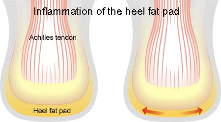
Symptoms: Pain when running and jumping, as well as when applying pressure at the attachment point of the hollow foot tendon directly under the heel bone. The pain is often most pronounced slightly on the inner side of the attachment.

Examination: Medical examination is not necessarily required in slight, early cases where the tenderness is slowly increasing without sudden worsening. A normal medical examination is usually sufficient in order to make the diagnosis, and in all cases when there is a sense of a “crack”, or sudden shooting pains in the tendon, medical attention should be sought as soon as possible to exclude a (partial) rupture of the hollow foot tendon and bone fracture. This situation is best determined by use of ultrasound scanning (or MRI examination). In cases where satisfactory progress is not in evidence, an ultrasound examination should be performed. Ultrasound scanning enables an evaluation of the extent of the change in the tendon; inflammation of the tendon (tendinitis), development of cicatricial tissue (tendinosis), calcification, Inflammation of the tissue surrounding the tendon (peritendinitis), as well as (partial) rupture (article) (Ultrasonic image).

Treatment: Rest from the painful activity (running). The injury can in some cases heal within a few weeks if treatment is commenced at an early stage (article). If the pain has been present for several months, and especially if ultrasound scanning reveals thickening and change in the tendon, a rehabilitation period of several months must be anticipated. It is imperative that the footwear is in order, with good running shoes including shock absorbing heels. Pressure on the hollow foot tendon can be relieved by walking in shoes with an arch support. Treatment with ice can be repeated every time the hollow foot tendon becomes tender during the rehabilitation phase. Injection of corticosteroid is not appropriate in the course of treatment (article).

Rehabilitation of children and adolescents: INSTRUCTION

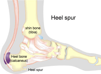
Heel spur: Inflammation of the hollow foot tendon is occasionally connected to a heel spur. A heel spur is a new bone development which is seen in connection with a prolonged Inflammation at the point of attachment of the hollow foot tendon on the heel bone due to overload. A heel spur is thus a consequence of an injury due to overload, and not the cause. If a heel spur has developed, it will never disappear. Heel spurs have no practical significance, and require no treatment. Many symptom free athletes have heel spurs without having had symptoms from the point of attachment of the hollow foot tendon. Surgical removal of heel spurs as performed in earlier days is now virtually no longer used


Bandage:
Taping to relieve problems with the hollow foot tendon is of questionable significance, but can be attempted as the tape will not invoke further injury if applied in the correct manner (tape-instruction).

Complications: If there is not a steady improvement in the condition a medical examination should be performed to exclude:

  • bone fracture in the foot
  • stress fracture
  • inflammation of the bursa
  • bone membrane tear (periosteal avulsion)
  • inflammation of the tendon sheath

Special: As prolonged overload or strain on the hollow foot tendon has a large risk of developing in to chronic Inflammation, which is extremely difficult to treat, it is important to prevent the injury from arising or recurring. The principles in “Rehabilitation, general” should be followed to ensure that quickly increasing training loads at the season start, or after an injury period, are avoided. It is important that running shoes fit well (tight heel cap, shock absorbing soles ). It is vital that the first signs of tenderness or pain are reacted upon, to enable the training to be adapted before the injury reaches the stage where continuing the sports activity may be at risk.