Alle indlæg af urtehave_com

Degenerative arthritis in the hip joint

DEGENERATIVE ARTHRITIS

Diagnosis: DEGENERATIVE ARTHRITIS
(Osteoarthritis)


Anatomy:
The hip joint consists of the hip socket and the femoral head (caput femoris). The articular surface is covered with a few mm thick cartilage layer, which reduces the load on the articular surfaces

Cause: In case of repeated loads, the cartilage primarily, and subsequently the bone below the cartilage, can be damaged (degenerative arthritis). The degenerative arthritis changes can in some cases cause an inflammation of the synovial membrane (synovitis) which causes fluid formation, swelling, movement restriction and pain in the hip joint.

Symptoms: Pain in the hip joint upon movement with load. There will often be movement restriction upon rotation in the hip joint.

Examination: Ordinary clinical examination is often sufficient to make the diagnosis. The examination can be supplemented with an X-ray examination. Ultrasound scan is the most suitable examination if you suspect a fluid accumulation in the hip joint.

Treatment: The treatment primarily comprises relief from the pain inducing activity until any swelling in the hip joint has decreased. Rehabilitation can subsequently be commenced with the primary goal to strengthen the muscles around the hip joint and preserve the joint mobility. There is no treatment that can restore the ruined cartilage (and bone). Cartilage transplants are not yet suitable for general degenerative arthritis changes. Upon swelling in the hip joint you can attempt to reduce the synovitis with rheumatic medicine (NSAID) or by attempting to drain the fluid and injecting corticosteroid, which should be conducted with ultrasound guidance to optimise the effect and minimize the risk. Pain without joint swelling is best treated with paracetamol. In cases of severe degenerative arthritis changes with pain when resting (at night) it may be necessary to replace the hip joint.

Complications: Degenerative arthritis which sits on the weight bearing parts of the joint is one of the most serious sports injuries, and often results in a termination of active sport. Cycling and swimming are significantly less stressful for the hip joint than running. In particular the following should be considered:

Special: Shock absorbing shoes or inlays will reduce the load.

Piriformis syndrom

MUSKELINFILTRATION I EN AF BALDEMUSKLERNE

Diagnosis: MUSCLE INFILTRATION (MYALGIA) IN 
THE BUTTOCK MUSCLES

(PIRIFORMIS SYNDROM)


Anatomy:
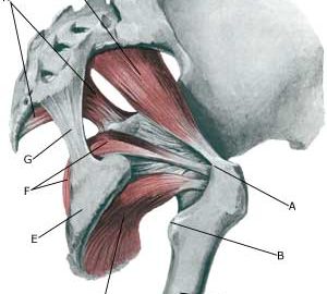
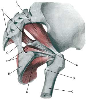
The piriformis muscle is a small muscle which origin from the sacrum and fastens on the outer femoral bone projection (tronchanter major). The muscle assists in rotating the thigh outwards.

  1. Trochanter major
  2. Trochanter minor
  3. Femur
  4. M. obturatorius externus
  5. Tuber ischiadicum
  6. M. obturatorius internus
  7. Lig. sacrotuberale
  8. Lig. sacrospinale et. m coccygeus
  9. M. piriformis

M. PIRIFORMIS

Cause: If the muscle is over-loaded, it will become taut and tender. In some cases, the muscle can become so taut that it jams against the ischias nerve (nervus ischiadicus).

Symptoms: Pain deep in the buttock, with periodic radiation into the leg.

Examination: The diagnosis will be rendered probable under clinical examinations by demonstrating tenderness by applying pressure on the muscle deep within the buttock, as well as provoking pain by stretching and activating the muscle. There is no suitable method to provide a pictorial image of the muscle (MR scanning can be attempted in the event of suspicions of nerve impingement) (article).

Treatment: The treatment usually comprises stretching and subsequent strength training of the muscles surrounding the lower back and buttocks. It is only in very rare cases involving nerve impingement that surgery is indicated. Uncomfortable pain can be treated medicinally in the form of paracetamol, or possibly rheumatic medicine (NSAID) . If this treatment does not provide the desired relief, ultrasound guided injection of corticosteroid in the most tender part of the muscle can be attempted (article 1), (article 2).

Special: Shock absorbing shoes or inner inlays will reduce the risk of various forms of muscle infiltrations. In the event of unsatisfactory progress, or relapse after successful rehabilitation, consideration must be given to performing an analysis of the patient’s running style to establish whether a correction of the running style should be recommended.

Complications: If progress is not smooth, the correctness of the diagnosis should be considered or whether complications have arisen. The following should be considered in particular:

Bursitis at the outer femoral bone projection (bursitis trochanterica)

BURSITIS AT THE OUTER FEMORAL BONE PROJECTION

Diagnosis: BURSITIS AT THE OUTER FEMORAL BONE PROJECTION
(Bursittis trochanterica)


Anatomy:
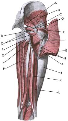
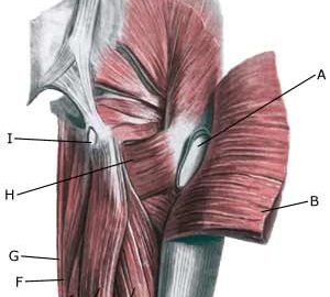
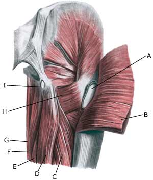
On the outside of the outer femoral bone projection (trochanter major) is a large bursa which reduces the load on muscles and tendons when these slide over the bone projection.

  1. M. piriformis
  2. M. gluteus minimus
  3. Bursa m. piriformis
  4. Bursae trochantericae m. glutei medii
  5. M. gluteus medius (resectus)
  6. Bursa trochanterica m. glutei maximi
  7. M. gluteus maximus
  8. Bursae intermusculares mm. gluteorum
  9. Tuberositas glutea
  10. Tractus iliotibialis
  11. M. biceps femoris
    (caput longum)
  12. M. biceps femoris
    (caput breve)
  13. M. adductor magnus
  14. B. subtendinea m. bicipitis femoris superior
  15. Tuber ischiadicum
  16. B. ischiadica m. glutei maximi
  17. Mm. gemilli sup. Et inf.
  18. B. ischiadica m. obturatorii interni

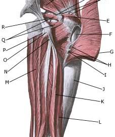
GLUTEAL MUSCLES FROM THE REAR

Cause: In case of repeated loads or blows the bursa can become inflamed, produce fluid, swell and become painful.

Symptoms: Pain when applying pressure on the thigh corresponding to the bursa, which sometimes (but far from always) can feel swollen. The pain can radiate down the thigh.

Acute treatment: Click here.

Examination: In light cases with only minimal tenderness medical examination is not necessarily required. In case of more pronounced pain or lack of progress, a medical examination should be carried out to ensure the diagnosis and commencement of any treatment. A normal medical examination is usually sufficient in order to make the diagnosis, however, if there is any doubt concerning the diagnosis an ultrasound scan can be performed (Ultrasonic image).

Treatment: The treatment primarily comprises relief. If the direct cause of the complaint is known, it should of course be removed. The treatment can be supplemented with rheumatic medicine (NSAID) or injection of corticosteroid in the bursa, which is best performed under the guidance of ultrasound.

Complications: If progress is not smooth, the correctness of the diagnosis should be considered or whether complications have arisen:

Special: Shock absorbing shoes or inlays will reduce the load. In case of lack of progress or recurrences after successful rehabilitation it can be considered to have a running style analysis, to evaluate whether correction of the running style is indicated.

Muscle rupture of the tendon fastening on the ischiatic bone

RUPTURE OF THE POSTERIOR THIGH MUSCLE FASTENING ON THE ISCHIATIC BONE

Diagnosis: RUPTURE OF THE POSTERIOR THIGH MUSCLE FASTENING ON THE ISCHIATIC BONE
(RUPTURA MUSCULI)


Anatomy:
The large posterior thigh muscles (hamstring muscles) have a common muscle tendon fastening on the ischiatic bone (tuber ischii). The posterior thigh muscles flex the knee and stretch the hip.


  1. Bursa trochanterica m. glutei maximi
  2. M. gluteus maximus
  3. M. biceps femoris (caput longum)
  4. M. semitendinosus
  5. M. semimembranosus
  6. M. adductor magnus
  7. M. gracilis
  8. M. quadratus femoris
  9. Bursa ischiadica m. glutei maximi

(Photo)

RIGHT GLUTEAL MUSCLES 
FROM THE REAR

Cause: When a muscle is subjected to a load beyond the strength of the muscle (typically sprinting), a rupture occurs. The vast majority of ruptures are partial muscle ruptures. Many of the ruptures at the muscle fastening on the ischiatic bone (tuber ischiadicum) er preceded by lengthy tendinitis (entesopatia tuber ischiadicum) at the same location. In rare cases, the muscle fastening can tear a piece of the ischiatic bone off (especially seen in children as the growth zone on the ischiatic bone (apophysis) increases the risk of tears).

Symptoms: In slight cases a local tenderness is felt after being subjected to load (“sprained muscle”, “imminent pulled muscle”). In severe cases sudden shooting pains are felt in the muscle (“partial muscle rupture”, “pulled muscle”) and in the worst case a sudden snap is felt rendering the muscle unusable (“total muscle rupture”). The following three symptoms are characteristic in connection with muscle injuries: pain upon applying pressure, stretching and activation of the muscle (flexing knee) against resistance. With total ruptures a defect and a swelling (the contracted muscle belly and bleeding) can often be seen and felt in the posterior thigh muscle just below the ischiatic bone and below. 

Acute treatment: Click here.

Examination: In very slight cases with only minimal tenderness and no discomfort when walking, medical examination is not necessarily required. The severity of the tenderness is however, not always a measure of the extent of the injury. In cases of more pronounced tenderness or pain, medical examination is required to ensure the diagnosis and treatment. The diagnosis is usually made following normal medical examination, however, if there is any doubt concerning the diagnosis, ultrasound scanning (or MRI scanning) can be performed, as these are the most suitable examinations to ensure the diagnosis (article). The larger the bleeding as assessed by ultrasound scanning, the longer the period needed to heal the injury. Tears on the ischiatic bone will usually be visible on x-rays.

Treatment: Treatment comprises relief and rehabilitation. The aim of the rehabilitation is to strengthen the posterior thigh muscles to enable the muscles to manage the loads which previously caused the rupture. It is only in very rare cases where there is a total rupture at the muscle fastening or tearing of a large piece of bone from the ischiatic bone that surgery is considered (article). Even large ruptures in the thigh muscles will usually be able to be healed and rehabilitated without giving functional disorder (but often cosmetic disfigurement with an irregular thigh muscle).

Complications: If steady progress is not experienced it should be considered if the diagnosis is correct or whether complications have arisen. The following should in particular be considered:

Bursitis at the ischiatic bone

Diagnosis: INFLAMMATION OF THE BURSA AT THE ISCHIATIC BONE
(BURSITTIS ISCHIADICA)


Anatomy:
The large posterior thigh muscles (hamstring muscles) have a common muscle tendon fastening on the ischiatic bone (tuber ischii). Below the tendon fastening there is a bursa which reduces the load on the tendon when the tendon slides against the bone. The posterior thigh muscles flex the knee and stretch the hip.

  1. Bursa trochanterica m. glutei maximi
  2. M. gluteus maximus
  3. M. biceps femoris (caput longum)
  4. M. semitendinosus
  5. M. semimembranosus
  6. M. adductor magnus
  7. M. gracilis
  8. M. quadratus femoris
  9. Bursa ischiadica m. glutei maximi

(Photo)

RIGHT GLUTEAL MUSCLES
FROM THE REAR

Cause: In case of repeated loads or blows the bursa can become inflamed, produce fluid, swell and become painful.

Symptoms: Pain upon applying pressure on the bursa (sitting position), which sometimes (but far from always) may feel swollen. Aggravated upon stretching and activation of the posterior thigh muscles (flexing of the knee against resistance) (article).

Acute treatment: Click here.

Examination: In slight cases with only minimal tenderness, medical examination is not necessarily required. In cases of more pronounced pain or lack of progress, a medical examination should be carried out to ensure a correct diagnosis and commencement of treatment. The diagnosis is usually made on the basis of a normal medical examination, however, if there is any doubt surrounding the diagnosis, it can easily and quickly be confirmed under an ultrasound scan.

Treatment: The treatment primarily consists of relief. The treatment can be supplemented with rheumatic medicine (NSAID) or the injection of corticosteroid in the bursa preceded by draining of the bursa, which can advantageously be done under ultrasound guidance.

Complications: If progress is not smooth, it should be considered if the diagnosis is correct or whether complications have arisen. The following should in particular be considered:

cause-article1

SportNetDoc

Deltoid and syndesmosis ligament injury of the ankle without fracture.

Miller CD, Shelton WR, Barrett GR, Savoie FH, Dukes AD. Am J Sports Med 1995 Nov-Dec;23(6):746-50.

Ankle diastasis without fracture is a rare injury with few examples reported. We report on four male patients, aged 16 to 18 years, who sustained this injury playing football. Swelling and tenderness over both the deltoid and syndesmosis ligaments are the most common physical findings. Plain ankle radiographs demonstrated lateral talus subluxation in three patients, and a stress radiograph demonstrated subluxation of the talus in one patient. Treatment consisted of reduction and fixation of the syndesmosis with a screw followed by 6 weeks of cast immobilization. Using the scale developed by Edwards and DeLee, three patients had excellent results and one had a good result. Diagnosis of tears of the deltoid and syndesmosis ligaments without fracture requires a high index of suspicion on the physician’s part. In patients whose mortise is more than 1 mm subluxated, reduction and screw fixation will produce good results.

prevention-article2

SportNetDoc

The prevention of ankle sprains in sports. A systematic review of the literature.

Thacker SB, Stroup DF, Branche CM, Gilchrist J, Goodman RA, Weitman EA. Am J Sports Med 1999 Nov-Dec;27(6):753-60.

To assess the published evidence on the effectiveness of various approaches to the prevention of ankle sprains in athletes, we used textbooks, journals, and experts in the field of sports medicine to identify citations. We identified 113 studies reporting the risk of ankle sprains in sports, methods to provide support, the effect of these interventions on performance, and comparison of prevention efforts. The most common risk factor for ankle sprain in sports is history of a previous sprain. Ten citations of studies involving athletes in basketball, football, soccer, or volleyball compared alternative methods of prevention. Methods tested included wrapping the ankle with tape or cloth, orthoses, high-top shoes, or some combination of these methods. Most studies indicate that appropriately applied braces, tape, or orthoses do not adversely affect performance. Based on our review, we recommend that athletes with a sprained ankle complete supervised rehabilitation before returning to practice or competition, and those athletes suffering a moderate or severe sprain should wear an appropriate orthosis for at least 6 months. Both coaches and players must assume responsibility for prevention of injuries in sports. Methodologic limitations of published studies suggested several areas for future research.

prevention-article1

SportNetDoc

The effect of preventive measures on the incidence of ankle sprains.

Verhagen EA, van Mechelen W, de Vente W. Clin J Sport Med 2000 Oct;10(4):291-6.

OBJECTIVE.
To critically review the current data concerning the efficacy of preventive measures described in the literature, on the incidence of lateral ankle ligament injuries.

DATA SOURCES.
MEDLINE, Sportdiscus, and EMBASE were searched for papers published between 1980 and December 1998. Keywords used in the search were “prevention” in combination with “ankle,” “ankle taping,” “ankle bracing,” “orthosis,” “shoes,” and “proprioception.” Additional references were reviewed from the bibliographies of the retrieved articles.

STUDY SELECTION.
A study was included if: 1) the study contained research questions regarding the prevention of lateral ankle ligament injuries; 2) the study was a randomized controlled trial, a controlled trail, or a time intervention; 3) the results of the study contained incidence rates of lateral ankle ligament injuries as study outcome; and 4) the study met the cut-off score set for quality.

DATA EXTRACTION AND SYNTHESIS.
Two reviewers reviewed relevant studies for strengths and weaknesses in design and methodology, according to a standardized set of predefined criteria. Eight relevant studies met the criteria for inclusion and were analyzed.

MAIN RESULTS.
Overall, all studies reported a significant decrease in incidence of ankle sprains using the studied preventive measure. There was a great variety in methodology and study design between the eight analyzed studies, and every study had one or more drawbacks. Therefore, between studies only general results could be compared.

CONCLUSIONS.
The use of either tape or braces reduces the incidence of ankle sprains. Next to this preventive effect, the use of tape or braces results in less severe ankle sprains. However, braces seem to be more effective in preventing ankle sprains than tape. It is not clear which athletes are to benefit more from the use of preventive measures: those with or those without previous ankle sprains. The efficacy of shoes in preventing ankle sprains is unclear. It is likely the newness of the footwear plays a more important role than shoe height in preventing ankle sprains. Proprioceptive training reduces the incidence of ankle sprains in athletes with recurrent ankle sprains to the same level as subjects without any history of ankle sprains.

examination-article1

SportNetDoc

Rupture of the tibiofibular syndesmosis without osseous fibular injury.

Ruf W, Friedl P, Frobenius H. Aktuelle Traumatol 1987 Aug;17(4):153-6.

Within 3 years 12 injuries of the anterior fibulo-tibial ligament without fracture of the fibula were recorded prospectively. In relation to the total number of the ankle joint fractures during the same period the incidence is 3.3%. The rupture of the ligament arose in all cases from a forced eversion combined with supination or pronation of the foot. Clinical characteristics are the circumscribed painful palpation of the area of the ligament together with eversion pain of the foot. Arthrography is the most sensitive diagnostic procedure, which, however, may be avoided if the clinical situation is absolutely clear. Differential diagnosis consists mainly in the rupture of the fibulo-talar ligament including a tear of the anterior capsule of the ankle joint. Treatment should always be surgical – suture of the ligament, reinforcement of the syndesmosis by means of a positioning screw. Aftercare is functional without external fixation.

KONDITION

STEP4

Training ladder for:
RUPTURE OF THE JOINT-CAPSULE AT THE FRONT OF THE ANKLE JOINT
(RUPTURA TRAUMATICA LIGAMENTI PEDIS)

STEP 4

KONDITION
Unlimited: Cycling. Swimming. Running with directional change.

UDSPÆNDING
(10 min)

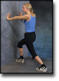
Stand with the injured leg stretched backwards with the toes facing front. Slowly bend the knee so that the calf muscles become increasingly stretched. Hold the position for 20 seconds and relax for 20 seconds before repeating.

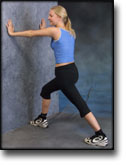
Stand with the injured leg outstretched with the ankle joint bent up against the wall. Press your abdomen against the wall so that the calf muscles become increasingly stretched. Hold the position for 20 seconds and relax for 20 seconds before repeating.

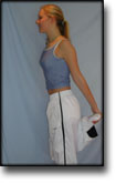
Stand on the good leg while gaining support on the wall with the hand on the same side. Bend the knee of the injured leg and draw the heel towards the buttocks. Take hold of the big toe side of the foot and draw slowly upwards so that the ankle joint is stretched to the maximum and drawn slightly outwards so that increased stretching is experienced on the inside of the shin bone. Hold the position for 20 seconds and relax for 20 seconds before repeating.

Stand on the good leg while gaining support from the wall with the opposite hand. Bend the knee of the injured leg and draw the heel towards the buttocks. Take hold of the little toe side of the foot and draw slowly upwards so that the ankle joint is stretched to the maximum and drawn over the good leg so that increased stretching is experienced on the outer side of the shin bone. Hold the position for 20 seconds and relax for 20 seconds before repeating.

KOORDINATION
(15 min)

Stand on the leg to be trained. Take-off and land on the same leg.

Seesaw. Balance on two legs, possibly using a hand as support against the wall, balancing subsequently on one leg without support. Look straight ahead and keep knees bent.

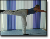
Stand on the injured leg with your upper body bent forwards at 90 degrees. Lift the good leg in a straight line behind you. When you feel comfortable with the exercise, it can be made more difficult by closing your eyes.

STYRKE
(35 min)

Sit on the floor. Tip the foot up and down with elastic under the forefoot so that the elastic becomes taut.

Sit on the floor. Tip the foot up and down with elastic on top of the forefoot so that the elastic becomes taut.

Sit on the floor. Tip the foot from side to side with elastic on the outer side of the foot, without moving the knee.

Sit on the floor. Tip the foot from side to side with elastic on the inner side of the foot, without moving the knee.

Stand behind a chair. Rise slowly up on tiptoe and go down again.

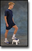
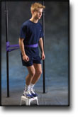
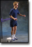
Up and down from the stool with load. Tie elastic around the hip and go up on the stool in a slow movement. The elastic should be fastened to the wall.

Go up and down from the stool. Go up with alternating right and left legs.

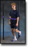
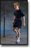
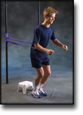
Stand on the healthy leg with elastic fixed around the hip. The elastic should be fixed to the wall or a wall bar. Take-off on the healthy leg and land on the leg to be trained and keep your balance. Remember that the elastic should be positioned so that it gives resistance at the moment of take-off. Change legs.

Stand with both legs on the stool with elastic around the hip. Take-off and land with feet together.

Stand with feet together. Using the ankle joint to take off, hop approx. 5 cm and land on both feet. The exercise should be done on one leg when you are able to do it without discomfort using both legs.

Stretching is carried out in the following way: stretch the muscle group for 3-5 seconds. Relax for 3-5 seconds. The muscle group should subsequently be stretched for 20 seconds. The muscle is allowed to be tender, but must not hurt. Relax for 20 seconds, after which the procedure can be repeated. The time consumed for stretching, coordination and strength training can be altered depending on the training opportunities available and individual requirements.