Kategoriarkiv: Hip

Stress fracture in the femoral neck

STRESS FRACTURE IN THE FEMORAL NECK

Diagnosis: STRESS FRACTURE IN THE FEMORAL NECK
(Stress fraktur)


Anatomy:
The femur and the hip bone form the hip joint.


  1. Caput femoris
  2. Collum femoris
  3. Trochanter minor
  4. Trochanter major

PELVIS AND THIGH BONE FROM THE FRONT


Cause: Repeated loads, especially when walking or running can cause cracks (stress fractures) in the femoral neck (collum femoris) (article-1) (article-2).

Symptoms: Pain in the hip when applying pressure (direct and indirect tenderness) and when under load (walking, running).

Examination: X-ray. Since many stress fractures cannot be seen early in the course of events, X-ray examination can be repeated after a few weeks. Scintigraphy, CT- and MRI and ultrasound scan can often diagnose stress fractures far earlier than X-rays (Ultrasonic image).
It is imperative for the result of the treatment that the diagnosis is made as early as possible (article).

Treatment: Relief. In some cases surgery is necessary (article).

Rehabilitation: The rehabilitation is completely dependent on the type of fracture and treatment (conservative or operative).
Also read rehabilitation, general.

Complications: If progress is not smooth, you should be re-examined to ensure that the fracture heals according to plan. In some cases a false joint can be formed (pseudoarthrosis), which requires surgical treatment.

Special: Shock absorbing shoes or inlays will reduce the load.

Fluid accumulation in the joint

FLUID ACCUMULATION IN THE JOINT

Diagnosis: FLUID ACCUMULATION IN THE JOINT
(Synovitis / coxitis)


Anatomy:
The hip joint consists of the hip socket (acetabulum) and the femoral head (caput femoris). The articular surfaces are coated with a cartilage layer a few mm thick, which reduces the load on the articular surfaces.

Cause: Many repeated loads or one violent load can cause an inflammation of the synovial membrane (synovitis), fluid formation, swelling, restriction of movement and pain in the hip joint. The condition is relatively often seen in children (Ultrasonic image).

Symptoms: Pain in the joint upon movement with load. Often there will be movement restriction with rotation of the hip joint.

Examination: It will often be necessary to supplement the ordinary clinical examination with an ultrasound scan, where the fluid in the hip joint can easily be seen (Ultrasonic image).

Treatment: The treatment primarily comprises relief from the pain inducing activity until the swelling has abated. Rehabilitation, within the pain threshold, can subsequently be commenced. In cases of lack of progress with relief the treatment can be supplemented with a medical treatment in the form of rheumatic medicine (NSAID) or by drainage of the joint fluid and the injection of corticosteroid, which should be done with ultrasound guidance.

Complications: In particular you should consider a bacterial infection in the joint (pyarthron), which is determined by ultrasound guided draining of the joint fluid, the child hip diseases (epifysiolysis capitis femoris and Calvé-Legg-Perthes disease), where the joint head on the femur slips or collapses (X-ray examination will determine the diagnosis), arthritis or:

Special: There are two childrens’ hip diseases that should always be considered in children with hip pain.

  • SLIPPING OF THE FEMORAL HEAD IN THE GROWTH ZONE (EPIFYSIOLYSIS CAPITIS FEMORIS), which mainly affects boys age 11-16 years (article). There will often be limping, groin pain, but sometimes the pain is sensed in the knee. It is important in order to obtain a good result from the treatment to be examined by a doctor and have the diagnosis made as soon as possible (with X-rays).

  • CALVÉ-LEGG-PERTHES DISEASE is a disease which mainly affects boys age 3-11 years (article). The bone core in the femoral head is dissolved and flattened. There will often be limping, tiredness and pain in the groin, but once in a while the pain is sensed in the knee instead. It is important in order for a good result of the treatment to be examined by a doctor and have the diagnosis made as soon as possible (by means of X-rays or ultrasound).

Inner snapping hip

INNER SNAPPING HIP

Diagnosis: INNER SNAPPING HIP
(Coxae saltans, intern)


Anatomy:
The deep hip flexor (M iliopsoas) consists of two muscles. The Psoas muscle originate from the lumbar vertebrae and the Iliacus muscle from the inside of the femur. The two muscles fuse and are both fastened on the inside of the femur (trochantes minor). The iliopsoas is the strongest flexor muscle of the hip.

  1. Origines m. psoatis
  2. M. psoas major
  3. M. iliacus
  4. M. psoas major
  5. M. psoas minor

THIGH FROM THE FRONT

Cause: In case of repeated loads the muscle tendon can become inflamed, swell and with some movements slip over the one of the bone projections at the pelvis or the hip joint. When the tendon slips over the bone projection, the uncomfortable symptoms are produced. There are three known causes of snapping hips: outer snapping hip (tractus iliotibialis), inner snapping hip (iliopsoas tendon) and joint conditioned (intraarticular) causes (article). The treatment is dependant on the cause.

Symptoms: With certain movements in the hip joint the deep hip flexor (M iliopsoas) can be made to slip over one of the bone projections on the pelvis or the hip joint, whereby a slipping sensation, often accompanied by discomfort, is felt and often heard.

Acute treatment: Click here.

Examination: In light cases with only minimal tenderness medical examination is not necessarily required. In case of more pronounced pain or lack of progress a medical examination should be carried out to ensure the diagnosis. A normal medical examination is usually sufficient in order to make the diagnosis, however, if there is any doubt concerning the diagnosis a dynamic ultrasound scan can be performed, where the tendon can be seen slipping as this triggers the symptoms (article 1) (article 2), (article 3).

Treatment: The treatment primarily comprises relief. If progress is not smooth the treatment can be supplemented with rheumatic medicine (NSAID) or the injection of corticosteroid, which should be done with ultrasound guidance to ensure the optimal effect and minimal risk. Only in very rare cases is surgical treatment necessary.

Complications: If progress is not smooth it should be considered whether the diagnosis is correct or if complications have arisen. In particular the following should be considered:

Outer snapping hip

OUTER SNAPPING HIP

Diagnosis: OUTER SNAPPING HIP
(Coxe saltans extern)


Anatomy:
Externally from the iliac crest to the shin bone runs a strong tendon (tractus iliotibialis), on which many of the thighs muscles are fastened. The tendon runs close above the outer femoral bone projection (trochanter major).


  1. M. gluteus medius
  2. M. gluteus maximus
  3. Trochanter major
  4. Tractus iliotibialis

(Drawing)

THIGH FROM THE REAR

Cause: With repeated movements in the knee and hip joint (running, dancing, gymnastics) the powerful tendon (tractus iliotibialis) slips over the outer bone projection (trochanter major) of the femur, which can cause inflammation in the tendon or in the underlying bursa. When the inflamed tendon slips over the bone projection, a sudden, slipping, and unpleasant sensation can be felt. There are three known causes of snapping hips: outer snapping hip (tractus iliotibialis), inner snapping hip (iliopsoas tendon) and joint conditioned (intraarticular) causes, Coxa Saltans: The Snapping Hip Revisited (article).

Symptoms: Upon certain movements in the hip joint, a sudden slipping, and unpleasant sensation can suddenly be produced on the outside of the thigh, which is often audible.

Acute treatment: Click here.

Examination: Usually the diagnosis can be made by an ordinary medical examination. You can often prevent the tendon from slipping over the outer bone projection by holding the tendon aside, while the movements provoking the condition are performed. The pain will decrease for approx. one hour after the injection of a local anaesthetic (diagnostic blockade) around the outer hip bone projection. If the diagnosis does not appear to be certain, ultrasound is recommended (Ultrasonic image), (article), or possibly a MRI scan.

Treatment: The treatment primarily comprises relief, stretching of the external tendon and rehabilitation. It is crucial that shoes have good shock absorbing soles. In cases of inappropriate foot stance, this should be corrected with shoes or inlays. In case of lack of progress the treatment can be supplemented with medical treatment in the form of rheumatic medicine (NSAID) or the injection of corticosteroid, which advantageously can be guided by ultrasound. In severe cases with no effect from relief, correct rehabilitation or medical treatment, you can operatively split the tendon.

Complications: In case of lack of progress it should be considered whether the diagnosis is correct. This will often require supplemental examinations (X-ray, ultrasound or MRI scan). In particular the following should be considered:

Special: Shock absorbing shoes or inlays will reduce the load. In case of lack of progress or recurrences after successful rehabilitation it can be considered to have a running style analysis performed, to evaluate whether correction of the running style is indicated.

Rupture of the deep hip flexor

RUPTURE OF THE DEEP HIP FLEXOR

Diagnosis: RUPTURE OF THE DEEP HIP FLEXOR
(Ruptura musculus iliopsoas)


Anatomy:
The deep hip flexor (M iliopsoas) consists of two muscles. The Psoas muscle originate from the lumbar vertebra and the Iliacus muscle from the inside of the hip bone. The two muscles fuse and are both fastened on the inside of the femur (trochanter minor). The iliopsoas is the strongest flexor muscle of the hip joint.


  1. Origines m. psoatis
  2. M. psoas major
  3. M. iliacus
  4. M. psoas major
  5. M. psoas minor

PELVIS FROM THE FRONT

Cause: When a muscle is subjected to a load beyond the strength of the muscle (jumping, kicking), a rupture occurs. The vast majority of ruptures are partial muscle ruptures.

Symptoms: In light cases a local tenderness is felt after the load (“sprained muscle”, “imminent pulled muscle”) e.g. kicking a ball with the instep. In severe cases a sudden shooting pain is felt in the muscle (“partial muscle rupture”, “pulled muscle”) and in the worst case a violent snap is felt, rendering the muscle unusable for e.g. walking up stairs (“total muscle rupture”). With muscle injuries the following three symptoms are characteristic: pain upon pressure, stretching and activation against resistance. In some cases the bleeding can be so great that it entraps the nerve to the bone (nervus femoralis) with increasing pains, reduction of power and symptoms into the leg (article).

Acute treatment: Click here.

Examination: In light cases with only minimal tenderness and no discomfort when walking normally (“sprained muscle”, “imminent pulled muscle”), medical examination is not necessarily required. The severity of the tenderness is, however, not always a measure of the extent of the injury. In case of more pronounced tenderness or pain medical examination is required to ensure the correct diagnosis and treatment. A normal medical examination is usually sufficient in order to make the diagnosis, however, if there is any doubt concerning the diagnosis an ultrasound scan can be performed. It is known that the larger the bleeding is assessed in the ultrasound scan, the longer the rehabilitation period (Ultrasonic image), (article).

Treatment: The treatment usually consists of relief and careful rehabilitation (article). Only in very rare cases is surgery indicated (e.g. in case of total ruptures or very heavy bleeding).

Complications: In case of lack of progress it should be considered if the diagnosis is correct or whether complications have arisen. In particular the following should be considered:

Rupture of the superficial hip flexor

BRISTNING AF DEN OVERFLADISKE HOFTEBØJER

Diagnosis: BRUPTURE OF THE SUPERFICIAL HIP FLEXOR
(Ruptura musculus rectus femoris)


Anatomy:
The superficial hip flexor (the forward straight thigh muscle, musculus quadriceps femoris) originate from the front edge of the hip (processus spinosus anterior inferior) and from the upper edge hip joint socket (acetabulum). The muscle is joined by three of the other thigh muscles and is attached in a common joint muscle tendon (quadriceps) on the upper edge of the kneecap (patella). The function of the superficial hip flexor is to stretch the knee and bend in the hip.

  1. Spina iliaca anterior superior
  2. M. iliopsoas
  3. Lig. inguinale
  4. Lig. lacunare
  5. Tuberculum pubicum
  6. M. pectineus
  7. M. adductor longus
  8. M. gracilis
  9. M. adductor magnus

THIGH FROM THE FRONT

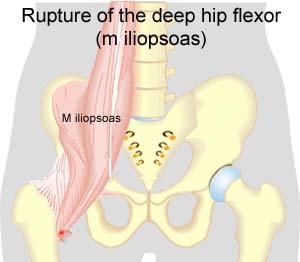
Cause: When a muscle is subjected to loads (repeated smaller loads or one very powerful load), beyond the strength of the muscle (jumping, kicking), a rupture occurs. The rupture can be microscopic and due to repeated loads in continuing sports activity, many small loads can trigger a chronic inflammation or a rupture. The vast majority of cases are partial muscle ruptures.

Symptoms: In light cases a local tenderness is felt after the load (“muscle strain”, “imminent pulled muscle”, “tendinitis”). The symptoms can often decrease after a thorough warm-up, only to return when the sports activity has ceased. In severe cases a sudden shooting pain is felt in the muscle (“partial muscle rupture”, “pulled muscle”) and in the worst case a sudden snap is felt rendering the muscle unusable (“total muscle rupture”). With muscle injuries the following three symptoms are characteristic: pain when applying pressure, stretching and activation against resistance. In total ruptures a defect in the muscle can often be seen and felt, and a swelling is felt above or below the rupture (the contracted muscular belly and the bleeding).

Acute treatment: Click here.

Examination: In light cases with only minimal tenderness and no discomfort with ordinary walking, medical examination is not necessarily required. The extent of the tenderness is, however, not always a mark of the degree of the injury. In cases of more pronounced pain or tenderness, medical examination is required to ensure the correct diagnosis and treatment. A normal medical examination is usually sufficient in order to make the diagnosis, however, if there is any doubt concerning the diagnosis an ultrasound scan can be performed (Ultrasonic image). X-ray examination is recommended when it is suspected that the bone on which the muscle tendon fastens has been torn off.

Treatment: The treatment usually comprises relief and rehabilitation. Only in very rare cases is surgery indicated (e.g. total ruptures in the muscle tendon, close to the fastening). Even large ruptures in the femoral muscle can usually be rehabilitated without resulting in functional harm (but often cosmetic damage, with an irregular femoral muscle). If the condition concerns tendinitis where there has been no sensation of a “snap” in the muscle, and smooth improvement has not been achieved after relief and gradually increasing rehabilitation, treatment can be supplemented with rheumatic medicine (NSAID) oand possibly injection of corticosteroid in the area surrounding the inflamed part of the muscle attachment point. If it concerns ruptures, (“total or partial muscle rupture”) the injection of corticosteroid is not indicated.

Complications: If the treatment does not progress according to plan, it should be considered whether the diagnosis is correct or whether complication have arisen. In particular the following should be considered:

Degenerative arthritis

DEGENERATIVE ARTHRITIS

Diagnosis: DEGENERATIVE ARTHRITIS
(Osteoarthritis)


Anatomy:
The hip joint consists of the hip socket and the femoral head (caput femoris). The articular surface is covered with a few mm thick cartilage layer, which reduces the load on the articular surfaces

Cause: In case of repeated loads, the cartilage primarily, and subsequently the bone below the cartilage, can be damaged (degenerative arthritis). The degenerative arthritis changes can in some cases cause an inflammation of the synovial membrane (synovitis) which causes fluid formation, swelling, movement restriction and pain in the hip joint.

Symptoms: Pain in the hip joint upon movement with load. There will often be movement restriction upon rotation in the hip joint.

Examination: Ordinary clinical examination is often sufficient to make the diagnosis. The examination can be supplemented with an X-ray examination. Ultrasound scan is the most suitable examination if you suspect a fluid accumulation in the hip joint.

Treatment: The treatment primarily comprises relief from the pain inducing activity until any swelling in the hip joint has decreased. Rehabilitation can subsequently be commenced with the primary goal to strengthen the muscles around the hip joint and preserve the joint mobility. There is no treatment that can restore the ruined cartilage (and bone). Cartilage transplants are not yet suitable for general degenerative arthritis changes. Upon swelling in the hip joint you can attempt to reduce the synovitis with rheumatic medicine (NSAID) or by attempting to drain the fluid and injecting corticosteroid, which should be conducted with ultrasound guidance to optimise the effect and minimize the risk. Pain without joint swelling is best treated with paracetamol. In cases of severe degenerative arthritis changes with pain when resting (at night) it may be necessary to replace the hip joint.

Complications: Degenerative arthritis which sits on the weight bearing parts of the joint is one of the most serious sports injuries, and often results in a termination of active sport. Cycling and swimming are significantly less stressful for the hip joint than running. In particular the following should be considered:

Special: Shock absorbing shoes or inlays will reduce the load.

Bursitis

BURSITIS

Diagnosis: BURSITIS
(Inflammation of the bursa)


Anatomy:
There are numerous bursas around the hip joint, serving the purpose of reducing the pressure on muscles, tendons and ligaments where these lie close to a bone projection.

Cause: In case of repeated loads or blows, the bursa can produce additional fluid, swell and become inflamed and painful.

Symptoms: Pain when applying pressure on the bursa, which sometimes (but far from always) can feel swollen. Pain is aggravated upon activation of the muscle closest to the bursa.

Acute treatment: Click here.

Examination: In light cases with only minimal tenderness, medical examination is not necessarily required. In case of more pronounced pain or lack of progress, a medical examination should be carried out to ensure the correctness of the diagnosis and the commencement of any treatment. The diagnosis is most easily and quickly made with ultrasound (which allows simultaneous treatment).

Treatment: The treatment primarily comprises relief. If the direct cause of the complaint is known, it should of course be removed. The treatment can be supplemented by rheumatic medicine (NSAID) or injection of corticosteroid in the bursa, preceded by draining of this, which can advantageously be performed under ultrasound guidance.

Rehabilitation: The treatment is dependant upon which bursa is inflamed, but sports activity can usually be cautiously resumed when pain has diminished, especially if the provoking factor has been identified and removed.
Also read rehabilitation, general.

Complications: If progress is not smooth, the correctness of the diagnosis or whether complications have arisen should be considered:

Special: Shock absorbing shoes or inlays will reduce the load.

Bursitis on the front of the hip joint

BURSITIS ON THE FRONT OF THE HIP JOINT

Diagnosis: BURSITIS ON THE FRONT OF THE HIP JOINT
(Bursitis iliopectinea)


Anatomy:
Between the deep hip flexor and the joint capsule is a large bursa (bursa iliopectinea), with the function to reduce the load on the muscle, when it slips over the hip joint. The bursa often communicates with the hip joint.

Cause: Upon repeated loads or blows the bursa can produce increased amounts of fluid, swell and become inflamed and painful.

Symptoms: Pain when applying pressure on the bursa, which occasionally (but far from always) may feel swollen. Pain is agravated upon activation of the deep flexor (flexing the hip joint).

Acute treatment: Click here.

Examination: In light cases with only minimal tenderness medical examination is not necessarily required. In case of more pronounced pain or lack of progress a medical examination should be carried out to ensure that the diagnosis is correct and commence any treatment. The diagnosis is most easily and quickly made with ultrasound (which allows simultaneous treatment) (article).

Treatment: The treatment primarily comprises relief. The treatment can be supplemented with rheumatic medicine (NSAID) or injection of corticosteroid in the bursa preceded by draining of the bursa, which can advantageously be done under ultrasound guidance (article).

Complications: If progress is not smooth the correctness of the diagnosis or whether complications have arisen, should be considered:

Special: Shock absorbing shoes or inlays will reduce the load.

Bursitis at the outer femoral bone projection

BURSITIS AT THE OUTER FEMORAL BONE PROJECTION

Diagnosis: BURSITIS AT THE OUTER FEMORAL BONE PROJECTION
(Bursittis trochanterica)


Anatomy:
On the outside of the outer femoral bone projection (trochanter major) is a large bursa which reduces the load on muscles and tendons when these slide over the bone projection.

  1. M. piriformis
  2. M. gluteus minimus
  3. Bursa m. piriformis
  4. Bursae trochantericae m. glutei medii
  5. M. gluteus medius (resectus)
  6. Bursa trochanterica m. glutei maximi
  7. M. gluteus maximus
  8. Bursae intermusculares mm. gluteorum
  9. Tuberositas glutea
  10. Tractus iliotibialis
  11. M. biceps femoris
    (caput longum)
  12. M. biceps femoris
    (caput breve)
  13. M. adductor magnus
  14. B. subtendinea m. bicipitis femoris superior
  15. Tuber ischiadicum
  16. B. ischiadica m. glutei maximi
  17. Mm. gemilli sup. Et inf.
  18. B. ischiadica m. obturatorii interni

GLUTEAL MUSCLES FROM THE REAR

Cause: In case of repeated loads or blows the bursa can become inflamed, produce fluid, swell and become painful.

Symptoms: Pain when applying pressure on the thigh corresponding to the bursa, which sometimes (but far from always) can feel swollen. The pain can radiate down the thigh.

Acute treatment: Click here.

Examination: In light cases with only minimal tenderness medical examination is not necessarily required. In case of more pronounced pain or lack of progress, a medical examination should be carried out to ensure the diagnosis and commencement of any treatment. A normal medical examination is usually sufficient in order to make the diagnosis, however, if there is any doubt concerning the diagnosis an ultrasound scan can be performed (Ultrasonic image).

Treatment: The treatment primarily comprises relief. If the direct cause of the complaint is known, it should of course be removed. The treatment can be supplemented with rheumatic medicine (NSAID) or injection of corticosteroid in the bursa, which is best performed under the guidance of ultrasound.

Complications: If progress is not smooth, the correctness of the diagnosis should be considered or whether complications have arisen:

Special: Shock absorbing shoes or inlays will reduce the load. In case of lack of progress or recurrences after successful rehabilitation it can be considered to have a running style analysis, to evaluate whether correction of the running style is indicated.