Kategoriarkiv: Adults injuries

Inflammation of the mucous fold

INFLAMMATION OF THE MUCOUS FOLD

Diagnosis: INFLAMMATION OF THE MUCOUS FOLD
(Plica synovialis)


Anatomy:
The joint cavity in the knee joint is covered with a thin synovial membrane (synovialis). Four mucous folds (plica synovialis) can develop in the membrane: one fold in the inner joint chamber (medial plica), outer joint chamber (lateral plica), upper (suprapatellar) and front (anterior plica). A mucous fold is found in the inner joint chamber (medial plica) in almost 50% of all people (article).

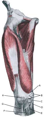
Cause: The mucous fold will become inflamed if an entrapment of the fold occurs, or if the fold suffers internal bleeding. This will result in thickening and subsequently cause pain.

Symptoms: The mucous fold in the inner joint chamber (medial plica) will most often give symptoms which are difficult to distinguish from the symptoms of a meniscus lesion. The pain is usually localised on the inner side of the knee cap, in front of the inner joint line. The pain often occurs quite suddenly following certain movements, and can be accompanied by swelling in the knee. The knee can lock if a flap of the mucous fold becomes entrapped (article).

Examination: A medical examination is always necessary to ensure the diagnosis if there is any suspicion of an inflamed mucous fold in the knee. A tender string inside the knee cap can occasionally give a slipping sensation, but often a normal clinical examination is not sufficient. It is often necessary to perform an arthroscopic examination (telescopic examination of the joint) or MR-scan to make the diagnosis (article).

Treatment: Treatment comprises relief and careful rehabilitation of the knee. If the discomfort does not slowly diminish, the treatment can be supplemented with rheumatic medicine (NSAID) or injection of corticosteroidi in the mucous fold. Medial synovial shelf plica syndrome. Treatment by intraplical steroid injection. If this does not give the desired results, the mucous fold can be removed by arthroscopy (telescopic examination of the knee).

Exerting load on the knee can commence as soon as the pain and swelling in the knee has diminished. In the best cases, full activity is possible after a period of a few weeks. In uncomplicated cases, it should be possible to resume a full level of sports activity during the course of a month. Rehabilitation must not be allowed to cause increased swelling (or pain) in the knee.

Complications: If insufficient progress is made prior to an operation it must be considered if the diagnosis is correct. Supplementary examinations will often be required (X-ray, ultrasound or MR scan). In particular the following should be considered:

Following an arthroscopic examination fluid accumulation in the joint (traumatic arthritis/synovitis), should be considered, as well as infection in the scar or knee joint, which will always require medical attention as soon as possible.

Meniscus lesion

MENISCUS LESION

Diagnosis: MENISCUS LESION


Anatomy:
The bones in the knee joint comprise the thigh bone (femur), shin bone (tibia) and the knee cap (patella). The articular surfaces on the thigh bone, shin bone and the knee cap are covered with a few mm thick cartilage coating which serves to reduce the strain on the articular surfaces. There is a ring-formed cartilage disk (meniscus) internally and externally in the joint. The knee joint is strengthened by a joint capsule which is in turn strengthened on the sides by an outer and an inner collateral ligament (ligamentum collaterale laterale (LCL) and ligamentum collaterale mediale (MCL)). The internal meniscus is attached to the inner collateral ligament, whereas the external meniscus is not attached to the outer collateral ligament.

  1. Ligamentum cruciatum posterius
  2. Ligamentum collaterale mediale/tibiale
  3. Meniscus medialis
  4. Insertio anterior menisci medialis
  5. Ligamentum transversum genus
  6. Tibiae
  7. Fibulae
  8. Ligamentum cruciatum anterius
  9. Ligamentum collaterale laterale/fibulare
  10. Meniscus lateralis
  11. Femur

KNEE JOINT FROM THE FRONT


  1. Ligamentum transversum genus
  2. Meniscus lateralis
  3. Meniscus medialis
  4. Insertio anterior menisci

KNEE JOINT FROM ABOVE

Cause: Meniscus lesions often occur when the knee is stretched at the same time as rotating. The meniscus will consequently be squeezed between the thigh bone and the shin bone. The inner meniscus is more often damaged than the external. Meniscus injuries in children is very rare (article).

Symptoms: Pain along the joint line when applying pressure and when rotating the knee. The pain often occurs suddenly when performing certain movements, and can be accompanied by swelling of the knee. In some instances the knee can lock due to a splinter from the meniscus becoming jammed. In other cases, a small and tender outpouching of fluid can be sensed at the joint line (meniscus cyst). Prolonged discomfort will often cause the thigh muscle to diminish.

Acute treatment: Click here.

Examination: A medical examination is always necessary if there is any suspicion of injury within the knee joint. A normal clinical examination is often not sufficient. The doctor can perform various knee tests (meniscus test), however, there is typically tenderness along the joint line which is aggravated when rotating the knee joint. It is often necessary to perform an arthroscopy (telescopic examination of the joint), MR-scan or ultrasound scan to make the diagnosis (article) (Ultrasonic image).

Treatment: In uncertain cases the treatment comprises relief and rehabilitation of the knee. If the discomfort does not slowly diminish, if there are instances of “locking” or if it is clear that the meniscus lesion is large (e.g. assessed from an ultrasound scan), surgery should be considered. Under an operation, it can be attempted to sew the torn piece of meniscus back (which will require a considerably longer rehabilitation period, but will probably in the long term reduce the risk of degenerative arthritis in the knee) (article). The torn piece of meniscus is usually removed, but in rare cases the whole meniscus is taken out.

Following an operation, exerting load on the knee can commence as soon as the pain and swelling in the knee has diminished. In the best cases, full activity is possible after a period of a few weeks. In uncomplicated cases, it should be possible to resume a full level of sports activity during the course of a month. Rehabilitation must not be allowed to cause increased swelling (or pain) in the knee.

Complications: If insufficient progress is made prior to an operation it must be considered if the diagnosis is correct. Supplementary examinations will often be required (X-ray, ultrasound or MR scan). In particular the following should be considered:

Following an arthroscopy, fluid accumulation in the joint (traumatic arthritis/synovitis) should be considered, and infection in the scar or knee joint, which will always require medical attention as soon as possible. Special: Since there is a risk that the injury can cause permanent disability, the injury should be reported to your insurance company.

Rupture of the posterior cruciate ligament

RUPTURE OF THE POSTERIOR CRUCIATE LIGAMENT

Diagnosis: RUPTURE OF THE POSTERIOR CRUCIATE LIGAMENT
(Ruptura ligamentum cruciatum posterius)


Anatomy:
The bones in the knee joint comprise the thigh bone (femur), shin bone (tibia) and the knee cap (patella). There is furthermore a small joint between the shin bone and the calf bone (fibula). The knee joint is strengthened by a joint capsule which is in turn strengthened on the sides by an outer and an inner collateral ligament (ligamentum collaterale laterale (LCL) and ligamentum collaterale mediale (MCL)). Inside the knee there are two ligaments, the anterior and posterior cruciate ligaments (ligamentum cruciatum anterius and ligamentum cruciatum posterius).

  1. Ligamentum cruciatum posterius
  2. Ligamentum collaterale mediale/tibiale
  3. Meniscus medialis
  4. Insertio anterior
    menisci medialis
  5. Ligamentum transversum genus
  6. Tibiae
  7. Fibulae
  8. Ligamentum cruciatum anterius
  9. Ligamentum collaterale laterale/fibulare
  10. Meniscus lateralis
  11. Femur

KNEE JOINT FROM THE FRONT

Cause: Rupture of the posterior cruciate ligament usually occurs following a blow or kick direct on the front of the shin bone just below the knee (Photo).

Symptoms: Usually a snap can be heard or felt and continued sports activity must be aborted. The knee can swell within the first few hours, after which the knee can not bend completely. You can subsequently often sense that the leg gives way (knee failure).

Acute treatment: Click here.

Examination: If a partial or complete rupture of the cruciate ligament is suspected, you should seek medical attention (casualty ward) immediately, to obtain a diagnosis. The doctor can perform various tests on the knee (rear drawer looseness) to examine the stability of the knee. It should be noted that the looseness in the knee can often only be demonstrated after two weeks. It is often necessary to supplement the examination with a MR-scan, ultrasound scan (Ultrasonic image) (article), or arthroscopy to make the diagnosis with certainty.

Treatment: Treatment of a rupture of the posterior cruciate ligament usually comprises relief and rehabilitation. It is only in cases of pronounced looseness, or if the rupture is combined with other ligament ruptures, that surgery is recommended (article).

An intensive rehabilitation period of at least six months is to be expected.

Bandage: Hinge bandages (Don-Joy) can be utilised the first few weeks. Tape treatment of cruciate ligament ruptures in the knee has no sure effect.

Complications: If smooth progress is not achieved, it should be considered whether the diagnosis is correct or whether complications have arisen. Amongst others the following should be considered:

Special: Since there is a risk that the injury can cause permanent disability, the injury should be reported to your insurance company.

Rupture of the anterior cruciate ligament

RUPTURE OF THE ANTERIOR CRUCIATE LIGAMENT

Diagnosis: RUPTURE OF THE ANTERIOR CRUCIATE LIGAMENT
(Ruptura ligamentum cruciatum anterius)


Anatomy:
The bones in the knee joint comprise the thigh bone (femur), shin bone (tibia) and the knee cap (patella). There is furthermore a small joint between the shin bone and the calf bone (fibula). The knee joint is strengthened by a joint capsule which is in turn strengthened on the sides by an outer and an inner collateral ligament (ligamentum collaterale laterale (LCL) and ligamentum collaterale mediale (MCL)). Inside the knee there are two ligaments, the anterior and posterior cruciate ligaments (ligamentum cruciatum anterius and ligamentum cruciatum posterius).

  1. Ligamentum cruciatum posterius
  2. Ligamentum collaterale mediale/tibiale
  3. Meniscus medialis
  4. Insertio anterior menisci medialis
  5. Ligamentum transversum genus
  6. Tibiae
  7. Fibulae
  8. Ligamentum cruciatum anterius
  9. Ligamentum collaterale laterale/fibulare
  10. Meniscus lateralis
  11. Femur

KNEE JOINT FROM THE FRONT

Cause: Rupture of the anterior cruciate ligament usually occurs during running with fast changes of direction or as the result of a fall while skiing. Most injuries occur without contact with an opponent.

Symptoms: Usually a snap can be heard or felt and continued sports activity must be aborted. The knee swells within the first few hours, after which the knee can not bend completely. You can subsequently often sense that the leg gives way (knee failure).

Acute treatment: Click here.

Examination: If a partial or complete rupture of the cruciate ligament is suspected, you should seek medical attention (casualty ward) immediately, to obtain a diagnosis. The doctor can perform various tests on the knee (front drawer looseness, Lachmann, Pivot shift) to examine the stability of the knee. It should be noted that the looseness in the knee can often only be demonstrated after two weeks. The fluid in the knee can be drained. Blood in the knee provides a very strong suspicion of a rupture of the anterior cruciate ligament. The bleeding that occurs after a rupture of the anterior cruciate ligament can usually be seen in an ultrasound scan (Ultrasonic image) (article) or MR-scan. Arthroscopy (a telescopic examination of the knee) is the best suited examination if there is any doubt in the diagnosis.

Treatment: Young athletes or others with physically demanding work will usually be advised surgery with the insertion of a new cruciate ligament (article).

An intensive rehabilitation period of at least six months is to be expected. It is important that the knee is stretched completely at least twice a day. The surgeon should be consulted if problems occur with stretching the knee completely.

Bandage: Hinge bandages (Don-Joy) can be utilised the first few weeks. Tape treatment of cruciate ligament ruptures in the knee has no sure effect.

Complications: In case of lack of progress you should consider various complications to the surgery:

In cases where the knee cap tendon is used as a new cruciate ligament an inflammation, is often seen where the knee cap tendon has been removed. With prolonged discomfort which does not diminish following relief, medical treatment in form of rheumatic medicine (NSAID) or injection of corticosteroid along the inflamed part of the knee cap tendon can be attempted (article).
Less than half the athletes suffering a rupture of the anterior cruciate ligament are able to resume sports activity on the same level as prior to the injury (article).

Special: Since there is a risk that the injury can cause permanent disability, the injury should be reported to your insurance company.

Outer collateral ligament rupture

OUTER COLLATERAL LIGAMENT RUPTURE

Diagnosis: OUTER COLLATERAL LIGAMENT RUPTURE
(Ruptura ligamentum collaterale laterale/fibulare)


Anatomy:
The bones in the knee joint comprise the thigh bone (femur), shin bone (tibia) and the kneecap (patella). There is furthermore a small joint between the shin bone and the calf bone (fibula). The knee joint is strengthened by a joint capsule which is in turn strengthened on the sides by an outer and an inner collateral ligament (ligamentum collaterale laterale (LCL) and ligamentum collaterale mediale (MCL)). Inside the knee there are two ligaments, the anterior and posterior cruciate ligaments (ligamentum cruciatum anterius and ligamentum cruciatum posterius).


  1. Ligamentum cruciatum posterius
  2. Ligamentum collaterale mediale/tibiale
  3. Meniscus medialis
  4. Insertio anterior menisci medialis
  5. Ligamentum transversum genus
  6. Tibiae
  7. Fibulae
  8. Ligamentum cruciatum anterius
  9. Ligamentum collaterale laterale/fibulare
  10. Meniscus lateralis
  11. Femur

KNEE FROM THE FRONT


  1. M. gastrocnemius
  2. Caput fibulae
  3. B. subtendinea m. bicipitis femoris inferior
  4. M. biceps femoris
  5. Ligamentum collaterale laterale/fibulare
  6. M. plantaris

OUTER LOWER LEG

Cause: When the shin bone is pressed inwards in relation to the thigh bone the outer side-ligament is stretched. If the load becomes sufficiently powerful (as is the case when the load is sudden, so that the thigh muscles do not have time to tighten and stabilize the joint) the ligament can rupture.

Symptoms: Sudden pain on the outside of the knee. Occasionally a snap is felt when the ligament ruptures. In severe cases the athlete complains about a sense of looseness in the knee.

Acute treatment: Click here.

Examination: If a total or partial rupture of the ligaments in the knee is suspected, you should seek medical attention for a diagnosis. The doctor can perform various knee tests to examine the stability of the knee (external side-instability-test) . If the knee is stable the injury is called a “sprain” of the outer side-ligament. If the knee is loose the injury is called a “rupture” of the ligament. The diagnosis is usually made from a normal medical examination. If there are any doubts surrounding the diagnosis, an ultrasound scan can be performed which will reveal the rupture and the bleeding along the ligaments (Ultrasonic image).

Treatment: The treatment of a partial or total rupture of the outer side-ligament involves relief and rehabilitation. If the knee is markedly loose, you can, for a short period, use a support splint (Don Joy). With pronounced sideways looseness surgery can be considered (article). (However if the rupture of the outer side-ligament is combined with other ligament ruptures, as is often the case, surgery is recommended).

Bandage: Hinge bandages can be used initially (Don Joy). Tape treatment of ligament ruptures in the knee have no sure effect.

Complications: If smooth progress is not achieved, it should be considered whether the diagnosis is correct or whether complications have arisen. Supplementary examinations (X-ray, ultrasound, MR scan or arthroscopy) will often be required. The following should be considered:

An inflammation by the ligament fastening is sometimes seen later in the course of the injury, and in some cases bursitis at the ligament fastening is seen. The inflammation and bursitis can possibly be treated with medication (rheumatic medicine (NSAID) or the injection of corticosteroid), if further relief has no effect (article).

Special: Since there is a risk that the injury can cause permanent disability, the injury should be reported to your insurance company.

Inner collateral ligament rupture

INNER COLLATERAL LIGAMENT RUPTURE

Diagnosis: INNER COLLATERAL LIGAMENT RUPTURE
(Ruptura ligamentum collaterale mediale/tibiale)


Anatomy:
The bones in the knee joint comprise the thigh bone (femur), shin bone (tibia) and the knee cap (patella). There is furthermore a small joint between the shin bone and the calf bone (fibula). The knee joint is strengthened by a joint capsule which is in turn strengthened on the sides by an outer and an inner collateral ligament (ligamentum collaterale laterale (LCL) and ligamentum collaterale mediale (MCL)). Inside the knee there are two ligaments, the anterior and posterior cruciate ligaments (ligamentum cruciatum anterius and ligamentum cruciatum posterius).

  1. Ligamentum cruciatum posterius
  2. Ligamentum collaterale mediale/tibiale
  3. Meniscus medialis
  4. Insertio anterior menisci medialis
  5. Ligamentum transversum genus
  6. Tibiae
  7. Fibulae
  8. Ligamentum cruciatum anterius
  9. Ligamentum collaterale laterale/fibulare
  10. Meniscus lateralis
  11. Femur

KNEE FROM THE FRONT

  1. Tendo m. adductoris magni
  2. Retinaculum patellae mediale
  3. Meniscus medialis
  4. Ligamentum collaterale mediale/tibiale
  5. Bursa anserina
  6. Bursa subtendinea m. sartori
  7. Ligamentum patellae
  8. Patella

KNEE FROM THE FRONT

Cause: When the shin bone is pressed outwards in relation to the thigh bone the inner side-ligament is stretched. If the load becomes sufficiently powerful (as is the case when the load is sudden, so that the thigh muscles do not have time to tighten and stabilize the joint) the ligament can rupture.

Symptoms: Sudden pain on the inside of the knee. Occasionally a snap is felt when the ligament ruptures. In severe cases the athlete complains about a sense of looseness in the knee.

Acute treatment: Click here.

Examination: If a total or partial rupture of the ligaments in the knee is suspected, you should seek medical attention for a diagnosis. The doctor can perform various knee tests (internal side-instability-test) to examine the stability of the knee. If the knee is stable the injury is called a “sprain” of the inner side-ligament. If the knee is loose the injury is called a “rupture” of the ligament. The diagnosis is usually made from a normal medical examination. If there are any doubts surrounding the diagnosis, an ultrasound scan can be performed which will reveal the rupture and the bleeding along the ligaments (Ultrasonic image) .

Treatment: The treatment of a partial or total rupture of the inner side-ligament involves relief and rehabilitation. If the knee is markedly loose, you can, for a short period, use a support splint (Don Joy). Surgical treatment previously considered has today largely been abandoned in uncomplicated ruptures of the inner side-ligament (article). (However if the rupture of the inner side-ligament is combined with other ligament ruptures, many would recommend surgery).

Bandage: Hinge bandages can be used initially (Don Joy). Tape treatment of ligament ruptures in the knee have no sure effect.

Complications: If smooth progress is not achieved, it should be considered whether the diagnosis is correct or whether complications have arisen. Supplementary examinations (X-ray, ultrasound, MR scan or arthroscopy) will often be required. The following should be considered:

An inflammation by the ligament fastening is sometimes seen later in the course of the injury, and in some cases bursitis at the ligament fastening is seen. The inflammation and bursitis can possibly be treated with medication (rheumatic medicine (NSAID) or injection of corticosteroid), if further relief has no effect.

Special: Since there is a risk that the injury can cause permanent disability, the injury should be reported to your insurance company.

Stress fracture in the femoral neck

STRESS FRACTURE IN THE FEMORAL NECK

Diagnosis: STRESS FRACTURE IN THE FEMORAL NECK
(Stress fraktur)


Anatomy:
The femur and the hip bone form the hip joint.


  1. Caput femoris
  2. Collum femoris
  3. Trochanter minor
  4. Trochanter major

PELVIS AND THIGH BONE FROM THE FRONT


Cause: Repeated loads, especially when walking or running can cause cracks (stress fractures) in the femoral neck (collum femoris) (article-1) (article-2).

Symptoms: Pain in the hip when applying pressure (direct and indirect tenderness) and when under load (walking, running).

Examination: X-ray. Since many stress fractures cannot be seen early in the course of events, X-ray examination can be repeated after a few weeks. Scintigraphy, CT- and MRI and ultrasound scan can often diagnose stress fractures far earlier than X-rays (Ultrasonic image).
It is imperative for the result of the treatment that the diagnosis is made as early as possible (article).

Treatment: Relief. In some cases surgery is necessary (article).

Rehabilitation: The rehabilitation is completely dependent on the type of fracture and treatment (conservative or operative).
Also read rehabilitation, general.

Complications: If progress is not smooth, you should be re-examined to ensure that the fracture heals according to plan. In some cases a false joint can be formed (pseudoarthrosis), which requires surgical treatment.

Special: Shock absorbing shoes or inlays will reduce the load.

Fluid accumulation in the joint

FLUID ACCUMULATION IN THE JOINT

Diagnosis: FLUID ACCUMULATION IN THE JOINT
(Synovitis / coxitis)


Anatomy:
The hip joint consists of the hip socket (acetabulum) and the femoral head (caput femoris). The articular surfaces are coated with a cartilage layer a few mm thick, which reduces the load on the articular surfaces.

Cause: Many repeated loads or one violent load can cause an inflammation of the synovial membrane (synovitis), fluid formation, swelling, restriction of movement and pain in the hip joint. The condition is relatively often seen in children (Ultrasonic image).

Symptoms: Pain in the joint upon movement with load. Often there will be movement restriction with rotation of the hip joint.

Examination: It will often be necessary to supplement the ordinary clinical examination with an ultrasound scan, where the fluid in the hip joint can easily be seen (Ultrasonic image).

Treatment: The treatment primarily comprises relief from the pain inducing activity until the swelling has abated. Rehabilitation, within the pain threshold, can subsequently be commenced. In cases of lack of progress with relief the treatment can be supplemented with a medical treatment in the form of rheumatic medicine (NSAID) or by drainage of the joint fluid and the injection of corticosteroid, which should be done with ultrasound guidance.

Complications: In particular you should consider a bacterial infection in the joint (pyarthron), which is determined by ultrasound guided draining of the joint fluid, the child hip diseases (epifysiolysis capitis femoris and Calvé-Legg-Perthes disease), where the joint head on the femur slips or collapses (X-ray examination will determine the diagnosis), arthritis or:

Special: There are two childrens’ hip diseases that should always be considered in children with hip pain.

  • SLIPPING OF THE FEMORAL HEAD IN THE GROWTH ZONE (EPIFYSIOLYSIS CAPITIS FEMORIS), which mainly affects boys age 11-16 years (article). There will often be limping, groin pain, but sometimes the pain is sensed in the knee. It is important in order to obtain a good result from the treatment to be examined by a doctor and have the diagnosis made as soon as possible (with X-rays).

  • CALVÉ-LEGG-PERTHES DISEASE is a disease which mainly affects boys age 3-11 years (article). The bone core in the femoral head is dissolved and flattened. There will often be limping, tiredness and pain in the groin, but once in a while the pain is sensed in the knee instead. It is important in order for a good result of the treatment to be examined by a doctor and have the diagnosis made as soon as possible (by means of X-rays or ultrasound).

Inner snapping hip

INNER SNAPPING HIP

Diagnosis: INNER SNAPPING HIP
(Coxae saltans, intern)


Anatomy:
The deep hip flexor (M iliopsoas) consists of two muscles. The Psoas muscle originate from the lumbar vertebrae and the Iliacus muscle from the inside of the femur. The two muscles fuse and are both fastened on the inside of the femur (trochantes minor). The iliopsoas is the strongest flexor muscle of the hip.

  1. Origines m. psoatis
  2. M. psoas major
  3. M. iliacus
  4. M. psoas major
  5. M. psoas minor

THIGH FROM THE FRONT

Cause: In case of repeated loads the muscle tendon can become inflamed, swell and with some movements slip over the one of the bone projections at the pelvis or the hip joint. When the tendon slips over the bone projection, the uncomfortable symptoms are produced. There are three known causes of snapping hips: outer snapping hip (tractus iliotibialis), inner snapping hip (iliopsoas tendon) and joint conditioned (intraarticular) causes (article). The treatment is dependant on the cause.

Symptoms: With certain movements in the hip joint the deep hip flexor (M iliopsoas) can be made to slip over one of the bone projections on the pelvis or the hip joint, whereby a slipping sensation, often accompanied by discomfort, is felt and often heard.

Acute treatment: Click here.

Examination: In light cases with only minimal tenderness medical examination is not necessarily required. In case of more pronounced pain or lack of progress a medical examination should be carried out to ensure the diagnosis. A normal medical examination is usually sufficient in order to make the diagnosis, however, if there is any doubt concerning the diagnosis a dynamic ultrasound scan can be performed, where the tendon can be seen slipping as this triggers the symptoms (article 1) (article 2), (article 3).

Treatment: The treatment primarily comprises relief. If progress is not smooth the treatment can be supplemented with rheumatic medicine (NSAID) or the injection of corticosteroid, which should be done with ultrasound guidance to ensure the optimal effect and minimal risk. Only in very rare cases is surgical treatment necessary.

Complications: If progress is not smooth it should be considered whether the diagnosis is correct or if complications have arisen. In particular the following should be considered:

Outer snapping hip

OUTER SNAPPING HIP

Diagnosis: OUTER SNAPPING HIP
(Coxe saltans extern)


Anatomy:
Externally from the iliac crest to the shin bone runs a strong tendon (tractus iliotibialis), on which many of the thighs muscles are fastened. The tendon runs close above the outer femoral bone projection (trochanter major).


  1. M. gluteus medius
  2. M. gluteus maximus
  3. Trochanter major
  4. Tractus iliotibialis

(Drawing)

THIGH FROM THE REAR

Cause: With repeated movements in the knee and hip joint (running, dancing, gymnastics) the powerful tendon (tractus iliotibialis) slips over the outer bone projection (trochanter major) of the femur, which can cause inflammation in the tendon or in the underlying bursa. When the inflamed tendon slips over the bone projection, a sudden, slipping, and unpleasant sensation can be felt. There are three known causes of snapping hips: outer snapping hip (tractus iliotibialis), inner snapping hip (iliopsoas tendon) and joint conditioned (intraarticular) causes, Coxa Saltans: The Snapping Hip Revisited (article).

Symptoms: Upon certain movements in the hip joint, a sudden slipping, and unpleasant sensation can suddenly be produced on the outside of the thigh, which is often audible.

Acute treatment: Click here.

Examination: Usually the diagnosis can be made by an ordinary medical examination. You can often prevent the tendon from slipping over the outer bone projection by holding the tendon aside, while the movements provoking the condition are performed. The pain will decrease for approx. one hour after the injection of a local anaesthetic (diagnostic blockade) around the outer hip bone projection. If the diagnosis does not appear to be certain, ultrasound is recommended (Ultrasonic image), (article), or possibly a MRI scan.

Treatment: The treatment primarily comprises relief, stretching of the external tendon and rehabilitation. It is crucial that shoes have good shock absorbing soles. In cases of inappropriate foot stance, this should be corrected with shoes or inlays. In case of lack of progress the treatment can be supplemented with medical treatment in the form of rheumatic medicine (NSAID) or the injection of corticosteroid, which advantageously can be guided by ultrasound. In severe cases with no effect from relief, correct rehabilitation or medical treatment, you can operatively split the tendon.

Complications: In case of lack of progress it should be considered whether the diagnosis is correct. This will often require supplemental examinations (X-ray, ultrasound or MRI scan). In particular the following should be considered:

Special: Shock absorbing shoes or inlays will reduce the load. In case of lack of progress or recurrences after successful rehabilitation it can be considered to have a running style analysis performed, to evaluate whether correction of the running style is indicated.