Kategoriarkiv: Hip

Cartilage damage (osteochondral lesions)

CARTILAGE DAMAGE IN THE JOINT

Diagnosis: CARTILAGE DAMAGE IN THE JOINT
(Osteochondral lesion)


Anatomy:
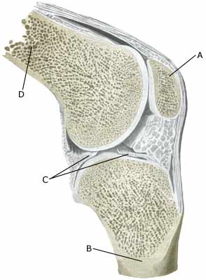
The bones in the knee joint comprise the thigh bone (femur), shin-bone (tibia) and the knee cap (patella). The articular surfaces of the thigh bone, shin-bone and knee cap are lined with a cartilage covering of a few millimetre’s thickness which serves to reduce the load or strain on the joint surfaces.

Cause: Twists in the knee joint causing the thigh bone and shin bone to collide can cause damage to the cartilage in the knee. In some cases a piece of cartilage can be shed which can wander in the joint (joint-mouse) and become inflamed and increase the production of synovial fluid.

Symptoms: Pain in the joint when under load or strain. Often, periodic swelling of the joint (synovitis).

Examination: Normal medical examination is often not sufficient. To make the diagnosis correctly it is therefore necessary to perform an arthroscopic examination (telescopic examination of the joint (Photo) or an MR-scanning  (Ultrasonic image).

Treatment: Treatment comprises relief from the painful activities until the pain is no longer experienced, after which gradual re-training can be commenced. There is no treatment that can restore the damaged cartilage, which has itself poor restorative ability. Different procedures to enhance the healing can be attempted using arthroscopy, however, the results are generally unsatisfactory. Results from experimental cartilage transplants are still not successful enough to warrant introduction as a routine treatment in the near future (article). Joint-mouse that provokes the symptoms must be surgically removed. Injection of corticosteroid in the knee joint, performed under ultrasound guidance, can be attempted to combat prolonged and pronounced cases of fluid accumulation in the knee joint (synovitis).

Rehabilitation: Rehabilitation is completely dependent upon the type of cartilage damage (size and position in the joint) and treatment (conservative or surgical). It is generally attempted to strengthen the musculature around the knee joint.
Also read rehabilitation, general.

Complications: Large cartilage damage which is positioned on the weight-bearing parts of the joint represents one of the most serious sports injuries, and often results in an end to the sporting career.

Special: As there is a risk that the injury can cause permanent disability, all cases should be reported to your insurance company.

Meniscus lesion

MENISCUS LESION

Diagnosis: MENISCUS LESION


Anatomy:
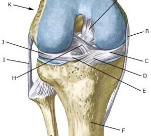
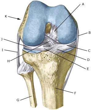
The bones in the knee joint comprise the thigh bone (femur), shin bone (tibia) and the knee cap (patella). The articular surfaces on the thigh bone, shin bone and the knee cap are covered with a few mm thick cartilage coating which serves to reduce the strain on the articular surfaces. There is a ring-formed cartilage disk (meniscus) internally and externally in the joint. The knee joint is strengthened by a joint capsule which is in turn strengthened on the sides by an outer and an inner collateral ligament (ligamentum collaterale laterale (LCL) and ligamentum collaterale mediale (MCL)). The internal meniscus is attached to the inner collateral ligament, whereas the external meniscus is not attached to the outer collateral ligament.

  1. Ligamentum cruciatum posterius
  2. Ligamentum collaterale mediale/tibiale
  3. Meniscus medialis
  4. Insertio anterior menisci medialis
  5. Ligamentum transversum genus
  6. Tibiae
  7. Fibulae
  8. Ligamentum cruciatum anterius
  9. Ligamentum collaterale laterale/fibulare
  10. Meniscus lateralis
  11. Femur

KNEE JOINT FROM THE FRONT


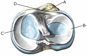
  1. Ligamentum transversum genus
  2. Meniscus lateralis
  3. Meniscus medialis
  4. Insertio anterior menisci

KNEE JOINT FROM ABOVE

Cause: Meniscus lesions often occur when the knee is stretched at the same time as rotating. The meniscus will consequently be squeezed between the thigh bone and the shin bone. The inner meniscus is more often damaged than the external. Meniscus injuries in children is very rare (article).

Symptoms: Pain along the joint line when applying pressure and when rotating the knee. The pain often occurs suddenly when performing certain movements, and can be accompanied by swelling of the knee. In some instances the knee can lock due to a splinter from the meniscus becoming jammed. In other cases, a small and tender outpouching of fluid can be sensed at the joint line (meniscus cyst). Prolonged discomfort will often cause the thigh muscle to diminish.

Acute treatment: Click here.

Examination: A medical examination is always necessary if there is any suspicion of injury within the knee joint. A normal clinical examination is often not sufficient. The doctor can perform various knee tests (meniscus test), however, there is typically tenderness along the joint line which is aggravated when rotating the knee joint. It is often necessary to perform an arthroscopy (telescopic examination of the joint), MR-scan or ultrasound scan to make the diagnosis (article) (Ultrasonic image).

Treatment: In uncertain cases the treatment comprises relief and rehabilitation of the knee. If the discomfort does not slowly diminish, if there are instances of “locking” or if it is clear that the meniscus lesion is large (e.g. assessed from an ultrasound scan), surgery should be considered. Under an operation, it can be attempted to sew the torn piece of meniscus back (which will require a considerably longer rehabilitation period, but will probably in the long term reduce the risk of degenerative arthritis in the knee) (article). The torn piece of meniscus is usually removed, but in rare cases the whole meniscus is taken out.

Following an operation, exerting load on the knee can commence as soon as the pain and swelling in the knee has diminished. In the best cases, full activity is possible after a period of a few weeks. In uncomplicated cases, it should be possible to resume a full level of sports activity during the course of a month. Rehabilitation must not be allowed to cause increased swelling (or pain) in the knee.

Complications: If insufficient progress is made prior to an operation it must be considered if the diagnosis is correct. Supplementary examinations will often be required (X-ray, ultrasound or MR scan). In particular the following should be considered:

Following an arthroscopy, fluid accumulation in the joint (traumatic arthritis/synovitis) should be considered, and infection in the scar or knee joint, which will always require medical attention as soon as possible.

Special: Since there is a risk that the injury can cause permanent disability, the injury should be reported to your insurance company.

Anterior collateral ligament rupture

RUPTURE OF THE ANTERIOR CRUCIATE LIGAMENT

Diagnosis: RUPTURE OF THE ANTERIOR CRUCIATE LIGAMENT
(Ruptura ligamentum cruciatum anterius)


Anatomy:
The bones in the knee joint comprise the thigh bone (femur), shin bone (tibia) and the knee cap (patella). There is furthermore a small joint between the shin bone and the calf bone (fibula). The knee joint is strengthened by a joint capsule which is in turn strengthened on the sides by an outer and an inner collateral ligament (ligamentum collaterale laterale (LCL) and ligamentum collaterale mediale (MCL)). Inside the knee there are two ligaments, the anterior and posterior cruciate ligaments (ligamentum cruciatum anterius and ligamentum cruciatum posterius).

  1. Ligamentum cruciatum posterius
  2. Ligamentum collaterale mediale/tibiale
  3. Meniscus medialis
  4. Insertio anterior menisci medialis
  5. Ligamentum transversum genus
  6. Tibiae
  7. Fibulae
  8. Ligamentum cruciatum anterius
  9. Ligamentum collaterale laterale/fibulare
  10. Meniscus lateralis
  11. Femur

KNEE JOINT FROM THE FRONT

Cause: Rupture of the anterior cruciate ligament usually occurs during running with fast changes of direction or as the result of a fall while skiing. Most injuries occur without contact with an opponent.

Symptoms: Usually a snap can be heard or felt and continued sports activity must be aborted. The knee swells within the first few hours, after which the knee can not bend completely. You can subsequently often sense that the leg gives way (knee failure).

Acute treatment: Click here.

Examination: If a partial or complete rupture of the cruciate ligament is suspected, you should seek medical attention (casualty ward) immediately, to obtain a diagnosis. The doctor can perform various tests on the knee (front drawer looseness, Lachmann, Pivot shift) to examine the stability of the knee. It should be noted that the looseness in the knee can often only be demonstrated after two weeks. The fluid in the knee can be drained. Blood in the knee provides a very strong suspicion of a rupture of the anterior cruciate ligament. The bleeding that occurs after a rupture of the anterior cruciate ligament can usually be seen in an ultrasound scan (Ultrasonic image) (article) or MR-scan. Arthroscopy (a telescopic examination of the knee) is the best suited examination if there is any doubt in the diagnosis.

Treatment: Young athletes or others with physically demanding work will usually be advised surgery with the insertion of a new cruciate ligament (article).

An intensive rehabilitation period of at least six months is to be expected. It is important that the knee is stretched completely at least twice a day. The surgeon should be consulted if problems occur with stretching the knee completely.

Bandage: Hinge bandages (Don-Joy) can be utilised the first few weeks. Tape treatment of cruciate ligament ruptures in the knee has no sure effect.

Complications: In case of lack of progress you should consider various complications to the surgery:

In cases where the knee cap tendon is used as a new cruciate ligament an inflammation, is often seen where the knee cap tendon has been removed. With prolonged discomfort which does not diminish following relief, medical treatment in form of rheumatic medicine (NSAID) or injection of corticosteroid along the inflamed part of the knee cap tendon can be attempted (article).
Less than half the athletes suffering a rupture of the anterior cruciate ligament are able to resume sports activity on the same level as prior to the injury (article).

Special: Since there is a risk that the injury can cause permanent disability, the injury should be reported to your insurance company.

Bursitis

DEGERATIVE ARTHRITIS

Diagnosis: DEGERATIVE ARTHRITIS
(Osteoarthritis)


Anatomy:
The bones in the knee joint comprise the thigh bone (femur), shin-bone (tibia) and the knee cap (patella). The articular surfaces of the thigh bone, shin-bone and knee cap are lined with a cartilage covering of a few millimetre’s thickness which serves to reduce the load or strain on the joint surfaces.

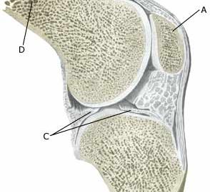
  1. Patella
  2. Tibiae
  3. Meniscus lateralis
  4. Femur

KNEE JOINT

Cause: Repeated and uniform loads can cause damage to the cartilage, and subsequently the bone beneath the cartilage (degenerative arthritis). of the synovial membrane (synovitis), which will cause formation of fluid, swelling, reduction in mobility and pain in the knee joint. Degenerative arthritis in the knee joint is often seen after a previous rupture of the anterior cruciate ligament, rupture of the posterior cruciate ligament eller meniscus lesion, where it has been necessary to remove the whole or part of the meniscus.

Symptoms: Pain in the joint with movement under load or strain. Difficulties in the start up phase are often experienced, alleviated after warm-up, but with pain again after a prolonged period under load. Swelling of the joint (synovitis) in occasional cases. With pronounced swelling, a fluid filled bursa can develop in the hollow (popliteal space) of the knee (Baker cyst).

Examination: Normal medical examination is usually sufficient to make the diagnosis, however, it is also often necessary to perform an x-ray (or ultrasound scan or MR scan) to make the diagnosis.

Treatment: Treatment comprises relief from the pain inducing activities until the swelling has gone down, after which training can commence with the primary aim to strengthen the muscles surrounding the joint and retain joint mobility (article). There is no treatment which can restore the damaged cartilage (and bone). Cartilage transplants are, as yet, not suitable for general degenerative arthritis. In cases of swelling in the joint (and popliteal space), inflammation of the synovial membrane (synovitis) can be attempted subdued by using rheumatic medicine (NSAID), or by draining the fluid and injecting corticosteroid. The injections can be performed to advantage by utilising an ultrasound guided method (Ultrasonic image). Pain without swelling of the joints is best treated with paracetamol. In severe cases of degenerative arthritis where there is pain when resting (at night), it may be necessary to replace the joint.

Rehabilitation: Rehabilitation is primarily aimed at strengthening the thigh muscles, whereby the joint can be stabilised and relieved to a certain extent.

Complications: Degenerative arthritis which is positioned on the weight-bearing parts of the joint surfaces represents one of the most serious sports injuries, and often results in an end to the sporting career. It is usually possible to participate in sports with lesser knee straining activity (i.e. swimming), whereas sports with great knee straining activity (i.e. running, football) should be treated with reservation.

Special: Shock absorbing shoes or inlays will reduce the load on the knee.

Fluid accumulation in the joint (traumatic arthritis/synovitis)

FLUID ACCUMULATION IN THE JOINT

Diagnosis: FLUID ACCUMULATION IN THE JOINT
(Traumatic arthritis/synovitis)


Anatomy:
The bones in the knee joint comprise the thigh bone (femur), shin bone (tibia) and the knee cap (patella). The joint cavity is coated with a very thin synovial membrane.

  1. Patella
  2. Tibiae
  3. Meniscus lateralis
  4. Femur

KNEE JOINT

Cause: An inflammation of the synovial membrane (synovialis) can occur following a twist in the knee joint, which subsequently thickens and produces fluid causing the joint to swell. The injury can occur when, for example, a soccer player strikes the ball with the outermost toe, and thereby twists in the foot and knee.

Symptoms: Swelling of the joint. Pain upon movement of the knee joint. Trouble flexing the knee completely.

Acute treatment: Click here.

Examination: Swelling of joints always requires medical examination. The diagnosis is usually made following a normal medical examination, where it is not possible to show damage to other structures (ligaments, meniscus). Smaller fluid accumulations in the knee can only be seen with ultrasound (article).

Treatment: Relief. If the swelling does not decrease despite relief, the treatment can be supplemented with medicinal treatment in the form of rheumatic medicine (NSAID) or the injection of corticosteroid in the joint, possibly preceded by drainage and evaluation of the joint fluid which can advantageously be performed under ultrasound guidance (article).

Complications: If smooth progress is not achieved, it should be considered whether the diagnosis is correct or whether complications have arisen. Amongst others the following should be considered:

Tendinitis

TENDINITIS

Diagnosis: TENDINITIS


Anatomy:
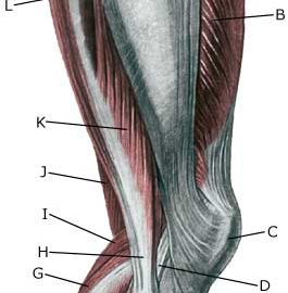
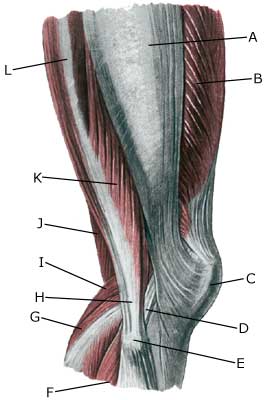
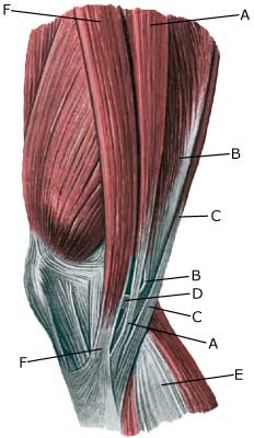
The tendons from the thigh to the shin bone pass the knee joint on the inner and outer sides, as well as to the front and rear of the knee.

  1. Tractus iliotibialis
  2. M. vastus lateralis
  3. Patella
  4. Ligamentum collaterale laterale/fibulare
  5. Caput fibulae
  6. M. soleus
  7. M. gastrocnemius
  8. Tendo m. biciptis femoris
  9. M. plantaris
  10. M. semimembranosus
  11. M. biceps femoris (caput breve)
  12. M. biceps femoris (caput longum)

OUTER KNEE

  1. M. gracilis
  2. M. semimembranosus
  3. M. semitendinosus
  4. Tendo m. semimembranosi
  5. M. gastrocnemius
  6. M. sartorius

MUSCLES AND TENDONS IN THE KNEE REGION
FROM THE MEDIAL POSITION

Cause: Tendinitis occurs as a result of repeated uniform loads causing microscopic ruptures in the tendon, and especially at the tendon fastening, which causes an inflammation. Tendinitis is a warning that the exercise performed is too strenuous for the particular muscle tendons, and if the load is not reduced a rupture can occur (“pulled muscle”) or a chronic inflammation with a substantially longer rehabilitation period as a consequence.

Symptoms: Pain upon applying pressure along the tendon, aggravated when stretching or activation of the muscle tendon.

Acute treatment: Click here.

Examination: Medical examination is not necessarily required in slight cases. Severe cases or cases that are not improved by relief should be medically evaluated to ensure a precise diagnosis. The diagnosis is usually made on the basis of a normal medical examination, however, if any doubts surround the diagnosis an ultrasound scan can be performed as this is the best type of examination to ensure the diagnosis.

Treatment: Relief, stretching and slowly increasing load within the pain threshold. If there is not sufficient progress with relief and regular rehabilitation, medical treatment in the form of rheumatic medicine (NSAID) or the injection of corticosteroid along the inflamed tendon can be considered. As the injection of corticosteroid is always a part of a long term rehabilitation of a chronic injury, it is often necessary for the rehabilitation period to stretch over several weeks to reduce the risk of relapse and ruptures. The tendon is naturally unable to accommodate maximum strain or load after a prolonged injury period after only a short rehabilitation period. If the diagnosis is made by use of ultrasound scanning and the injections are performed under ultrasound guidance, and the rehabilitation is progressed in accordance with the guidelines mentioned, then the treatment involving corticosteroid injections has very few risks connected. In cases of lack of progress with rehabilitation and medicinal treatment, surgery can be considered. However, this is very rarely necessary after a regular and strict rehabilitation period.

The rehabilitation is dependant upon which tendons are involved, but it is usually the tendons in the popliteal space of the knee (tendons contributing to the flexing of the knee).

Complications: If smooth progress is not achieved, it should be considered whether the diagnosis is correct or whether complications have arisen. Amongst others the following should be considered:

Special: Shock absorbing shoes or inlays will reduce the load. In case of lack of progress or recurrence after successful rehabilitation, a running style analysis can be considered to evaluate whether correction of the running style is indicated.

KONDITION

week15+

GENOPTRÆNING

TRAINING LADDER FOR CHILDREN AND ADOLESCENTS:
FOR RUPTURE OF THE POSTERIOR CRUCIATE LIGAMENT
(RUPTURA LIGAMENTUM CRUCIATUM POSTERIUS)

Uge 15 +

The following exercises can only be considered as a supplement to the guidelines furnished by the doctor which performed the operation. Specific precautions are necessary as the operation can be complicated. The training must not bring about swelling or pain in the knee.
KONDITION
Unlimited: Cycling. Swimming. Running.

UDSPÆNDING
(10 min):

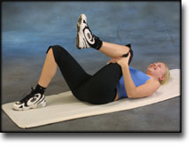
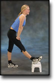
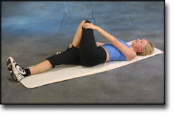
Lie on your back. Draw the injured leg up towards your head so that the muscles in the back of the thigh become increasingly stretched. Perform the exercise with outstretched as well as bent knee. Hold the position for 20 seconds and relax for 20 seconds before repeating. The exercise can also be performed standing with the injured leg outstretched on a chair while the upper body is bent slightly forwards.

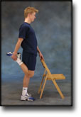
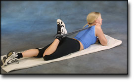
Stand with support from the back of a chair or the wall. Using your hand, bend the knee and draw the foot up and your knee slightly backwards so that the muscles in the front of the thigh become increasingly stretched. Hold the position for 20 seconds and relax for 20 seconds before repeating. The exercise can also be done lying down. If you lie on your stomach you can draw the foot up by using a towel.

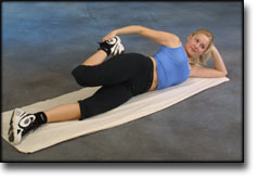
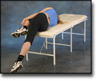
Lie on your side on a table. Bend one leg up under your body and let the other hang over the edge of the table so that the muscles in the outer side of the thigh become increasingly stretched. Hold the position for 20 seconds and relax for 20 seconds before repeating. The exercise can also be done standing by placing the outstretched injured leg behind the good leg at the same time as bending over the injured leg.

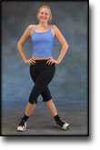
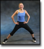
Stand with one leg outstretched and the other slightly bent. Thrust your weight to the side over the bent leg so that the inner side of the opposite thigh becomes increasingly stretched. Hold the position for 20 seconds and relax for 20 seconds before repeating.

Lie on your back with one leg outstretched and the other bent with the foot on the other side of the outstretched leg. Draw the knee up towards the opposite shoulder so that the buttocks become increasingly stretched. Hold the position for 20 seconds and relax for 20 seconds before repeating.

KOORDINATION
(10 min):

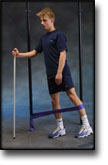
Seesaw. Balance on two legs, possibly using a hand as support against the wall, balancing subsequently on one leg without support. Look straight ahead and keep knees bent.

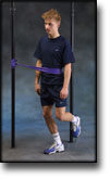
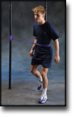
Stand on the healthy leg with elastic fixed around the hip. The elastic should be fixed to the wall or a wall bar. Take-off on the healthy leg and land on the leg to be trained and keep your balance. Remember that the elastic should be positioned so that it gives resistance at the moment of take-off. Change legs.

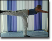
Stand on the injured leg with your upper body bent forwards at 90 degrees. Lift the good leg in a straight line behind you. When you feel comfortable with the exercise, it can be made more difficult by closing your eyes.

STYRKE
(40 min):

Up and down from the stool with load. Tie elastic around the hip and go up on the stool in a slow movement. The elastic should be fastened to the wall.

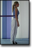
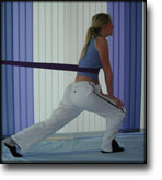
Stand with elastic around the hip. Step forward over one knee and hold the front foot firmly against the floor. Bend the rear leg and go forward onto your toes. Remember to change leg.

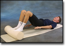
Lie on your back with a ball or firm round cushion under both feet. Roll the ball backwards and forwards in a steady pace while lifting your backside.

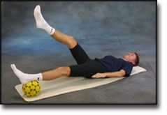
Lie on your back with a ball or firm round cushion under the injured leg. Lift your backside up from the floor and stretch the healthy leg. Hold the position for a few seconds.

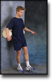
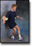
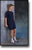
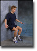
Stand with your back against a wall with a ball or firm round cushion between the wall and your back. Slowly go down to bend your knee 90 degrees before slowly rising up again.

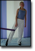
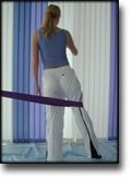
Stand on the healthy leg with the elastic around the inside of the injured leg. Move the injured leg from side to side in a slow smooth movement. Moving the position of the elastic lower down the leg can increase the load.

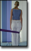
Stand on the healthy leg with the elastic around the outside of the injured leg. Move the injured leg from side to side in a slow smooth movement. Moving the position of the elastic lower down the leg can increase the load.

Stretching is carried out in the following way: stretch the muscle group for 3-5 seconds. Relax for 3-5 seconds. The muscle group should subsequently be stretched for 20 seconds. The muscle is allowed to be tender, but must not hurt. Relax for 20 seconds, after which the procedure can be repeated.

The time consumed for stretching, coordination and strength training can be altered depending on the training opportunities available and individual requirements.

KONDITION

week13-15

GENOPTRÆNING

TRAINING LADDER FOR CHILDREN AND ADOLESCENTS:
FOR RUPTURE OF THE POSTERIOR CRUCIATE LIGAMENT
(RUPTURA LIGAMENTUM CRUCIATUM POSTERIUS)

Uge 13-15


The following exercises can only be considered as a supplement to the guidelines furnished by the doctor which performed the operation. Specific precautions are necessary as the operation can be complicated. The training must not bring about swelling or pain in the knee.
KONDITION
Unlimited: Cycling. Swimming. Light running.

UDSPÆNDING
(10 min):

Lie on your back. Draw the injured leg up towards your head so that the muscles in the back of the thigh become increasingly stretched. Perform the exercise with outstretched as well as bent knee. Hold the position for 20 seconds and relax for 20 seconds before repeating. The exercise can also be performed standing with the injured leg outstretched on a chair while the upper body is bent slightly forwards.

Stand with support from the back of a chair or the wall. Using your hand, bend the knee and draw the foot up and your knee slightly backwards so that the muscles in the front of the thigh become increasingly stretched. Hold the position for 20 seconds and relax for 20 seconds before repeating. The exercise can also be done lying down. If you lie on your stomach you can draw the foot up by using a towel.

Lie on your side on a table. Bend one leg up under your body and let the other hang over the edge of the table so that the muscles in the outer side of the thigh become increasingly stretched. Hold the position for 20 seconds and relax for 20 seconds before repeating. The exercise can also be done standing by placing the outstretched injured leg behind the good leg at the same time as bending over the injured leg.

Stand with one leg outstretched and the other slightly bent. Thrust your weight to the side over the bent leg so that the inner side of the opposite thigh becomes increasingly stretched. Hold the position for 20 seconds and relax for 20 seconds before repeating.

Lie on your back with one leg outstretched and the other bent with the foot on the other side of the outstretched leg. Draw the knee up towards the opposite shoulder so that the buttocks become increasingly stretched. Hold the position for 20 seconds and relax for 20 seconds before repeating.

KOORDINATION
(10 min):

Seesaw. Balance on two legs, possibly using a hand as support against the wall, balancing subsequently on one leg without support. Look straight ahead and keep knees bent.

Stand on the healthy leg with elastic fixed around the hip. The elastic should be fixed to the wall or a wall bar. Take-off on the healthy leg and land on the leg to be trained and keep your balance. Remember that the elastic should be positioned so that it gives resistance at the moment of take-off. Change legs.

Stand on the injured leg with your upper body bent forwards at 90 degrees. Lift the good leg in a straight line behind you. When you feel comfortable with the exercise, it can be made more difficult by closing your eyes.

STYRKE
(40 min):

Stand with your back to the wall with your weight on both feet. Slowly go down and bend the knee to 90 degrees, and slowly rise again.

Stand with elastic around the hip. Step forward over one knee and hold the front foot firmly against the floor. Bend the rear leg and go forward onto your toes. Remember to change leg.

Lie on your back with a ball or firm round cushion under both feet. Roll the ball backwards and forwards in a steady pace while lifting your backside.

Lie on your back with a ball or firm round cushion under the injured leg. Lift your backside up from the floor and stretch the healthy leg. Hold the position for a few seconds.

Stand on the healthy leg with the elastic around the inside of the injured leg. Move the injured leg from side to side in a slow smooth movement. Moving the position of the elastic lower down the leg can increase the load.

Stand on the healthy leg with the elastic around the outside of the injured leg. Move the injured leg from side to side in a slow smooth movement. Moving the position of the elastic lower down the leg can increase the load.

Stretching is carried out in the following way: stretch the muscle group for 3-5 seconds. Relax for 3-5 seconds. The muscle group should subsequently be stretched for 20 seconds. The muscle is allowed to be tender, but must not hurt. Relax for 20 seconds, after which the procedure can be repeated.

The time consumed for stretching, coordination and strength training can be altered depending on the training opportunities available and individual requirements.

KONDITION

week10-12

GENOPTRÆNING

TRAINING LADDER FOR CHILDREN AND ADOLESCENTS:
FOR RUPTURE OF THE POSTERIOR CRUCIATE LIGAMENT
(RUPTURA LIGAMENTUM CRUCIATUM POSTERIUS)

Uge 10-12

The following exercises can only be considered as a supplement to the guidelines furnished by the doctor which performed the operation. Specific precautions are necessary as the operation can be complicated. The training must not bring about swelling or pain in the knee.
KONDITION
Unlimited: Cycling with raised saddle. Swimming. Light jogging.

UDSPÆNDING
(10 min):

Lie on your back. Draw the injured leg up towards your head so that the muscles in the back of the thigh become increasingly stretched. Perform the exercise with outstretched as well as bent knee. Hold the position for 20 seconds and relax for 20 seconds before repeating. The exercise can also be performed standing with the injured leg outstretched on a chair while the upper body is bent slightly forwards.

Stand with support from the back of a chair or the wall. Using your hand, bend the knee and draw the foot up and your knee slightly backwards so that the muscles in the front of the thigh become increasingly stretched. Hold the position for 20 seconds and relax for 20 seconds before repeating. The exercise can also be done lying down. If you lie on your stomach you can draw the foot up by using a towel.

Lie on your side on a table. Bend one leg up under your body and let the other hang over the edge of the table so that the muscles in the outer side of the thigh become increasingly stretched. Hold the position for 20 seconds and relax for 20 seconds before repeating. The exercise can also be done standing by placing the outstretched injured leg behind the good leg at the same time as bending over the injured leg.

Stand with one leg outstretched and the other slightly bent. Thrust your weight to the side over the bent leg so that the inner side of the opposite thigh becomes increasingly stretched. Hold the position for 20 seconds and relax for 20 seconds before repeating.

Lie on your back with one leg outstretched and the other bent with the foot on the other side of the outstretched leg. Draw the knee up towards the opposite shoulder so that the buttocks become increasingly stretched. Hold the position for 20 seconds and relax for 20 seconds before repeating.

KOORDINATION
(10 min):

Stand on the healthy leg with elastic fixed around the hip. The elastic should be fixed to the wall or a wall bar. Take-off on the healthy leg and land on the leg to be trained and keep your balance. Remember that the elastic should be positioned so that it gives resistance at the moment of take-off. Change legs.

Seesaw. Balance on two legs, possibly using a hand as support against the wall, balancing subsequently on one leg without support. Look straight ahead and keep knees bent.

Stand on the injured leg with your upper body bent forwards at 90 degrees. Lift the good leg in a straight line behind you. When you feel comfortable with the exercise, it can be made more difficult by closing your eyes.

STYRKE
(40 min):

Up and down from the stool with load. Tie elastic around the hip and go up on the stool in a slow movement. The elastic should be fastened to the wall.

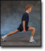
Go forward on the injured leg until the knee is bent to max. 90 degrees. Stand up on the same leg and return to the starting position.

Lie on your back with a ball or firm round cushion under both feet. Roll the ball backwards and forwards in a steady pace while lifting your backside.

Lie on your back with a ball or firm round cushion under the injured leg. Lift your backside up from the floor and stretch the healthy leg. Hold the position for a few seconds.

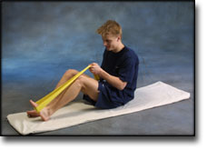
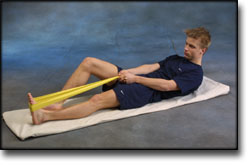
Lie on the floor with slightly bent knees. Put the elastic under the foot and hold firmly with your hands. Stretch the knee so that the elastic is drawn tight and slowly bend the knee again.

Stand on the good leg with the elastic around the inner side of the injured leg above the knee. Move the injured leg from side to side in a slow movement.

Stand on the good leg with the elastic around the outer side of the injured leg above the knee. Move the injured leg from side to side in a slow movement.

Stretching is carried out in the following way: stretch the muscle group for 3-5 seconds. Relax for 3-5 seconds. The muscle group should subsequently be stretched for 20 seconds. The muscle is allowed to be tender, but must not hurt. Relax for 20 seconds, after which the procedure can be repeated.

The time consumed for stretching, coordination and strength training can be altered depending on the training opportunities available and individual requirements.

>