Kategoriarkiv: Hip

KONDITION

step1

Training ladder for:
DEGENERATIVE ARTHRITIS
(ARTROSE)

STEP 1

The indications of time after stretching, coordination training and strength training show the division of time for the respective type of training when training for a period of one hour. The time indications are therefore not a definition of the daily training needs, as the daily training is determined on an individual basis.

KONDITION
Unlimited: Cycling with raised saddle. Swimming. Running in deep water.

UDSPÆNDING
(10 min)

Lie on your back. Draw the injured leg up towards your head so that the muscles in the back of the thigh become increasingly stretched. Perform the exercise with outstretched as well as bent knee. Hold the position for 20 seconds and relax for 20 seconds before repeating. The exercise can also be performed standing with the injured leg outstretched on a chair while the upper body is bent slightly forwards.

Stand with support from the back of a chair or the wall. Using your hand, bend the knee and draw the foot up and your knee slightly backwards so that the muscles in the front of the thigh become increasingly stretched. Hold the position for 20 seconds and relax for 20 seconds before repeating. The exercise can also be done lying down. If you lie on your stomach you can draw the foot up by using a towel.

Lie on your side on a table. Bend one leg up under your body and let the other hang over the edge of the table so that the muscles in the outer side of the thigh become increasingly stretched. Hold the position for 20 seconds and relax for 20 seconds before repeating. The exercise can also be done standing by placing the outstretched injured leg behind the good leg at the same time as bending over the injured leg.

Stand with one leg outstretched and the other slightly bent. Thrust your weight to the side over the bent leg so that the inner side of the opposite thigh becomes increasingly stretched. Hold the position for 20 seconds and relax for 20 seconds before repeating.

KOORDINATION
(5 min)

Seesaw. Balance on two legs, possibly using a hand as support against the wall, balancing subsequently on one leg without support. Look straight ahead and keep knees bent.

STYRKE
(45 min)

Sit on a chair with elastic around the ankle, facing the elastic. Lift the leg and slowly bend and stretch the knee.

Sit on a chair with elastic attached to the ankle. Raise the leg and slowly stretch and bend the knee.

Pull yourself forward on a chair by use of your heel against the floor. Increase the resistance by having someone hold the chair.

Sit on a chair and lift the knee to a horizontal position. Hold for 1 minute, lower the leg to approx. 45 degrees for 30 seconds. Lower again to the starting position.

Stand on the healthy leg with the elastic around the inside of the injured leg. Move the injured leg from side to side in a slow smooth movement. Moving the position of the elastic lower down the leg can increase the load.

Stand on the healthy leg with the elastic around the outside of the injured leg. Move the injured leg from side to side in a slow smooth movement. Moving the position of the elastic lower down the leg can increase the load.

Lie on your back with the healthy leg bent and the injured leg against the wall. Place a ball between the foot and the wall. Move the injured leg up and down the wall while applying a slight pressure on the ball.

Stretching is carried out in the following way: stretch the muscle group for 3-5 seconds. Relax for 3-5 seconds. The muscle group should subsequently be stretched for 20 seconds. The muscle is allowed to be tender, but must not hurt. Relax for 20 seconds, after which the procedure can be repeated. The time consumed for stretching, coordination and strength training can be altered depending on the training opportunities available and individual requirements.

treatment-article

SportNetDoc

Effects of muscle-strength training on the functional status of patients with osteoarthritis of the knee joint.

Schilke JM, Johnson GO, Housh TJ, O’Dell JR. Nurs Res 1996 Mar-Apr;45(2):68-72.

This study was designed to determine whether an 8-week isokinetic muscle-strength-training program improved the functional health status of patients with osteoarthritis of the knee joint. Twenty volunteers with osteoarthritis of the knee joint were randomly assigned to either an experimental (n=10) or control (n=10) group. The experimental group completed six sets of five maximal contractions three times per week for 8 weeks on a Cybex II dynamometer at 90 degrees per second. Both groups were pre- and posttest for extension and flexion strength of the right and left legs, the 50-foot walk time, range of motion at the knee joint, the Osteoarthritis Screening Index (OASI), and the Arthritis Impact Measurement Scale (AIMS). There was a significant decrease in pain and stiffness, and a significant increase in mobility. There was also a significant decline in arthritis activity in the experimental group as measured by the OASI and AIMS. The experimental group significantly increased in all strength measures, while the control group increased in only right leg flexion and left leg extension across the training period.

Baker cyst

SYNOVIAL FLUID IN THE POPLITEAL SPACE

Diagnosis: SYNOVIAL FLUID IN THE POPLITEAL SPACE
(Baker’s cyst)


Anatomy:
If an excess of fluid is formed in the knee joint, the synovial fluid will be pressed through the weakest point of the rear of the joint-capsule, and accumulate in an outpouching in the hollow (popliteal space) of the knee (Baker’s cyst).

Cause: Injuries in the knee that bring about inflammation of the synovial membrane (synovitis), causing formation of an excess of synovial fluid in the knee. The fluid is pressed out through the joint-capsule and accumulates in the popliteal space (Baker cyst). The Baker cyst is consequently a symptom of something not right in the knee. The connection from the joint to the Baker cyst can in some cases become strangulated, meaning that the Baker cyst can still be present even thought he injury in the knee has healed.

Symptoms: A sensation of the popliteal space being filled up, and difficulty in flexing the knee completely. There is often also discomfort from the changes in the knee that provoked the Baker cyst.

Examination: As the presence of a Baker cyst is usually a symptom of an injury in the knee, anyone with a Baker cyst or discomfort in the knee should undergo a medical examination. It will often prove difficult to diagnose even a large Baker cyst from a normal examination, and the diagnosis is made easiest and swiftest from an ultrasound scan (Ultrasonic image).

Treatment: Treatment is naturally dependant upon the injury in the knee joint that has provoked the Baker cyst. The accumulation in the knee and the Baker cyst can be treated with rheumatic medicine (NSAID) or more effectively by injection of corticosteroid in the knee (or the Baker cyst), preceded by draining of the fluid which can advantageously be performed under ultrasound guidance. Synovial fluid can be drained from the Baker cyst if there is a connection between the knee joint and the Baker cyst, and ultrasound scanning will show the injected corticosteroid spread through the knee joint as well as the Baker cyst (article). The content of the Baker cyst is often quite thick (gelatinous) if the Baker cyst does not communicate with the knee joint.

Rehabilitation: Treatment is completely dependant upon the provoking cause of the Baker cyst.
Also read rehabilitation, general.

Complications: If smooth progress is not achieved, it should be considered whether the diagnosis is correct. Amongst others, the following should be considered:

The bursa can become infected with bacteria in rare cases. This is a serious condition where the bursa becomes red, warm and increasingly swollen and tender. This condition requires immediate medical examination and treatment. If relief and medicinal treatment (including ultrasound guided injection of corticosteroid) does not produce any progress, a surgical removal of the bursa can be attempted.

Outer collateral ligament rupture

OUTER COLLATERAL LIGAMENT RUPTURE

Diagnosis: OUTER COLLATERAL LIGAMENT RUPTURE
(Ruptura ligamentum collaterale laterale/fibulare)


Anatomy:
The bones in the knee joint comprise the thigh bone (femur), shin bone (tibia) and the kneecap (patella). There is furthermore a small joint between the shin bone and the calf bone (fibula). The knee joint is strengthened by a joint capsule which is in turn strengthened on the sides by an outer and an inner collateral ligament (ligamentum collaterale laterale (LCL) and ligamentum collaterale mediale (MCL)). Inside the knee there are two ligaments, the anterior and posterior cruciate ligaments (ligamentum cruciatum anterius and ligamentum cruciatum posterius).

  1. Ligamentum cruciatum posterius
  2. Ligamentum collaterale mediale/tibiale
  3. Meniscus medialis
  4. Insertio anterior menisci medialis
  5. Ligamentum transversum genus
  6. Tibiae
  7. Fibulae
  8. Ligamentum cruciatum anterius
  9. Ligamentum collaterale laterale/fibulare
  10. Meniscus lateralis
  11. Femur

KNEE FROM THE FRONT

  1. M. gastrocnemius
  2. Caput fibulae
  3. B. subtendinea m. bicipitis femoris inferior
  4. M. biceps femoris
  5. Ligamentum collaterale laterale/fibulare
  6. M. plantaris

OUTER LOWER LEG

Cause: When the shin bone is pressed inwards in relation to the thigh bone the outer side-ligament is stretched. If the load becomes sufficiently powerful (as is the case when the load is sudden, so that the thigh muscles do not have time to tighten and stabilize the joint) the ligament can rupture.

Symptoms: Sudden pain on the outside of the knee. Occasionally a snap is felt when the ligament ruptures. In severe cases the athlete complains about a sense of looseness in the knee.

Acute treatment: Click here.

Examination: If a total or partial rupture of the ligaments in the knee is suspected, you should seek medical attention for a diagnosis. The doctor can perform various knee tests to examine the stability of the knee (external side-instability-test) . If the knee is stable the injury is called a “sprain” of the outer side-ligament. If the knee is loose the injury is called a “rupture” of the ligament. The diagnosis is usually made from a normal medical examination. If there are any doubts surrounding the diagnosis, an ultrasound scan can be performed which will reveal the rupture and the bleeding along the ligaments (Ultrasonic image).

Treatment: The treatment of a partial or total rupture of the outer side-ligament involves relief and rehabilitation. If the knee is markedly loose, you can, for a short period, use a support splint (Don Joy). With pronounced sideways looseness surgery can be considered (article). (However if the rupture of the outer side-ligament is combined with other ligament ruptures, as is often the case, surgery is recommended).

Bandage: Hinge bandages can be used initially (Don Joy). Tape treatment of ligament ruptures in the knee have no sure effect.

Complications: If smooth progress is not achieved, it should be considered whether the diagnosis is correct or whether complications have arisen. Supplementary examinations (X-ray, ultrasound, MR scan or arthroscopy) will often be required. The following should be considered:

An inflammation by the ligament fastening is sometimes seen later in the course of the injury, and in some cases bursitis at the ligament fastening is seen. The inflammation and bursitis can possibly be treated with medication (rheumatic medicine (NSAID) or the injection of corticosteroid), if further relief has no effect (article).

Special: Since there is a risk that the injury can cause permanent disability, the injury should be reported to your insurance company.

Inner collateral ligament rupture

INNER COLLATERAL LIGAMENT RUPTURE

>

Diagnosis: INNER COLLATERAL LIGAMENT RUPTURE
(Ruptura ligamentum collaterale mediale/tibiale)


Anatomy:
The bones in the knee joint comprise the thigh bone (femur), shin bone (tibia) and the knee cap (patella). There is furthermore a small joint between the shin bone and the calf bone (fibula). The knee joint is strengthened by a joint capsule which is in turn strengthened on the sides by an outer and an inner collateral ligament (ligamentum collaterale laterale (LCL) and ligamentum collaterale mediale (MCL)). Inside the knee there are two ligaments, the anterior and posterior cruciate ligaments (ligamentum cruciatum anterius and ligamentum cruciatum posterius).

  1. Ligamentum cruciatum posterius
  2. Ligamentum collaterale mediale/tibiale
  3. Meniscus medialis
  4. Insertio anterior menisci medialis
  5. Ligamentum transversum genus
  6. Tibiae
  7. Fibulae
  8. Ligamentum cruciatum anterius
  9. Ligamentum collaterale laterale/fibulare
  10. Meniscus lateralis
  11. Femur

KNEE FROM THE FRONT

  1. Tendo m. adductoris magni
  2. Retinaculum patellae mediale
  3. Meniscus medialis
  4. Ligamentum collaterale mediale/tibiale
  5. Bursa anserina
  6. Bursa subtendinea m. sartori
  7. Ligamentum patellae
  8. Patella

KNEE FROM THE FRONT

Cause: When the shin bone is pressed outwards in relation to the thigh bone the inner side-ligament is stretched. If the load becomes sufficiently powerful (as is the case when the load is sudden, so that the thigh muscles do not have time to tighten and stabilize the joint) the ligament can rupture.

Symptoms: Sudden pain on the inside of the knee. Occasionally a snap is felt when the ligament ruptures. In severe cases the athlete complains about a sense of looseness in the knee.

Acute treatment: Click here.

Examination: If a total or partial rupture of the ligaments in the knee is suspected, you should seek medical attention for a diagnosis. The doctor can perform various knee tests (internal side-instability-test) to examine the stability of the knee. If the knee is stable the injury is called a “sprain” of the inner side-ligament. If the knee is loose the injury is called a “rupture” of the ligament. The diagnosis is usually made from a normal medical examination. If there are any doubts surrounding the diagnosis, an ultrasound scan can be performed which will reveal the rupture and the bleeding along the ligaments (Ultrasonic image) .

Treatment: The treatment of a partial or total rupture of the inner side-ligament involves relief and rehabilitation. If the knee is markedly loose, you can, for a short period, use a support splint (Don Joy). Surgical treatment previously considered has today largely been abandoned in uncomplicated ruptures of the inner side-ligament (article). (However if the rupture of the inner side-ligament is combined with other ligament ruptures, many would recommend surgery).

Bandage: Hinge bandages can be used initially (Don Joy). Tape treatment of ligament ruptures in the knee have no sure effect.

Complications: If smooth progress is not achieved, it should be considered whether the diagnosis is correct or whether complications have arisen. Supplementary examinations (X-ray, ultrasound, MR scan or arthroscopy) will often be required. The following should be considered:

An inflammation by the ligament fastening is sometimes seen later in the course of the injury, and in some cases bursitis at the ligament fastening is seen. The inflammation and bursitis can possibly be treated with medication (rheumatic medicine (NSAID) or injection of corticosteroid), if further relief has no effect.

Special: Since there is a risk that the injury can cause permanent disability, the injury should be reported to your insurance company.

Inflammation of the bursa

INFLAMMATION OF THE BURSA

Diagnosis: INFLAMMATION OF THE BURSA
(Bursitis)


Anatomy:
There are numerous bursas around the knee for the purpose of reducing the pressure on the muscles, tendons and ligaments which lie close to bone projections. Bursas can be present at all muscle fastenings around the knee, externally (i.e. bursa pes anserinus, bursa subtendinea m bicipitis), internally, to the front (i.e. bursae prepatellaris, bursa infrapatellaris profunda) and to the rear of the knee (i.e. bursae m semimembranosus, bursa subtendinea m gastrocnemii medialis & lateralis, Baker cyst).

  1. M. gastrocnemius
  2. M. plantaris
  3. M. soleus
  4. Tendo m. gastrocnemii
  5. Tendo calcaneus (Achillis)
  6. M. popliteus
  7. Bursa m. semimembranosi
  8. M. semimembranosus
  9. Bursa subtendinea m. gastrocnemii medialis

KNEE FROM THE REAR

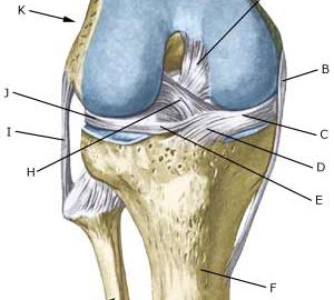
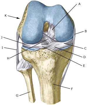
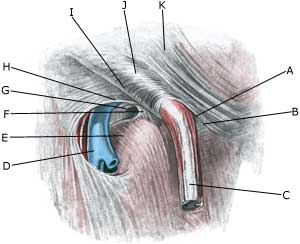
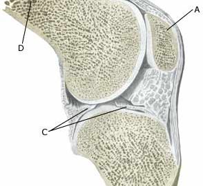
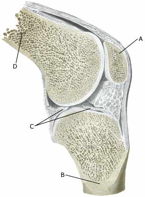
Cause: The bursas can become inflamed, produce fluid, swell and become painful with repeated over-load or due to blows. Although the condition is termed inflammation of the bursa, there is not often an infection in the bursa.

Symptoms: Pain when applying pressure to the bursa, which sometimes, but far from always, can give the impression of being swollen. The pain is aggravated when the muscle above the bursa is activated.

Acute treatment: Click here.

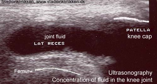
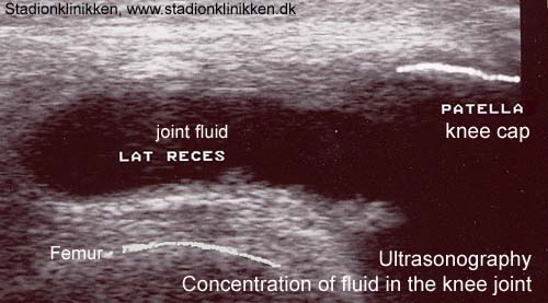
Examination: Medical examination is usually not required in light cases with only minimal tenderness. With more pronounced pain, or lack of improvement, medical examination should always be performed to confirm the diagnosis and commencement of treatment if required. The diagnosis is usually made from a normal medical examination, however, if any doubts arise an ultrasound scan can be performed which is most well suited to confirm the diagnosis.

Treatment: Treatment is primarily concentrated on providing rest. Treatment can be supplemented with rheumatic medicine (NSAID) or injection of corticosteroid in the bursa preceded by draining, which can be best performed under ultrasound guidance (article).

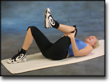
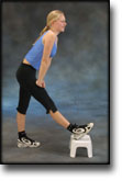
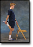
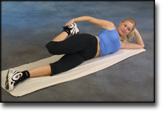
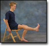
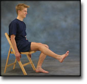
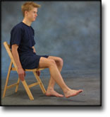
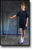
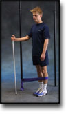
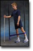
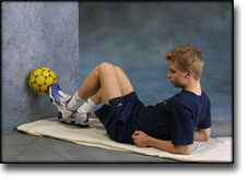
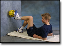
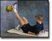
Rehabilitation: Treatment is completely dependent upon which bursa is inflamed, but the sports activity can usually be cautiously resumed when the pain has diminished, especially if the provoking factor has been identified and removed.
Also read rehabilitation, general.

Complications: If there is not a steady improvement in the condition consideration must be given as to whether the diagnosis is correct, or if complications have arisen:

In rare cases, the bursa can be infected with bacteria. This is a serious condition if the bursa becomes red, warm and increasingly swollen and tender. This condition requires immediate examination and treatment. If relief and medicinal treatment (including ultrasound guided injection of corticosteroid) does not produce any progress, a surgical removal of the bursa can be attempted.

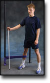
Special: Shock absorbing shoes or inlays will reduce the load. If there is a lack of progress or a relapse after successful rehabilitation, consideration must be given to performing a running style analysis to establish whether a correction of the running style should be recommended.

Sportsman’s hernia

SPORTSMANS HERNIA

Diagnosis: “SPORTSMAN’S HERNIA”


Anatomy:
The various muscles of the abdominal wall are penetrated by the inguinal canal, which contains nerves (N ilioinguinalis and the genital branch of N genitofemoralis) and in men the spermatic cord (funilicus spermaticus). In women the inguinal canal instead contains a small fibrous ligament. Where the inguinal canal penetrates the abdominal wall weak spots arise (anulus inguinalis superficialis and anulus inguinalis profundus).

  1. M. recti abdominis
  2. Funiculus spermaticus
  3. Ligamentum inguinale
  4. Spina iliaca anterior superior
  5. M. obliquus externus abdominis

INGUINAL CANAL

  1. Anulus inguinalis superficialis
  2. Crus mediale
  3. Funiculus spermaticus
    et m. cremaster
  4. V. femoralis
  5. Hiatus saphenus
  6. Lig. lacunare
  7. Anulus femoralis
  8. Margo falciformis
    (cornu superius)
  9. Lig inguinale
  10. Fibrae intercrurales
  11. M. obliquus externus abdominis

RIGHT INGUINAL CANAL

Cause: The existence of “sportsman’s hernia” is debated amongst professionals (article 1). Generally it involves a inguinal hernia (direct hernia), that can not be detected with certainty neither before nor after surgery. Some consider the condition to be an early stage of an imminent inguinal hernia, while others believe it to be small ruptures in the muscles and tendons around the inguinal canal (article 2) (article 3).

Symptoms: Pain in the groin without any detectable swelling in the groin or any other explanation for the pain.

Examination: There are no examinations (X-ray, ultrasound, MRI-scan, scintigraphy), that can detect the sportsman’s hernia (article). Previously used attempts with X-ray contrast in the abdominal cavity (herniography) showed “sportsman’s hernia” in 49% of healthy subjects, which naturally renders the examination unusable.

Treatment: Before you choose to be operated for “sportsman’s hernia”, all non-operative possibilities should be attempted, including sufficient relief and rehabilitation of the most tender structures and muscles.

Training of the muscles around the groin, stomach and loin before possible surgery.

Complications: Since it may be difficult to make a correct diagnosis in athletes with long-term groin pain (article). A multidisciplinary approach, it should be supplemented with, amongst other things, ultrasound scan and consideration of X-ray scintigraphy and possibly MRI-scan. You should of course consider the correctness of the diagnosis and amongst other things consider the following:

Inflamed mucous fold (plica synovialis)

INFLAMMATION OF THE MUCOUS FOLD

Diagnosis: INFLAMMATION OF THE MUCOUS FOLD
(Plica synovialis)


Anatomy:
The joint cavity in the knee joint is covered with a thin synovial membrane (synovialis). Four mucous folds (plica synovialis) can develop in the membrane: one fold in the inner joint chamber (medial plica), outer joint chamber (lateral plica), upper (suprapatellar) and front (anterior plica). A mucous fold is found in the inner joint chamber (medial plica) in almost 50% of all people (article).

Cause: The mucous fold will become inflamed if an entrapment of the fold occurs, or if the fold suffers internal bleeding. This will result in thickening and subsequently cause pain.

Symptoms: The mucous fold in the inner joint chamber (medial plica) will most often give symptoms which are difficult to distinguish from the symptoms of a meniscus lesion. The pain is usually localised on the inner side of the knee cap, in front of the inner joint line. The pain often occurs quite suddenly following certain movements, and can be accompanied by swelling in the knee. The knee can lock if a flap of the mucous fold becomes entrapped (article).

Examination: A medical examination is always necessary to ensure the diagnosis if there is any suspicion of an inflamed mucous fold in the knee. A tender string inside the knee cap can occasionally give a slipping sensation, but often a normal clinical examination is not sufficient. It is often necessary to perform an arthroscopic examination (telescopic examination of the joint) or MR-scan to make the diagnosis (article).

Treatment: Treatment comprises relief and careful rehabilitation of the knee. If the discomfort does not slowly diminish, the treatment can be supplemented with rheumatic medicine (NSAID) or injection of corticosteroidi in the mucous fold. Medial synovial shelf plica syndrome. Treatment by intraplical steroid injection. If this does not give the desired results, the mucous fold can be removed by arthroscopy (telescopic examination of the knee).

Exerting load on the knee can commence as soon as the pain and swelling in the knee has diminished. In the best cases, full activity is possible after a period of a few weeks. In uncomplicated cases, it should be possible to resume a full level of sports activity during the course of a month. Rehabilitation must not be allowed to cause increased swelling (or pain) in the knee.

Complications: If insufficient progress is made prior to an operation it must be considered if the diagnosis is correct. Supplementary examinations will often be required (X-ray, ultrasound or MR scan). In particular the following should be considered:

Following an arthroscopic examination fluid accumulation in the joint (traumatic arthritis/synovitis), should be considered, as well as infection in the scar or knee joint, which will always require medical attention as soon as possible.

Bone membrane tear (periosteal avulsion)

BONE MEMBRANE TEAR

Diagnosis: BONE MEMBRANE TEAR
(Perisotael avulsion)


Anatomy:
The bones in the knee joint comprise the thigh bone (femur), shin-bone (tibia) and the knee cap (patella). A small joint is also formed between the shin-bone and the calf bone (fibula).

  1. Patella
  2. Tibiae
  3. Meniscus lateralis
  4. Femur

KNEE JOINT

Cause: A violent twist in the knee joint can stretch the ligaments and tear a small piece of bone membrane from the ligament attachment point. In the majority of cases the symptoms will diminish after a few weeks, however, in some cases the tears will provoke an inflammation and can be of a longer duration. Bone membrane tears in the knee joint area are common, and can be seen in most cases where the person has played football for many years (“football-knee”). Bone membrane tears can occur on all the bones of the knee where tendons or ligaments are attached, but is most commonly seen on the inner and outer part of the knee joint as a consequence of previous spraining of the collateral ligaments.

Symptoms: Pain when applying pressure, and when stretching the tendon or ligaments which are attached to the bone where the tear has occurred.

Acute treatment: Click here.

Examination: Normal clinical examination is often sufficient. Larger tears can be seen on an X-ray. Many lesser tears can be best seen via an ultrasound scan, from which an inflammation surrounding the tear can also be seen (Ultrasonic image).

Treatment: Minor tears merely require relief from the pain inducing activities. Larger tears can require surgery. Some cases can cause prolonged discomfort with pain that does not recede despite relief. This can be due to the tear causing chronic inflammation in the tissue. In such cases, rheumatic medicine (NSAID) or injection of corticosteroid in the area surrounding the tear can be recommended.

Rehabilitation: Rehabilitation is totally dependent upon the type of tear, and the treatment (conservative or surgical). The tears on the inner side of the knee are usually re-trained in the manner of inner collateral ligaments ruptures, whilst tears externally to the knee are re-trained as outer collateral ligament ruptures.
Also read rehabilitation, general.

Complications: If smooth progress is not achieved it should be considered whether the diagnosis is correct, which will often require supplementary examination (X-ray, ultrasound scanning or MR scanning). The following should especially be considered: