Kategoriarkiv: Adults injuries

Chronic compartment syndrome

CHRONIC COMPARTMENT SYNDROME

Diagnosis: CHRONIC COMPARTMENT SYNDROME


Anatomy:
The calf muscles are divided into three groups (muscle compartments) of powerful muscle membranes (fascias) that are partially unremitting. A forward, an exterior and a rear muscle group (which is divided into a superficial and a deep part). Each muscle group has its own blood and nerve supply.

  1. Tendo calcaneus (Achillis)
  2. M. soleus
  3. M. gastrocnemius

LOWER LEG

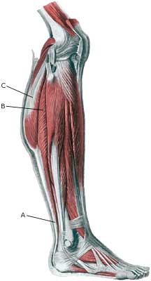
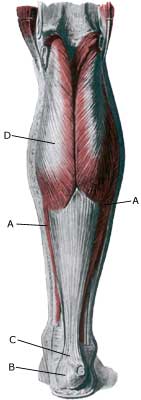
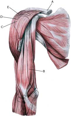
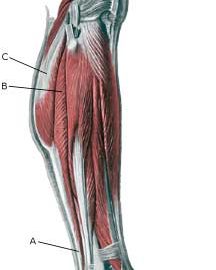
Cause: With rapidly increasing training of the calf muscles, the muscles can grow so fast that the muscle membranes surrounding the muscles can not keep up with the expansion, thereby increasing the pressure in the muscle group. The pressure can become so high that an impingement of blood vessels and nerves can occur. In some cases chronic compartment syndrome is due to scar tissue formation in the muscle (after earlier muscle ruptures).

Symptoms: With the chronic compartment syndrome, there is slowly insetting pain in the muscle after a few minutes activity. There is a sensation that the muscle is “tightened” and becomes hard, which is accompanied by discomfort. If the activity is stopped the discomfort diminishes, but returns a short period after the resumption of sports activity.

Examination: The diagnosis is made on the basis of the characteristic history and possibly with a pressure measurement in the muscle compartment (article).

Treatment: With the chronic muscle compartment syndrome the treatment comprises relief and slowly increasing training intensity, rheumatic medicine (NSAID) and massage (article). If there is scar tissue in the muscle, ultrasound guided injection of corticosteroid around the scar tissue formation can be attempted. In cases where there is a lack of progress a surgical splitting of the muscle membranes can be performed, which is usually a minor procedure with good results
(article-1) (article-2).

Rehabilitation: INSTRUCTION

The rehabilitation is completely dependant on which muscle groups are affected. The sports activity can generally be slowly resumed once pain has diminished, under the principles mentioned under rehabilitation, general.

Acute compartment syndrome

ACUTE COMPARTMENT SYNDROME

Diagnosis: ACUTE COMPARTMENT SYNDROME


Anatomy:
The calf muscles are divided into three groups (muscle compartments) of powerful muscle membranes (fascias) that are partially unremitting. A forward, an exterior and a rear muscle group (which is divided into a superficial and a deep part). Each muscle group has its own blood and nerve supply.

  1. Tendo calcaneus (Achillis)
  2. M. soleus
  3. M. gastrocnemius

LOWER LEG

Cause: The pressure in a muscle compartment can rise so fast (due to bleeding or fluid extraction) that the muscle membranes cannot keep up. Therefore the pressure in the muscle compartment can increase so greatly that impingement of blood vessels and nerves can occur.

Symptoms: With the acute muscle compartment syndrome there is increasing pain, which is often more powerful than expected from the primary evaluation of the extent of the injury. At the same time sensory disturbances can occur in the toes.

Acute treatment: Click here.

Acute treatment: Off course compression bandage should not be applied.

Examination: The diagnosis is made on the basis of the characteristic history, increased circumference of the calf muscles, which are tight and hard and by a pressure measurement in the muscle compartment (article).

Treatment: With the acute muscle compartment syndrome the treatment in severe cases comprises acute splitting of the muscle membrane. It is imperative for the continuing function of the muscle that this operation be acute, which is, of course, only possible if the athlete seeks acute medical attention (article-1) (article) (article-3)

Rehabilitation: The rehabilitation is completely dependant on which muscle groups are affected, the provoking cause (blow to the muscle, muscle rupture or over training) and which treatment that has been performed (relief, surgical splitting). The sports activity can generally be slowly resumed, once pain has decreased according to the principles as mentioned under rehabilitation, general.

Complications: Muscles and nerves can suffer permanent damage if the treatment is not started as soon as possible.

Special: Since there is a chance of permanent injury, the injury should be reported to your insurance agency.

Bone fracture

BONE FRACTURE

Diagnosis: BONE FRACTURE


Anatomy:
The bones in the lower leg comprise the shin bone (tibia) and the calf bone (fibula).

  1. Tibia
  2. Fibula

SHIN BONE FROM THE FRONT

Cause: Violent loads can cause a fracture on the shin-bone as well as the calf bone. Fracture of the fibula is often seen after a kick on the outer side of the shin bone.

Symptoms: Pain upon applying pressure (direct and indirect tenderness) and when under load (walking and running).

Acute treatment: Click here.

Examination: X-rays.

Treatment: The treatment comprises relief and bandaging. In some cases surgery is necessary depending on the type of fracture and any displacement of the fracture-surfaces.

Rehabilitation: The rehabilitation is completely dependant on the type of fracture and the treatment (relief, bandaging or surgical). Approximately six months’ rehabilitation must be expected before shin bone fractures allow resumption of maximum load, and approximately 3 months’ rehabilitation before calf bone fractures allow maximum load.
Also read rehabilitation, general.

Plastbandage: Individual plastic bandages can be manufactured for use during sports activity after bone fractures. Individual plastic bandages are particularly well-suited after a fracture of the fibula.

Complications: If progress is not smooth you should be medically re-examined to ensure that the fracture is healing according to plan. In some cases a false joint can be formed (pseudoarthrosis), requiring surgical treatment.

Stress fracture

STRESS FRACTURE

Diagnosis: STRESS FRACTURE


Anatomy:
The bones of the lower leg comprise the shin bone (tibia) and the calf bone (fibula).


  1. Tibia
  2. Fibula

SHIN BONE FROM THE FRONT

Cause: Repeated uniform loads, particularly when walking or running, can cause such great stress that cracks (fractures) appear in the bone (article).

Symptoms: Pain upon applying pressure (direct and indirect tenderness) and applying load (walking, running).

Examination: X-ray. Since many stress fractures are not visible early in the course, x-ray examination can be repeated after a few weeks, if stress fractures are still suspected. Scintigraphy, MRI and ultrasound scans can often diagnose stress fractures far earlier than x-rays (Ultrasonic image) (Photo). It is crucial for the result of the treatment that the diagnosis is made as early as possible (article).

Treatment: The treatment primarily comprises relief and possibly bandaging. Only in special cases is surgery necessary. It is imperative that there are good shock-absorbing soles in the shoes (article).

Rehabilitation: The rehabilitation is completely dependant on the type of fracture and the treatment (relief or surgical).
Also read rehabilitation, general.

Complications: If progress is not smooth, you should be medically re-evaluated to ensure that the fracture is healing according to plan. In some cases a false joint is formed (pseudoarthrosis), which requires surgical treatment.

Special: Shock absorbing shoes or inlays will reduce the load.

Inflammation of the outer shin bone

INFLAMMATION OF THE OUTER SHIN BONE

Diagnosis: INFLAMMATION OF THE OUTER SHIN BONE
(Lateral tibial periostitis)


Anatomy:
The calf muscles are divided in three muscle groups (compartments) by powerful muscle membranes (fascie). A forward, an exterior and a rear muscle group (which is divided in a superficial and a deep part). The large rear muscle groups’ muscle membrane (fascie) is fastened on the inner edge of the shin bone, while the forward muscle group is fastened on the external edge of the shin bone (Photo).

Cause: In cases of repeated uniform loads, a stress related inflammation can occur where the muscle membrane fastens on the edge of the shin bone. The condition is most often seen in athletes who jump a lot and with frequent change of running surface or shoes.

Symptoms: Pain upon applying pressure, particularly on the exterior edge of the shin bone and upon straining, and is aggravated when the foot is bent upwards against resistance (extension). Occasionally an irregular bone edge can be felt on the shin bone.

Acute treatment: Click here.

Examination: In light cases medical examination is not necessarily required. In cases with more pronounced pain or lack of progress despite relief, a medical examination is recommended to ensure the diagnosis and rule out amongst other things a stress fracture. A normal medical examination is usually sufficient in order to make the diagnosis. It can be necessary to supplement with X-ray, scintigraphy or ultrasound scanning.

Treatment: The treatment involves relief, stretching and slow rehabilitation. It is imperative that there are good shock absorbing soles in the shoes. In cases where there is a lack of progress the treatment can be supplemented with medical treatment in the form of rheumatic medicine (NSAID) or the injection of corticosteroid (which is always a part of a longer lasting rehabilitation). In severe cases with no effect from relief, correct rehabilitation and medical treatment you can surgically split the membranes (fasciotomy) which fasten on the edge of the shin bone (article).

Complications: If the course does not progress smoothly, you should be medically re-examined to ensure that the diagnosis is correct, and that complications have not arisen in the form of stress fracture.

Special: Shock absorbing shoes or inlays will reduce the load. In cases with lack of progress or relapse after successful rehabilitation, a running style analysis can be considered to evaluate whether correction of the running style is indicated.

Inflammation of the inner shin bone

INFLAMMATION OF THE INNER SHIN BONE

Diagnosis: INFLAMMATION OF THE INNER SHIN BONE
(Periostitis tibialis medialis, “Shin splint”)


Anatomy:
The calf muscles are divided in three muscle groups (compartments) by powerful muscle membranes (fascie). A forward, an exterior and a rear muscle group (which is divided into a superficial and a deep part). The large rear muscle groups’ muscle membrane (fascie) is fastened on the inner edge of the shin bone (tibia), while the forward muscle group is fastened on the external edge of the shin bone (Photo).

Cause: In cases of repeated uniform loads, a stress related inflammation can occur where the muscle membrane fastens on the edge of the shin bone. The condition is most often seen in athletes with a tendency to rotate the foot outwards (hyperpronation) or with a high foot arch. Frequent change of running surface or shoes increases the risk.

Symptoms: Pain on the inner edge of the shin bone, aggravated upon applying pressure, load (running) and stretching in the foot joint against resistance (flexion). The pain is often localized to the lower part of the shin bone. Sometimes an irregular bone edge can be felt on the shin bone.

Acute treatment: Click here.

Examination: In light cases medical examination is not necessarily required. In cases with more pronounced pain or lack of progress despite relief, medical examination is recommended to ensure the diagnosis and rule out amongst other things a stress fracture. A normal medical examination is usually sufficient in order to make the diagnosis. In some cases it may be necessary to supplement with X-rays, scintigraphy or ultrasound scanning.

Treatment: The treatment comprises relief, stretching and slow rehabilitation. It is imperative that there are good shock absorbing soles in the shoes. In cases where there is a lack of progress the treatment can be supplemented with a medical treatment in the form of rheumatic medicine (NSAID) or the injection of corticosteroid (which is always a part of a longer lasting rehabilitation). In severe cases with no effect from relief, correct rehabilitation and medical treatment, you can surgically split the membranes (fasciotomy) which fasten on the edge of the shin bone (article).

Complications: If the course does not progress smoothly, you should be medically re-examined to ensure that the diagnosis is correct, and that complications have not arisen in the form of stress fracture.

Special: Shock absorbing shoes or inlays will reduce the load. In case of lack of progress or relapses after successful rehabilitation, a running style analysis can be considered to evaluate whether correction of the running style is indicated.

Muscular bleeding in the calf muscle

MUSCULAR BLEEDING IN THE CALF MUSCLE

Diagnosis: MUSCULAR BLEEDING IN THE CALF MUSCLE
(Haematoma musculi)


Anatomy:
The calf muscle (M Gastrocnemicus) is comprised of two muscle heads which gather in a wide tendinous ligament and continue in to the Achilles tendon. Another of the larger calf muscles (M Soleus) is attached to the front side of the Achilles tendon and thus forms a part of the Achilles tendon. It is these muscles that are most frequently subjected to ruptures and bleeding. On the lower leg there are many other muscles, where bleeding is relatively rare.


  1. M. soleus
  2. Tuber calcanei
  3. Tendo calcaneus (Achillis)
  4. M. gastrocnemius

LOWER LEG FROM THE REAR

Cause: If a muscle is subjected to a blow the muscle belly, which contains blood vessels, is pressed against the bones, causing an injury and rupture of the muscle fibres and blood vessels. The rupture usually occurs deep in the muscle. In other cases the bleeding can occur after a larger or smaller muscle rupture in the calf. The bleeding can either penetrate the muscle membrane and spread over a large area, or it can accumulate in the muscle.

Symptoms: Pain and swelling in the muscle. In some cases a hard, tender accumulation can be felt (accumulated bleeding in the muscle). In other cases a bluish discoloration of the subcutis (the bleeding has penetrated the muscle membrane and spread into the subcutis). The pain is aggravated upon activation and stretching of the muscle.

Acute treatment: Click here.

Examination: In light cases with only minimal tenderness and no discomfort when walking normally, medical examination is not necessarily required. The severity of the tenderness is, however, not always a measure of the extent of the injury. In case of more pronounced pain medical examination is required to ensure the diagnosis and treatment. A normal medical examination is usually sufficient in order to make the diagnosis, however, if there is any doubt concerning the
diagnosis an ultrasound scan can be performed for ensuring the diagnosis
(Ultrasonic image) (article). The larger the bleeding, as evaluated by ultrasound, the longer the healing process.

Treatment: The treatment primarily consists of relief, as with muscle rupture in the calf. In cases of large accumulated bleeding the blood accumulation can be drained under ultrasound guidance. Some recommend treatment with rheumatic medicine (NSAID) and advise caution with massage to reduce the risk of myositis ossificans (formation of bony bars within the muscle).

Complications: If the course does not progress smoothly, you should be medically re-examined to ensure that the diagnosis is correct, and that complications to muscle ruptures have not arisen.

Muscle rupture in the calf

MUSCLE RUPTURE IN THE CALF

Diagnosis: MUSCLE RUPTURE IN THE CALF
(Ruptura musculi)


Anatomy:
The calf muscle (M Gastrocnemicus) is comprised of two muscle heads which gather in a wide tendinous ligament and continue in to the Achilles tendon. Another of the larger calf muscles (M Soleus) is attached to the front side of the Achilles tendon and thus forms a part of the Achilles tendon. It is these muscles that are most frequently subjected to ruptures. On the lower leg there are many other muscles, where ruptures are relatively rare.


  1. M. soleus
  2. Tuber calcanei
  3. Tendo calcaneus (Achillis)
  4. M. gastrocnemius

LOWER LEG FROM THE REAR

Cause: When a muscle is subjected to a load beyond the strength of the muscle, a rupture occurs. The vast majority of ruptures are partial muscle ruptures. The weakest point is often between the fascia and the meaty part of the muscle.

Symptoms: In light cases a local tenderness is felt after being subject to load (“sprained muscle”, “imminent pulled muscle”). In severe cases sudden shooting pains are felt in the muscle (“partial muscle rupture”, “pulled muscle”) and in the worst case a sudden snap is felt rendering the muscle unusable (“total muscle rupture”). With muscle injuries the following three symptoms are characteristic: pain upon applying pressure, stretching and activation of the calf muscle against resistance. With total ruptures a defect can often be seen and felt in the muscle, and above and below the rupture a swelling can be felt (the contracted muscle belly and bleeding). The most frequent place for ruptures on the lower leg is in the inner (medial) part of the calf muscle on the transition between the muscle belly and the tendon part of the muscle (“Tennis leg”).

Acute treatment: Click here.

Examination: In light cases (“sprained muscle”) with only minimal tenderness and no discomfort when walking normally, medical examination is not necessarily required. The severity of the tenderness is however, not always a measure of the extent of the injury. In cases of more pronounced tenderness or pain, medical examination is required to ensure the diagnosis and treatment. A normal medical examination is usually sufficient in order to make the diagnosis, however, if there is any doubt concerning the diagnosis an ultrasound scan can be performed to ensure the diagnosis (article) (Ultrasonic image).

Treatment: The treatment of the vast majority of muscle injuries today involves relief and rehabilitation (article). In some cases with total ruptures, surgery can be considered.

Complications: If the course does not progress smoothly, you should be medically re-examined to ensure that the diagnosis is correct, and that complications to muscle ruptures have not arisen.

Nerve entrapment on the back of the shoulder blade

NERVEAFKLEMNING BAG PÅ SKULDERBLADET

Diagnosis: NERVE ENTRAPMENT ON THE BACK OF THE SHOULDER BLADE
(Entrapment nervus suprascapularis)


Anatomy:
Behind the upper edge of the shoulder blade in a small channel (incisura scapulae) closed by a ligament (ligamentus transversum), runs a nerve (N suprascapularis) that distributes nerve fibres to parts of the shoulder joint-capsule and the upper and lower shoulder blade muscle (M supraspinatus and M infraspinatus)

  1. Acromion
  2. Cavitas glenoidalis
  3. Fossa supraspinata
  4. Spina scapulae
  5. Incisura scapulae
  6. Processus coracoideus

RIGHT SHOULDER BLADE SEEN
FROM THE REAR

Cause: In case of repeated strain with movement in the shoulder joint, an entrapment of the nerve (article). Entrapment of the nerve may also occur as the result of a blow.

Symptoms: Usually a burning or cutting pain in the shoulder. Possibly a sense of weakness in the shoulder. In case of long-term symptoms degradation of the shoulder blade muscles will occur.

Examination: Uncharacteristic shoulder symptoms, not improved by relief, should be examined by a physician. If the results of the examination indicate nerve entrapment the diagnosis is made by a nerve conduction velocity test (EMG).

Treatment: The treatment involves relief from the pain inducing activity, stretching and rehabilitation of the muscles around the shoulder. If no progress is made with relief and rehabilitation, surgical intervention may be attempted (article-1) (article-2).

Relief is the primary element, and it is particularly important that the rehabilitation does not cause any discomfort. Fitness training should not be resumed until the pain has stopped

Complications: If satisfactory progress is not made, a physician should be consulted to ensure that the diagnosis is correct and that no complications have arisen. Amongst others the following should be considered:

Impigment syndrome in the shoulder

IMPINGEMENT SYNDROME IN THE SHOULDER

Diagnosis: IMPINGEMENT SYNDROME IN THE SHOULDER
(Impingement)


Anatomy:
Four muscles are part of the rotator cuff surrounding the shoulder joint, and controls, coordinates and assists movement of the shoulder: M supraspinatus (the upper shoulder blade muscle), M infraspinatus (the lower shoulder blade muscle), M subscapularis and M teres minor. When the arm is moved away from the body and above the head (abducated) the supraspinatus muscle slips under the upper bone projection of the shoulder blade (acromion).


  1. Clavicula
  2. M. biceps brachii
  3. Tuberculum majus
  4. M. deltoideus
  5. Acromion

(Photo)

SHOULDER AND UPPER ARM MUSCLES
FROM THE FRONT

Cause: There are two main causes for impingement: One external and one internal.
The external impingement is usually caused by repeated loads with the arm above the head (tennis, swimming) and causes an inflammation in the upper shoulder blade muscle (M supraspinatus) and the above-lying bursa (bursae subacromialis), which swells and may become squeezed between the head of the humerus (caput humeri) and the upper bone projection of the shoulder blade (acromion), when the arm is raised above the head (see inflammation of the bursa).
The internal impingement is caused by the supraspinatus muscle tendon scraping against the edge of the shoulder’s meniscus (posterior-superior edge of the labrum glenoidale) when throwing (90 degrees abduction and maximal extension and outwards rotation), causing damage to both the muscle tendon and the meniscus (article).

Symptoms: Slowly insetting local tenderness after the load, externally and to the front of the shoulder. Sometimes radiating down the upper arm. Pain deteriorates when external pressure is applied to the supraspinatus muscle, on the front of the shoulder and when the muscle is brought into contact with the meniscus in the shoulder (throwing exercises) or when the arm is brought approx. 80-120 degrees away from the body (abduction).

Acute treatment: Click here.

Examination: If no progress is made with relief and discontinuance of the pain inducing activity, a medical examination should be carried out to ensure that the diagnosis is correct and to commence the correct treatment. It is important to make the diagnosis as fast as possible, since the injury may otherwise slowly deteriorated (article). Ordinary medical examination is usually sufficient to make the diagnosis (internal impingement-test: Chicken-wing test), but if there is uncertainty concerning the diagnosis, it should be supplemented with an ultrasound scan, which is the most suitable examination for shoulder injuries (article). Though it is not rare for the diagnosis only being made after a arthroscopic examination is carried out (telescopic examination of the shoulder joint).

Treatment: The treatment primarily involves relief from the pain inducing activity, stretching and rehabilitation of the muscles around the shoulder, Impingement syndrome in athletes. In case of lack of progress a medical treatment can be considered, rheumatic medicine (NSAID) or injection of corticosteroid (usually in the subacromiale bursa and possibly in the shoulder joint itself as well (internal impingement with signs of synovitis)). Since the injection of corticosteroid is part of a long-term rehabilitation of a long-term injury, it is often necessary for the rehabilitation period to stretch over several weeks to months in order to reduce the risk of recurrences. Ultrasound guided injections offer the optimal effect with a minimum of risk, (article). In case of lack of progress in the rehabilitation and medical treatment, an operative treatment can be attempted.

Complications: If progress is not smooth, it should be considered whether the diagnosis is correct or whether complications have arisen. Amongst others the following should be considered: