Kategoriarkiv: Adults injuries

Tendon luxation, outer ankle knuckle

Diagnosis: TENDON LUXATION – OUTER ANKLE KNUCKLE
(Peroneus luxation)


Anatomy:
The tendons surrounding the ankle joint are connected to the bones in several places by connective tissue strings (retinaculum). Two peroneus muscles are positioned behind the outer ankle knuckle, which are used to stabilise and flex the ankle joint.


  1. M. peroneus brevis
  2. M. peroneus longus
  3. Tendo calcaneus (Achillis)
  4. Bursa subcutanea malleoli lateralis
  5. Retinaculum mm. peroneorum inferius
  6. Vagina synovialis mm. peroneorum communis
  7. Retinaculum mm. peroneorum superius

OUTER FOOT

Cause: Discomfort can be produced if a rupture of the connective tissue (retinaculum musculorum peroneorum superius & inferius) behind the outer ankle knuckle (malleolus lateralis) occurs. A rupture of the connective tissue (retinaculum) is relatively often combined with outer ligament injuries in the ankle joint.

Symptoms: Pain at the outer ankle knuckle (malleolus lateralis), where certain movements of the ankle joint can give rise to a painful sense of “slipping”.

Acute treatment: Click here.

Examination: When the painful movement of the ankle joint is performed, normal examination can detect the tendon slip over the outer ankle knuckle. A normal medical examination is usually sufficient in order
to make the diagnosis, however, if there is any doubt concerning the
diagnosis it can be made with certainty by use of ultrasound scanning while the ankle joint is in motion (dynamic ultrasound scanning). Ultrasound scanning will reveal whether there is bleeding in the acute stage. Inflammation of the tendon sheath can develop at a later stage.

Treatment: Treatment is primarily concentrated on providing rest from the painful activities (running). If steady progress from rest and rehabilitation is not achieved, and ultrasound scanning reveals inflammation of the tendon sheath, the treatment can be supplemented by medicinal treatment in the form of rheumatic medicine (NSAID) Alternatively, draining and evaluating fluid can be performed, and injection of corticosteroid into the tendon sheath. Injection into the tendon sheath is best performed if ultrasound guided. Surgical intervention can be attempted in certain cases, if resting, rehabilitation and medicinal treatment do not provide the desired result (article).

Complications: If there is not a steady improvement in the condition consideration must be given as to whether the diagnosis is correct, or if complications have arisen:

Possibly supplement with further examinations (x-ray, ultrasound scanning).

Bone membrane tear

Diagnosis: BONE MEMBRANE TEAR
(Perisotael avulsion)


Anatomy:
The foot bones comprise the 7 tarsal bones (ossa tarsi), the 5 metatarsal bones (ossa metatarsi) and the 14 bones in the toes (phalanx). Muscles or ligaments are attached or anchored to all the bones. The outer surface of the bone is called bone membrane (periosteum), (Ultrasonic image).

Cause: A violent twist can stretch the ligaments and tear a small piece of bone membrane (periosteum) from the ligament anchor point. In other cases, bone membrane tears can occur when the bones impact upon each other (for example if the foot is bent or flexed backwards with great force so that one of the tarsal bones (talus) impacts against the front edge of the shin bone). Bone membrane tears in the ankle joint area are common, and can be seen in most cases where the person has played football for many years (“football-ankle”). Bone membrane tears can occur on all the bones of the foot where tendons or ligaments are anchored.

Symptoms: Pain when applying pressure, and when stretching the tendon or ligament which is attached to the bone.

Acute treatment: Click here.

Examination: Normal clinical examination is often sufficient. Larger tears can be seen on an x-ray. Many lesser tears can be best seen via an ultrasound scan or MRI examination, (article).

Treatment: Relief from the pain inducing activities. Larger tears can require surgical operation. Lesser tears do not require treatment. Some cases can cause prolonged discomfort with pain which does not recede despite relief. This can be due to the tear causing inflammation in the tissue. In such cases, rheumatic medicine (NSAID) or injection of corticosteroid in the area surrounding the tear can be recommended.

Rehabilitation: Rehabilitation is totally dependent upon the type of tear, and the treatment (conservative or surgical).
Also read rehabilitation, general.

Complications: If there is not a steady improvement in the condition consideration must be given as to whether the diagnosis is correct, or if complications have arisen:

Stress fracture

STRESS FRACTURE

Diagnosis: STRESS (FATIGUE) FRACTURE


Anatomy:
The foot bones comprise the 7 tarsal bones (ossa tarsi), the 5 metatarsal bones (ossa metatarsi) and the 14 bones in the toes (phalanx).

  1. Phalanx media
  2. Tuberositas ossis metatarsalis V
  3. Os cuboideum
  4. Calcaneus
  5. Talus
  6. Os naviculare
  7. Os cuneiforme laterale
  8. Os cuneiforme intermedium
  9. Os cuneiforme mediale
  10. Os metatarsalei
  11. Os sesamoideum
  12. Phalanx proximalis
  13. Phalanx distalis

THE FOOT FROM ABOVE

Cause: Repeated load or strain (walking or running) can in some cases entail the load exceeding the strength of the bone tissue, thus resulting in a stress (or fatigue) fracture. Stress fractures are most often seen in the metatarsal bones, (article) (article).

Symptoms: Pain when applying pressure (direct or indirect tenderness), and when applying load or strain.

Examination: X-ray examination will usually, but not always, reveal a stress fracture. The x-ray examination can be repeated after a few weeks as a number of stress fracture are not easily discernible in the early stages. Bone scintigraphy, ultrasound scanning and MRI examination can often diagnose a stress fracture much earlier than x-ray examination (Ultrasonic image), (Scintigraphy-image).

Treatment: Treatment is primarily relief and rest, and possible bandaging. Surgical intervention is only required in very special cases. It is imperative that shoes are equipped with impact absorbing soles (article).

Rehabilitation:
Rehabilitation is totally dependent upon the type of fracture, and the treatment (conservative or surgical). Until the pain has subsided, the guidelines under rehabilitation, general should be followed.

Complications: If there is not a steady improvement in the condition a medical examination should be performed once more to ensure that the fracture is healing according to plan. In some cases, a false joint can develop which will require surgical treatment.

Bone fracture in the ankle

BONE FRACTURE IN THE ANKLE

Diagnosis: BONE FRACTURE IN THE ANKLE


Anatomy:
The foot bones comprise the 7 tarsal bones (ossa tarsi), the 5 metatarsal bones (ossa metatarsi) and the 14 bones in the toes (phalanx). Furthermore, the lower part of the shin bone (tibia) and calf bone (fibula) form a part of the ankle joint.

  1. Phalanx media
  2. Tuberositas ossis metatarsalis V
  3. Os cuboideum
  4. Calcaneus
  5. Talus
  6. Os naviculare
  7. Os cuneiforme laterale
  8. Os cuneiforme intermedium
  9. Os cuneiforme mediale
  10. Os metatarsalei
  11. Os sesamoideum
  12. Phalanx proximalis
  13. Phalanx distalis

THE FOOT FROM ABOVE

Cause: A blow or violent twist can cause a fracture of the bone (X-ray picture).

Symptoms: Pain when applying pressure (direct or indirect tenderness), and when applying load or strain.

Acute treatment: Click here.

Examination: X-ray examination will usually reveal the fracture. The fracture can in some cases first be seen after 14 days, thus the x-ray examination should be repeated if there is a continued suspicion of a fracture.

Treatment: Treatment is completely dependent upon which bones are broken, and whether there is a dislocation of the fracture. In some cases relief and rest without bandaging can be opted for, whereas other types of fracture require bandaging and possibly surgical intervention (article) (X-ray picture).

Rehabilitation: Rehabilitation is totally dependent upon the type of fracture, and the treatment (conservative or surgical).
Also read rehabilitation, general.

Complications: If there is not a steady improvement in the condition a medical examination should be performed once more to ensure that the fracture is healing according to plan. In some cases, a false joint can develop which will require (renewed) surgical treatment (X-ray picture).

Special: As there is a risk that the injury can cause permanent disability, all cases should be reported to your insurance company.

Rupture of the ligament between shin and calf bones

RUPTURE OF THE LIGAMENT BETWEEN SHIN AND CALF BONES

Diagnosis: RUPTURE OF THE LIGAMENT BETWEEN SHIN AND CALF BONES
(Syndesmosis rupture)


Anatomy:
The shin bone (tibia) and the calf bone (fibula) are held together by a connective tissue membrane (membrana interossea cruris) which is particularly strong at the ankle joint and forms a false joint. This, together with the membrane, is termed syndesmosis tibiofibularis which is strengthened in front and behind with two strong ligaments (ligamentum tibiofibulare anterius & posterius).

  1. Tibiae
  2. Malleolus medialis
  3. Malleolus lateralis
  4. Lig. tibiofibulare anterius
  5. Membrana interossea cruris
  6. Fibulae

LOWER LEG FROM THE FRONT

Cause: The syndesmosis rupture is most often caused by twisting the foot. The rupture is almost always combined with a fracture in the ankle joint, Injuries of the interior tibiofibular syndesmosis. An isolated syndesmosis rupture is quite rare (article-1), (article-2).

Symptoms: Pain in front of the ankle joint between the shin and calf bones. The pain is aggravated when the foot is turned in relation to the shin.

Acute treatment: Click here.

Examination: As the injury is almost always combined with a fracture of the bones in the ankle, an x-ray examination will always be indicative. The x-ray will show the fracture and a possible increase in the distance between the shin and calf bones.

Treatment: Dressing with a bandage and possible surgical intervention dependant upon the presence of a bone fracture.

Rehabilitation: Rehabilitation is dependent upon the course of treatment (conservative/surgical operation), and of possible fractures and their treatment.
Also read rehabilitation, general.

Complications: If there is not a steady improvement in the condition consideration must be given as to whether the diagnosis is correct, or if complications have arisen:

Rupture of the joint-capsule at the front of the ankle joint

RUPTURE OF THE JOINT-CAPSULE AT THE FRONT OF THE ANKLE JOINT

Diagnosis: RUPTURE OF THE JOINT-CAPSULE
AT THE FRONT OF THE ANKLE JOINT


Anatomy:
The ankle joint is stabilised by a joint-capsule as well as a wide fan shaped ligament on the inside (ligamentum deltoideum/mediale), and a set of outer ligaments (ligamentum talofibulare anterius fore, ligamentum calcaneofibulare centre, and ligamentum talofibulare posterius at the rear). There is also a strengthening of the ligaments fore and rear (ligamentum tibiofibulare anterius & posterius). The joint-capsule and ligaments stabilise the ankle joint, especially when twisting and running with sudden directional changes.

Cause: A rupture of the joint-capsule at the front of the ankle joint arises if the foot is over-stretched (plantar flexion), resulting in the joint-capsule over-streching and rupturing. This is often seen when a football player kicks the ground, or strikes the ball on the toe when trying to kick with the instep. In slight cases the injury can be termed a strain or sprain, and in more serious instances as full or partial rupture or tear.

Symptoms: Pain in the ankle joint which is worsened when stretching the ankle joint.

Acute treatment: Click here.

Examination: Medical examination is not necessarily required for very minor cases (slight sprain) with only minimal swelling and no discomfort when walking. The extent of the swelling is, however, not always a mark of the degree of the injury. Medical examination is recommended with more extensive swelling or pain, in order to eliminate bone fracture, bone membrane tear (periosteal avulsion), outer ligament injury in the ankle joint, inner ligament injury in the ankle joint and rupture of the ligament between shin and calf bones (syndesmosis rupture) (article). A normal medical examination is usually sufficient in order to make the diagnosis. X-ray examination will confirm or exclude any suspicion of fracture. Small bone membrane tears (periosteal avulsions) will be best seen using ultrasound scanning.

Treatment: Treatment of uncomplicated joint-capsule ruptures will usually be conservative (rehabilitation).

Bandage: It is recommended to use tape in the course of rehabilitation when starting to run on an uneven surface, with sudden directional change, or kicking balls. Taping does, however, not have the same importance as with injuries to the outer or inner ligament in the ankle (tape-instruction).

Prevention: Seesaw exercise is important in the rehabilitation phase, as well as in a preventive capacity. As a preventive measure, seesaw exercises should be performed frequently throughout the rest of the active sporting career if ligament injuries in the ankle joint have previously been experienced. Begin by standing with both feet on the seesaw and use hands for support on the wall. Gradually let go of the support to finally train by standing on only one leg (article). Special bandages have in some studies been shown to reduce the risk of ligament injuries (article).

Complications: If there is not a steady improvement in the condition consideration must be given as to whether the diagnosis is correct, or if complications have arisen:

Ligament injury in the ankle joint, inner ligament

LIGAMENT INJURY IN THE ANKLE JOINT, INNER LIGAMENT

Diagnosis: LIGAMENT INJURY IN THE ANKLE JOINT, INNER LIGAMENT
(Ruptura traumatica ligamenti medialis pedis)


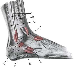
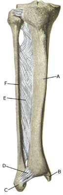
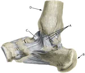
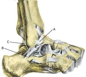
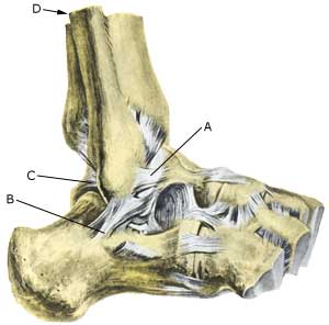
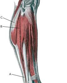
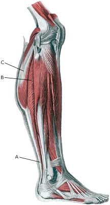
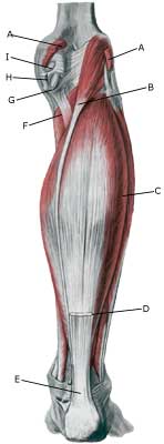
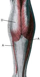
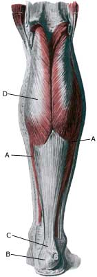
Anatomy:
The ankle joint is stabilised by a wide fan shaped ligament on the inside (ligamentum deltoideum), and a set of outer ligaments (fore, centre, and at the rear). The ligaments stabilise the ankle joint, especially when twisting and running with sudden directional changes (Photo).


  1. Ligamentum mediale/deltoideum
  2. Calcaneus
  3. Talus
  4. Tibia

INNER ANKLE JOINT

Cause: A rupture of the of the inner ligaments in the ankle joint arises if the foot is twisted such that the ligaments are overstretched and finally rupture. In slight cases the injury can be termed a strain or sprain, and in more serious instances as full or partial rupture or tear. Consequential injuries are often associated with ligament ruptures, amongst others concentration of fluid in the joint (traumatic arthritis/synovitis)and inflammation of the ankle joint tendon sheath, (article), however, these are often unfortunately overlooked (article).

Symptoms: Pain on and under the inner ankle bone (malleolus medialis), swelling due to bleeding, pain when walking.

Acute treatment: Click here.

Examination: Medical examination is not necessarily required for very minor cases (slight sprain) with only minimal swelling and no discomfort when walking. The extent of the swelling is, however, not always a mark of the degree of the injury. Medical examination is recommended with more extensive swelling or pain, in order to eliminate bone fracture in the ankle and a rupture of the ligament between the shin and calf bones (syndesmosis rupture). A normal medical examination is usually sufficient in order to make the diagnosis. X-ray examination should be performed in all cases where there is a suspicion of a bone fracture or syndesmosis rupture. Vigorous twisting of the ankle in the acute stage to appraise the degree of looseness is not indicative, as this has no influence on the choice of treatment (article). Ultrasound examination will give valuable information when the ligament is ruptured (Ultrasonic image).

Treatment: Treatment of ligament injuries is today conservative (rehabilitation). Many patients have earlier undergone operations and setting of plaster cast, however, this course of treatment has practically been abandoned in relation to uncomplicated ligament ruptures (article).

Bandage: It is recommended to use tape in the course of rehabilitation when starting to run on an uneven surface, or with sudden directional change. Nerve cells (proprioreceptors) in the ligaments transmit information to the brain on the position of the ankle joint. The brain sends information to the muscles, which are activated, ensuring that the ankle is held correctly. The nerve paths do not function in an optimal manner when the ligament is injured, and the risk of a new twist of the foot is increased thereby. Use of tape is primarily designed to stimulate the small nerve cells in the skin, thus enabling these to act as a substitute for the nerve cells temporarily damaged in the ligament. The function of the tape is thus not a pure mechanical stabilising of the ankle joint (tape-instruction). In some cases, certain forms of bandaging around the ankle can be used to advantage.

Complications: If there is not a steady improvement in the condition consideration must be given as to whether the diagnosis is correct, or if complications have arisen:

It is extremely rare that the injury results in a chronically loose ankle joint if rehabilitation is handled sensibly. If the injury does result in a chronically loose ankle joint, intensive co-ordination training must be recommended. Bandages can be tried if this is not sufficient, and if this still does not give the desired effects, surgical intervention to tighten the ligaments can be attempted.

Special: Seesaw exercise is important in the rehabilitation phase, as well as in a preventive capacity. As a preventive measure, seesaw exercises should be performed frequently throughout the rest of the active sporting career if ligament injuries in the ankle joint have previously been experienced. Begin by standing with both feet on the seesaw and use hands for support on the wall. Gradually let go of the support to finally train by standing on only one leg (article). Special bandages have in some studies been shown to reduce the risk of ligament injuries.
(article-1), (article-2).

Ligamant injury in the ankle joint, outer ligament

LIGAMENT INJURY IN THE ANKLE JOINT, OUTER LIGAMENT

Diagnosis: LIGAMENT INJURY IN THE ANKLE JOINT, OUTER LIGAMENT
(Ruptura traumatica ligamenti lateralis pedis)


Anatomy:
The ankle joint is stabilised by a joint-capsule as well as a wide fan shaped ligament on the inside (ligamentum deltoideum/mediale), and a set of outer ligaments (ligamentum talofibulare anterius fore, ligamentum calcaneofibulare centre, and ligamentum talofibulare posterius at the rear). There is also a strengthening of the ligaments in front and behind (ligamentum tibiofibulare anterius & posterius). The surrounding tendons are often enclosed by tendon sheaths and joined to the bones with connective tissue strings. The ligaments stabilise the ankle joint, especially when twisting and running with sudden directional changes (Photo).


  1. Lig. talofibulare anterius
  2. Lig. calcaneofibulare
  3. Lig. talofibulare posterius
  4. Fibula

OUTER ANKLE JOINT

Cause: A rupture of the of the outer lateral ligaments in the ankle joint arises if the foot is twisted such that the ligaments are over-stretched and finally rupture. In slight cases the injury can be termed a strain or sprain, and in more serious instances as full or partial rupture or tear. Consequential injuries are often associated with ligament ruptures, amongst others concentration of fluid in the joint (traumatic arthritis/synovitis) and inflammation of the ankle joint tendon sheath (article), however, these are often unfortunately overlooked (article).

Symptoms: Pain on and under the outer ankle bone (malleolus lateralis), swelling due to bleeding, pain when walking.

Acute treatment: Click here.

Examination: Medical examination is not necessarily required for very minor cases (slight sprain) with only minimal swelling and no discomfort when walking. The extent of the swelling is, however, not always a mark of the degree of the injury. Medical examination is recommended with more extensive swelling or pain, in order to eliminate bone fracture in the ankle and a rupture of the ligament between the shin and calf bones (syndesmosis rupture). A normal medical examination is usually sufficient in order to make the diagnosis. X-ray examination should be performed in all cases where there is a suspicion of a fracture or syndesmosis rupture. Vigorous twisting of the ankle in the acute stage to appraise the degree of looseness is not indicative, as this has no influence on the choice of treatment (article). Ultrasound examination will give valuable information when the ligament is ruptured (Ultrasonic image).

Treatment: Treatment of ligament injuries is today conservative (rehabilitation). Many patients have earlier undergone operations and setting of plaster cast, however, this course of treatment has practically been abandoned in relation to uncomplicated ligament ruptures, although there is not definitive agreement on this subject (article-1) (article-2).

Bandage: It is recommended to use tape in the course of rehabilitation when starting to run on an uneven surface, or with sudden directional change. Nerve cells (proprioreceptors) in the ligaments transmit information to the brain on the position of the ankle joint. The brain sends information to the muscles, which are activated, ensuring that the ankle is held correctly. The nerve paths do not function in an optimal manner when the ligament is injured, and the risk of a new twist of the foot is increased thereby. Use of tape is primarily designed to stimulate the small nerve cells in the skin, thus enabling these to act as a substitute for the nerve cells temporarily damaged in the ligament. The function of the tape is thus not a pure mechanical stabilising of the ankle joint (tape-instruction). In some cases, certain forms of bandaging around the ankle can be used to advantage. Special bandages have in some studies been shown to reduce the risk of ligament injuries.
(article-1), (article-2).

Complications: If there is not a steady improvement in the condition consideration must be given as to whether the diagnosis is correct, or if complications have arisen:

It is extremely rare that the injury results in a chronically loose ankle joint if rehabilitation is handled sensibly. If the injury does result in a chronically loose ankle joint, intensive co-ordination training must be recommended. Bandages can be tried if this is not sufficient, and if this still does not give the desired effects, surgical intervention to tighten the ligaments can be attempted. The results are usually quite acceptable (article).

Special: Seesaw exercise is important in the rehabilitation phase, as well as in a preventive capacity. As a preventive measure, seesaw exercises should be performed frequently throughout the rest of the active sporting career if ligament injuries in the ankle joint have previously been experienced. Begin by standing with both feet on the seesaw and use hands for support on the wall. Gradually let go of the support to finally train by standing on only one leg (article).

Rupture of the soleus muscle

Diagnosis: RUPTURE OF THE SOLEUS MUSCLE
(Ruptura M soleus)


Anatomy:
The calf muscle (M Gastrocnemicus) is comprised of two muscle heads which gather in a wide tendinous ligament and continue in to the Achilles tendon. Another of the larger calf muscles (M Soleus) is attached to the front side of the Achilles tendon and thus forms a part of the Achilles tendon. The Achilles is attached to the heel bone (calcaneus).

  1. Tendo calcaneus (Achillis)
  2. M. soleus
  3. M. gastrocnemius

LOWER LEG

  1. M. gastrocnemius
  2. M. plantaris
  3. M. soleus
  4. Tendo m. gastrocnemii
  5. Tendo calcaneus (Achillis)
  6. M. popliteus
  7. Bursa m. semimembranosi
  8. M. semimembranosus
  9. Bursa subtendinea m. gastrocnemii medialis

KNEE FROM THE REAR

Cause: Full or partial rupture of the soleus muscle usually occurs when the calf muscle becomes stretched while it is contracting (eccentric contraction). Partial ruptures represent the majority of the ruptures. The rupture occurs in many instances at the point of attachment of the soleus muscle to the Achilles tendon, which will often trigger an inflammation of the Achilles tendon as a result of the soleus rupture.

Symptoms: Pain when activating the calf muscle (running and jumping), when applying pressure on the Achilles tendon approx. 4 cm. above the anchor point on the heel bone or higher up in the calf muscle, and when stretching the tendon. Walking on tip-toe will aggravate the pain.

Acute treatment: Click here.

Examination: In all cases when there is a sense of a “crack”, or sudden shooting pains in the Achilles tendon, medical attention should be sought as soon as possible. Ultrasound scanning or MRI examination is used to advantage when making the diagnosis, as even full ruptures can easily be overlooked by normal clinical examination.

Treatment: Treatment of the rupture can comprise rest, stretching and training.

Complications: If there is not a steady improvement in the condition an ultrasound scan should be performed to exclude:

Inflammation of the Achilles tendon attachment

BETÆNDELSE VED VOKSEZONEN PÅ HÆLKNOGLEN


Diagnosis: INFLAMMATION OF THE ACHILLES TENDON ATTACHMENT
(Achilles enthesopathy)


Anatomy:
The calf muscle (M Gastrocnemicus) is comprised of two muscle heads which gather in a wide tendinous ligament and continue in to the Achilles tendon. Another of the larger calf muscles (M Soleus) is attached to the front side of the Achilles tendon and thus forms a part of the Achilles tendon. The Achilles is attached to the heel bone (calcaneus) where a growth zone is found in children represents the weakest area of the Achilles tendon and calf muscle in non-adult persons.


  1. M. soleus
  2. Tuber calcanei
  3. Tendo calcaneus (Achillis)
  4. M. gastrocnemius

LOWER LEG FROM THE REAR

Cause: Inflammation at the point of attachment of the Achilles tendon at the heel bone occurs with continued overload in the form of running and jumping.

Symptoms: Pain when activating the Achilles tendon (running and jumping) and with stretching of the tendon. Tenderness is experienced when applying pressure at the rear of the heel bone.

Acute treatment: Click here.

Examination: Medical examination is not necessarily required in slight, early cases where the tenderness is slowly increasing without sudden worsening. In all cases when there is a sense of a “crack”, or sudden shooting pains in the Achilles tendon, medical attention should be sought as soon as possible to exclude a (partial) rupture of the Achilles tendon. This situation is best determined by use of ultrasound scanning, as a number of injuries requiring treatment can easily be overlooked during a medical examination. A normal medical examination is usually sufficient in order to make the diagnosis, however, in all cases where satisfactory progress is not in evidence, an ultrasound examination should be performed as early as possible. Ultrasound scanning enables an evaluation of the extent of the change in the tendon; inflammation of the tendon (tendinitis), development of cicatricial tissue (tendinosis), calcification, inflammation of the tissue surrounding the tendon (peritendinitis), inflammation of the bursa (bursitis), as well as (partial) rupture and fraying of the bone membrane, as is often the case in children with inflammation at the point of attachment of the Achilles tendon at the growth zone on the heel bone (Photo) (article).

Treatment: Treatment is primarily comprised of relief from the painful activity (running, jumping). If the treatment is commenced early, the injury can in some cases heal within a few weeks. When the pain has subsided, the sports activity can be resumed preceded by stretching and strength training of the calf muscle. Unfortunately the Achilles problems at the attachment (enthesopathy) are more difficult to treat by (eccentric) training than mid-portion Achilles tendinitis (article). It is naturally crucial that footwear is in good condition (good running shoes with shock absorbing heel and close fitting heel cap). Pressure on the Achilles tendon can be relieved by using shoes with an elevated heel, whilst a heel cushion in the shoe is of less significance since the heightening achieved by this method is greatly limited. If experiencing tenderness at the point of attachment of the Achilles tendon during the rehabilitation period, treatment with ice for a period of at least 20 minutes is recommended. To relieve pronounced pain whilst walking, medicinal treatment in the form of rheumatic medicine (NSAID) (gel or crème) can be considered in the acute stage but is seldom indicated in the chronic stage. Medicine must not be (mis)used in order to continue the sports activity. Injection of corticosteroid is not appropriate in the course of treatment in children (article) but can be used in adults.

Complications: If there is not a steady improvement in the condition an ultrasound scan should be performed to exclude: