Kategoriarkiv: Adults injuries

Crooked back

Diagnosis: CROOKED BACK
(SCOLIOSIS)


Anatomy:
The back is constructed of box-like vertebrae which are held in place partly with the help of the shape of the bones, partly by ligaments and partly by the large and small back and stomach muscles.


  1. Vertebra prominens
  2. Vertebra coccygea I
  3. Promontorium
  4. L I
  5. Th I
  6. Axis

THE BACK SEEN FROM THE SIDE

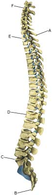
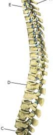
Cause: The cause of scoliosis in children and adolescents is unknown in the majority of cases (idiopathic). Scoliosis is most commonly seen during the growing years, and is more often seen in girls than boys (Photo).

Symptoms: Scoliosis does not necessarily cause pain or other symptoms.

Examination: It is important that the diagnosis is made as soon as possible as better results are achieved if the treatment is commenced as soon as it is necessary. One should therefore always be aware of the early signs of scoliosis (uneven shoulder or hip level, one or both shoulder blades being prominent, slanting waist). A normal medical examination will usually be sufficient to make the diagnosis. X-rays will reveal the degree of severity of the scoliosis. In some cases, CT or MRI scanning is recommended (article).

Treatment: Treatment is dependant upon the degree of severity. The majority of cases will normally be able to be controlled without treatment (article). It will normally be possible to take part in sports activities without any problems (article). Strength training and stretching of the stomach and back muscles is recommended. Supportive bandaging can be used if the scoliosis becomes worse (> 25-30 degrees) and the young person is still growing. It is normally possible to take part in sport at the usual level despite the bandaging (article). An operation may become necessary if the scoliosis becomes pronounced (> 40-50 degrees), and even earlier in some special cases. Certain forms of sport can be resumed 6-9 months after the operation (article).

Complications: In some cases the presence of scoliosis can have other causes (infection, nerve disease, inborn bone change, rheumatic illness, bone disease, metabolic disorder).

Fracture of the vertebral arch

FRACTURE OF THE VERTEBRAL ARCH (SPONDYLOLYSIS)

Diagnosis: FRACTURE OF THE VERTEBRAL ARCH
(SPONDYLOLYSIS)


Anatomy:
The back is constructed of box-like vertebrae which are held in place partly with the help of the shape of the bones, partly by ligaments and partly by the large and small back and stomach muscles. (Sketch)

  1. Vertebra prominens
  2. Vertebra coccygea I
  3. Promontorium
  4. L I
  5. Th I
  6. Axis

THE BACK SEEN FROM THE SIDE

Cause: There are several causes for looseness of the lumbar vertebrae (spondylolyse): inborn (dysplastic), fracture of the vertebral arch and degenerative. Fracture of the vertebral arch is the most frequent cause amongst adolescents and athletes, and is seen in 5% of the normal population. It is most often seen in athletes who repeatedly bend the back backwards (high-jumpers, gymnasts, pole vaulters, footballers) (article) (article). In some cases, the looseness can cause one of the vertebrae to slide forwards and apply pressure on the spinal cord (spondylolisthesis).

Symptoms: Pain and stiffness in the lower back (lumbago) and buttocks, occasionally radiating to the leg (“sciatica”).

Examination: Slight back discomfort does not necessarily require medical examination, however, all cases with strong or repeated back pain should be examined. The doctor will be able to evaluate whether further examination is required, i.e. x-ray (including lateral projection) (X-Ray) ,(Scintigraphy), CT or MRI scanning (article).

Treatment: Treatment is dependant upon the degree of severity. The majority of cases will normally be able to be treated with relief (article-1) (article-2) (which in sever cases must last for several months). In approximately 10% of cases where a vertebra has slid significantly forwards (spondylolisthesis), it will be necessary to operate (article).


Complications:
If the pain does not decline under the treatment, medical (re)examination by a doctor should be performed. Special consideration should be given to:

however, many other causes of lumbago are found (infection, tumour), of which some will require further examination.

Special: Training should be performed on a “lifelong” basis to reduce the risk of relapse after a successful rehabilitation.

Slipped disc

SLIPPED DISC

Diagnosis: SLIPPED DISC
(PROLAPSUS DISCI INTERVERTEBRALIS LUMBALIS)


Anatomy:
The back is constructed of box-like vertebrae which are held in place partly with the help of the shape of the bones, partly by ligaments and partly by the large and small back and stomach muscles. Cartilage discs (disci) are located between the vertebrae which act as shock absorbers. The discs lie close to the spinal canal from where nerves depart to the legs.

  1. Ligamentum longitudinale posterius
  2. Foramen intervertebrale
  3. Ligamentum flavum
  4. Ligamentum interspinale
  5. Processus spinosus
  6. Ligamentum supraspinale
  7. Corpus vertebrae
  8. Ligamentum longitudinale anterius
  9. Discus intervertebralis
  10. Nucleus pulposus

BACK VERTEBRAE

(Photo)

Cause: If the back is subjected to a load which exceeds its capabilities, a crack in one of the discs may occur so that the liquid content in the centre of the disc (nucleus pulposus) can be squeezed out and apply pressure on a nerve root.

Symptoms: Pain and stiffness in the lower back (lumbago), radiating to one of the legs. There can be sensory interference and reduction in strength of the leg. Symptoms are often aggravated by coughing. In rare cases the nerves can be so severely compressed that problems can arise in control of bladder and bowels, requiring acute surgical treatment.

Examination: If a slipped disc is suspected medical attention should be sought at once to establish the diagnosis and which treatment should be initiated.

Treatment: If examination reveals signs of a slipped disc without alarming symptoms (problems in control of bladder and bowels or substantial deterioration of muscle/paralysis), treatment will primarily be directed at altering the imbalance between the load the back is subjected to, opposed to the level the back is trained to manage. It is therefore recommended that you are instructed (possibly by a physiotherapist) in the appropriate way to put a strain on the back, and which loads and movements should be avoided (“ergonomic guidance”). A few days’ rest and relief may be needed to subdue the pain, after which steadily increasing training should be started with back and stomach stabilising and strengthening exercises. If painkillers are required, paracetamol can be recommended, possibly combined with rheumatic medicine (NSAID). Chronic back pain may suggest stronger painkillers, however, stronger medicine should be used with extreme caution as it can quickly lose its effect and there is a risk of increased dependence on the medicine. By far the majority of slipped discs can be managed through correct training (article 1). In cases where the above treatment does not produce progress in the condition, a CT or MRI scan will be considered with a view to possible operation. CT and MRI scan and operation is therefore first considered if the rehabilitation programme does not succeed (article 2). In cases with alarming symptoms (problems in control of bladder and bowels or substantial deterioration of muscle/paralysis) acute hospitalisation is recommended for evaluation of the need for acute surgery.

Special: Training should be performed on a “lifelong” basis to reduce the risk of relapse after a successful rehabilitation. Smoking causes increased risk of lumbago by reducing the flow of blood to the cartilage discs (disci), implying that daily small injuries do not heal so well. Stopping smoking is therefore an important part of the treatment. Shock absorbing shoes or insoles will reduce the load on the back.

Lumbago

LUMBAGO

Diagnosis: LUMBAGO
(Insufficientia dorsi)


Anatomy:
The back is constructed of box-like vertebrae which are held in place partly with the help of the shape of the bones, partly by ligaments and partly by the large and small back and stomach muscles.

  1. Vertebra prominens
  2. Vertebra coccygea I
  3. Promontorium
  4. L I
  5. Th I
  6. Axis

SIDE VIEW OF BACK

Cause: If the back is subjected to a load which exceeds its capabilities, over-load conditioned pain will be triggered from the muscles, tendons, ligaments, and possibly from the cartilage discs (disci) and bones. Pain in the lower back is one of the most frequent sports injuries (comprises approx. 10% of injuries in a top flight football club).

Symptoms: Pain and stiffness in the lower back (lumbago), occasionally radiating to the leg (“sciatica”).

Examination: Slight back discomfort does not necessarily require medical examination, however, all cases with strong or repeated back pain should be examined. The doctor will be able to evaluate whether further examination is required, i.e. x-ray, CT or MRI scan.

Treatment: Treatment will primarily be directed at altering the imbalance between the load the back is subjected to, opposed to the level the back is trained to manage. It is therefore recommended that you are instructed (possibly by a physiotherapist) in the appropriate way to put a strain on the back, and which loads and movements should be avoided (“ergonomic guidance”). A few days’ rest and relief may be needed to subdue the pain, after which steadily increasing training should be started with back and stomach stabilising and strengthening exercises. If painkillers are required, paracetamol can be recommended, possibly combined with rheumatic medicine (NSAID). Chronic back pain may suggest stronger painkillers, however, stronger medicine should be used with extreme caution as it can quickly lose its effect and there is a risk of increased dependence on the medicine. With acute lumbago without signs of a slipped disc, assistance can be sought from manipulative treatment by a doctor, physiotherapist or chiropractor.

Complications: If the pain does not decline under the treatment, clinical (re)examination by a doctor should be performed. Special consideration should be given to:

However, many other causes of lumbago are found, of which some will require further examination. In the majority of cases, the treatment will be identical. It will not be possible for some to be completely free of discomfort (i.e. with heavy degenerative arthritis in the lumbar region), and the aim of the treatment will often be to reduce the frequency and degree of pain.

Special: Training should be performed on a “lifelong” basis to reduce the risk of relapse after a successful rehabilitation. Smoking causes increased risk of lumbago by reducing the flow of blood to the cartilage discs (disci), implying that daily small injuries do not heal so well. Stopping smoking is therefore an important part of the treatment. Shock absorbing shoes or insoles will reduce the load on the back.

Muscular bleeding on the upper arm

MUSCULAR BLEEDING ON THE UPPER ARM

Diagnosis: MUSCULAR BLEEDING ON THE UPPER ARM
(Haematoma musculi)


Anatomy:
The muscles on the back of the upper arm (brachium) comprise the 3-headed arm stretcher (M triceps brachii). The muscle primarily stretches the elbow joint (and shoulder joint). The muscles on the front of the upper arm comprise 3 muscles. The function of two of the muscles, amongst other functions, is to bend the elbow joint (M biceps brachii, M brachialis), while the third muscle (M coracobrachialis) brings the arm towards the body (adduction).


  1. M. biceps brachii
  2. M. triceps brachii
  3. Epicondylus medialis
  4. Aponeurosis
    m. bicipitis brachii
  5. M. brachioradialis
  6. M. brachialis
  7. M. coracobrachialis
  8. Tuberculum majus
  9. M. deltoideus
  10. Acromion
  11. Processus coracoideus
  12. Clavicula

SHOULDER AND UPPER ARM MUSCLES FROM THE FRONT


  1. Clavicula
  2. Bursa subcutanea acromialis
  3. M. deltoideus
  4. M. triceps brachii
    (caput laterale)
  5. M. brachioradialis
  6. Epicondylus lateralis
  7. Olecranon
  8. Epicondylus medialis
  9. M. triceps brachii
    (caput mediale)
  10. M. triceps brachii (caput longum)

SHOULDER AND UPPER ARM MUSCLES FROM THE REAR

Cause: If a muscle is subjected to a blow the muscle belly, which contains blood vessels, is pressed against the bones, causing an injury and rupture of the muscle fibres and blood vessels. The rupture usually occurs deep in the muscle. In other cases the bleeding can occur after a larger or minor rupture of a muscle on the front of the upper arm or rupture of a muscle on the back of the upper arm. The bleeding can either penetrate the muscle membrane and spread over a large area or it can accumulate in the muscle.

Symptoms: Pain and swelling in the muscle. In some cases a hard, tender accumulation (accumulated bleeding in the muscle) can be felt. In other cases a bluish discolouration of the subcutis (the bleeding has penetrated the muscle membrane and spread into the subcutis) occurs after a few days. The pain is aggravated upon activation and stretching of the muscle.

Acute treatment: Click here.

Examination: In light cases with only minimal tenderness and no discomfort when walking normally, medical examination is not necessarily required. Although the extent of the pain is not always a measure of the extent of the injury. In cases of more pronounced pain or difficulty using the arm, medical examination is required to ensure the correct diagnosis and treatment. Ultrasound is the most suited examination to ensure the diagnosis (Ultrasonic image). The larger the bleeding, as revealed in the ultrasound scan, the longer the healing process.

Treatment: The treatment involves relief and rehabilitation as with a rupture of a muscle on the front of the upper arm or rupture of a muscle on the back of the upper arm, depending on whether the bleeding is located on the front or the back of the upper arm. In large bleedings the accumulated blood can be drained under ultrasound guidance. Some recommend treatment with rheumatic medicine (NSAID) and advise caution with regard to massage to reduce the risk of calcification in the muscle (myositis ossificans) .

Rehabilitation: The rehabilitation follows the principles with muscle ruptures. See therefore “rupture of muscle on the front of the upper arm” or “rupture of muscle on the back of the upper armdepending on whether the bleeding is on the front or back of the upper arm.

Complications: If progress is not smooth, you should be (re)examined and consider if the diagnosis is correct or whether complications to the muscle bleeding have added.

Rupture of the muscle on the back of the upper arm

RUPTURE OF THE MUSCLE ON THE BACK OF THE UPPER ARM

Diagnosis: RUPTURE OF THE MUSCLE ON THE BACK OF THE UPPER ARM
(Ruptura M triceps brachii)


Anatomy:
The muscles on the back of the upper arm (brachium) comprise the 3-headed arm stretcher (M triceps brachii). The muscle primarily stretches the elbow joint (and the shoulder joint). The triceps muscle has 3 muscle heads around the shoulder joint which join and fasten on the back of the bone of the forearm by the elbow (olecranon).

  1. Clavicula
  2. Bursa subcutanea acromialis
  3. M. deltoideus
  4. M. triceps brachii
    (caput laterale)
  5. M. brachioradialis
  6. Epicondylus lateralis
  7. Olecranon
  8. Epicondylus medialis
  9. M. triceps brachii
    (caput mediale)
  10. M. triceps brachii
    (caput longum)

SHOULDER AND UPPER ARM MUSCLES FROM THE REAR

Cause: When a muscle is suddenly subjected to a load beyond the strength of the muscle, a rupture occurs. The vast majority of triceps ruptures are partial muscle ruptures. Ruptures most often occur if the muscle is contracting while being stretched, e.g. a fall on a bent arm (eccentric contraction).

Symptoms: In light cases a local tenderness is felt after the load (“strained muscle”, “imminent pulled muscle”). In severe cases sudden shooting pains are felt in the muscle (“partial muscle rupture”, “pulled muscle”) and in the worst case a violent snap is felt, after which bending of the elbow against resistance has been severely reduced (“total muscle rupture”). With muscle injuries the following three symptoms are characteristic: pain upon applying pressure, stretching (stretching the elbow) and activation against resistance (bending the elbow).

Acute treatment: Click here.

Examination: In light cases with only minimal tenderness, medical examination is not necessarily required. In cases of more pronounced pain or difficulty using the arm (stretching the elbow), a medical examination should be carried out to ensure a correct diagnosis and treatment. It can be necessary to supplement the normal clinical examination with an ultrasound scan (article) or an MRI scan.

Treatment: Most partial ruptures are treated with relief and rehabilitation. Only in cases with total or near-total ruptures, particularly in young people, is surgery advised (article).

Complications: If smooth progress is not achieved, it should be considered whether the diagnosis is correct or whether complications have arisen. Amongst others the following should be considered: complication for muscular bleeding.

Rupture of the muscle on the front of the upper arm

RUPTURE OF MUSCLE ON THE FRONT OF THE UPPER ARM

Diagnosis: RUPTURE OF MUSCLE ON THE FRONT OF THE UPPER ARM
(Ruptura M biceps brachii)


Anatomy:
The muscles on the front of the upper arm comprise three muscles. Two of the muscles bend the elbow joint (M biceps brachii, M brachialis), while the third muscle (M coracobrachialis) brings the arm towards the body (adduction). The biceps muscle also rotates the hand (suppination).

  1. M. biceps brachii
  2. M. triceps brachii
  3. Epicondylus medialis
  4. Aponeurosis m. bicipitis brachii
  5. M. brachioradialis
  6. M. brachialis
  7. M. coracobrachialis
  8. Tuberculum majus
  9. M. deltoideus
  10. Acromion
  11. Processus coracoideus
  12. Clavicula

SHOULDER AND UPPER ARM MUSCLES FROM THE FRONT

  1. Scapula
  2. M. brachialis
  3. Epicondylus medialis
  4. Ulna
  5. Radius
  6. Tendo bicipitio brachii
  7. Epicondylus lateralis
  8. M. coracobrachialis
  9. Caput breve
    (m. bicipitis brachi)
  10. Caput humeri
  11. Processus coracoideus

DEEP MUSCLES OF THE UPPER ARM FROM THE FRONT

Cause: When a muscle is suddenly subjected to a load beyond the strength of the muscle, a rupture occurs. The vast majority of ruptures are partial muscle ruptures. Ruptures occur most often if the muscle is contracting at the same time as it is being stretched (eccentric contraction). Full ruptures are most frequently seen in elderly persons and generally localised to the long head of the biceps tendon at the shoulder joint (caput longum biceps brachii) or at the tendon fastening in the elbow (aponeurosis musculi bicipitis brachii).

Symptoms: In light cases a local tenderness is felt after the load (“strained muscle”, “imminent pulled muscle”). In severe cases sudden shooting pains are felt in the muscle (“partial muscle rupture”, “pulled muscle”) and in the worst case a violent snap is felt, after which bending the elbow against resistance is severely reduced (“total muscle rupture”). With muscle injuries the following three symptoms are characteristic: pain upon applying pressure, stretching (stretch the elbow) and activation against resistance (bending the elbow). With many elderly the rupture has not produced symptoms.

Acute treatment: Click here.

Examination: In light cases with only minimal tenderness medical examination is not necessarily required. In cases of more pronounced pain or difficulty using the arm (bending the elbow and rotate the hand (suppination)) a medical examination should be carried out to ensure the correct diagnosis and treatment. It can be necessary to supplement the normal clinical examination with an ultrasound scan (article 1) or an MRI scan (article 2).

Treatment: Most partial ruptures are treated with relief and rehabilitation. Only in cases with total or near-total ruptures, especially in young people, is surgery advised (article 1) (article 2) (article 3).

Complications: If progress is not smooth, you should be (re)examined and consider whether the diagnosis is correct or whether complications have arisen. Ruptures of the long biceps head can be combined with other injuries in the shoulder, for instance inflammation of the biceps muscle and complications for muscular bleeding.

Inflammation of the bursa

INFLAMMATION OF THE BURSA

Diagnosis: INFLAMMATION OF THE BURSA
(Bursitis)


Anatomy:
On the upper arm there are numerous bursas, reducing the pressure on muscles and tendons, where these lie close to the bone.

  1. M. biceps brachii
  2. Epikondylus mediale
  3. Aponeurosis m. bicipitis brachii
  4. M. pronator teres
  5. M. flexor carpi radialis
  6. M. palmaris longus
  7. M. flexor digitorum superficialis
  8. M. flexor carpi ulnaris

FLEXORS OF THE FOREARM

Cause: In case of repeated loads or blows the bursas can become inflamed, produce fluid, swell and become painful. Even though the condition is called bursitis the bursa is usually not infected. One of the most frequent bursitis forms, on the upper arm, is inflammation of the bursa located between the biceps tendon and the fastening on the radius (tuberositas radii).

Symptoms: Pain upon applying pressure on the bursa, which sometimes (but far from always) may feel swollen. Aggravated upon activation of the muscle located immediately above the bursa.

Acute treatment: Click here.

Examination: In light cases with only minimal tenderness, medical examination is not necessarily required. In cases of more pronounced pain or lack of progress, a medical examination should be carried out to ensure a correct diagnosis and treatment. A normal medical examination is usually sufficient in order to make the diagnosis, however, if there is any doubt concerning the diagnosis an ultrasound scan can be performed.

Treatment: The treatment primarily consists of relief. Removal of the provoking cause, if such is known. The treatment can be supplemented with rheumatic medicine (NSAID) or the injection of corticosteroid in the bursa preceded by draining of the bursa, which can advantageously be done under ultrasound guidance.

Rehabilitation: The treatment is completely dependant on which bursa is inflamed, but sports activity can usually be cautiously resumed once pain has decreased, particularly if it has been possible to remove the provoking cause.

Complications: If smooth progress is not achieved, it should be considered whether the diagnosis is correct or whether complications have arisen. In rare cases the bursa can become infected with bacteria. This is a serious condition where the bursa becomes red, warm and increasingly swollen and tender. This condition requires immediate medical examination and treatment. 
If there is no progress with relief, medical treatment rheumatic medicine (NSAID) and the ultrasound guided injection of corticosteroid, surgical removal of the bursa may be attempted.

Fracture of the upper arm

FRACTURE OF THE UPPER ARM

Diagnosis: FRACTURE OF THE UPPER ARM
(Fractura humeri)


Anatomy:
The upper arm (humerus) consists of the head (caput), the neck (collum), the long tubular bone (corpus) and the epicondyle.

  1. Caput humeri
  2. Collum chirurgicum
  3. Epicondylus medialis
  4. Epicondylus lateralis
  5. Tuberculum minus
  6. Sulcus intertubercularis
  7. Tuberculum majus
  8. Collum anatomicum

UPPER ARM FROM THE FRONT

Cause: A fracture of the humerus can occur in cases of a direct fall on the shoulder or outstretched arm. The fracture may occur anywhere on the humerus, but a fracture through the neck of the humerus (collum) and the middle of the long tubular bone (corpus) are the most common locations.

Symptoms: Sudden pain and pain induced constriction of movement of the arm and shoulder after a fall.

Acute treatment: Click here.

Examination: Sudden, powerful pains in the arm with constriction of movement after a fall, should always lead to acute medical examination. The fracture is usually visible on x-rays, and on the basis of the type of fracture, the correct treatment can be determined.

Treatment: Depending on the type of fracture, rest and possible use of bandaging until pain decreases and the fracture is fixed (which usually takes approx. 6 weeks). Most fractures are treated non-operatively (article). In certain types of fractures, an operative fixation is an option.

Rehabilitation: When pain has decreased (after 2-3 weeks) physical training in the form of cycling may be started along with retraining as specified under rehabilitation, general. After approximately 4-6 weeks running can be commenced, and subsequently careful training of the upper arm and shoulder muscles. Participation in contact sports will be possible after approx. three months have elapsed.

Bandage: Special plastic bandages can be made for use when contact sports are resumed.

Complications: In the vast majority of cases the fracture heals without complications, although in some cases a delayed healing occurs, possibly with the development of a false joint (pseudoartrosis) requiring (renewed) surgery. In some cases, the fracture can affect the nerve supply to the arm (N radialis), which can cause sensory disturbances in the hand (and is usually treated with (renewed) surgery).

Inflammation of the bursa

INFLAMMATION OF THE BURSA

Diagnosis: INFLAMMATION OF THE BURSA
(Bursitis)


Anatomy:
On the forearm there are numerous bursas reducing the pressure on muscles and tendons, where these lie close to the bone.

Cause: In case of repeated loads or blows the bursas can become inflamed, produce fluid, swell and become painful. One of the most frequent bursitis forms on the forearm, is inflammation of the bursa located between the biceps tendon and the fastening on the radius (tuberositas radii) (article).

Symptoms: Pain upon applying pressure on the bursa, which sometimes (but far from always) may feel swollen. Aggravated upon activation of the muscle located immediately above the bursa.

Acute treatment: Click here.

Examination: In slight cases with only minimal tenderness, medical examination is not necessarily required. In cases of more pronounced pain or lack of progress, a medical examination should be carried out to ensure a correct diagnosis and treatment. The diagnosis is usually made on the basis of a normal medical examination, however, if there is any doubt surrounding the diagnosis, it can easily and quickly be confirmed under an ultrasound scan.

Treatment: The treatment primarily consists of relief, and removal of the provoking cause (if identified). The treatment can be supplemented with rheumatic medicine (NSAID) or the injection of corticosteroid in the bursa preceded by draining of the bursa, which can advantageously be done under ultrasound guidance.

Rehabilitation: The treatment is completely dependant on which bursa is inflamed, but sports activity can usually be cautiously resumed once pain has decreased, particularly if it has been possible to remove the provoking cause.
Also read rehabilitation, general.

Complications: If progress is not smooth, it should be considered if the diagnosis is correct or whether complications have arisen. In rare cases the bursa can become infected with bacteria. This is a serious condition where the bursa becomes red, warm and increasingly swollen and tender, and requires immediate medical examination and treatment.

If there is no progress with relief, medical treatment (rheumatic medicine (NSAID) and the ultrasound guided injection of corticosteroid), surgical removal of the bursa may be attempted.