Kategoriarkiv: Back

Scheuermans decease

Diagnosis: SCHEUERMANN’S DISEASE


Anatomy:
The back is constructed of box-like vertebrae which are held in place partly with the help of the shape of the bones, partly by ligaments and partly by the large and small back and stomach muscles.


  1. Vertebra prominens
  2. Vertebra coccygea I
  3. Promontorium
  4. L I
  5. Th I
  6. Axis

THE BACK SEEN FROM THE SIDE

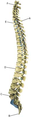
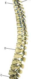
Cause: Scheuermann’s disease occurring in approximately 4% of the population (article). A curvature of the back occurs (bending over forwards) due to the vertebrae becoming wedge shaped. There are also characteristic x-ray finds. The cause of the condition is unknown, but evidence tends to suggest that the condition is hereditary (article).

Symptoms: Back curvature localised high in the back (thoracal Scheuermann) gives often only few, if any, symptoms. Back curvature localised in the lower back (thoracolumbal or lumbal Scheuermann) does entail back pain for the majority (article).

Examination: The diagnosis is usually made following a medical examination supplemented with an x-ray (at least 3 adjacent vertebrae with at least 5 degrees wedge form, Schmorlske impressions, flattening of discs, irregular end plates) (article). The crooked back is often mistaken in the beginning for “bad posture”. In some cases, CT or MRI scanning is recommended.

Treatment: The vast majority of cases can be treated with training, attempting to maintain the mobility of the back, counteract the curvature tendency and strengthen the stomach and back muscles. A corset can in some cases be used until the young person is fully grown. An operation can be performed only in very rare cases. The condition has a good prognosis (article), and even after an operation it is still possible to take part in many different forms of sport (article).

Rehabilitation of children and adolescents: INSTRUCTION

Complications: In some cases a crooked back can have other causes (infection, nerve disease, inborn bone change, rheumatic illness, bone disease, metabolic disorder).

Crooked back

Diagnosis: CROOKED BACK
(SCOLIOSIS)


Anatomy:
The back is constructed of box-like vertebrae which are held in place partly with the help of the shape of the bones, partly by ligaments and partly by the large and small back and stomach muscles.


  1. Vertebra prominens
  2. Vertebra coccygea I
  3. Promontorium
  4. L I
  5. Th I
  6. Axis

THE BACK SEEN FROM THE SIDE

Cause: The cause of scoliosis in children and adolescents is unknown in the majority of cases (idiopathic). Scoliosis is most commonly seen during the growing years, and is more often seen in girls than boys (Photo).

Symptoms: Scoliosis does not necessarily cause pain or other symptoms.

Examination: It is important that the diagnosis is made as soon as possible as better results are achieved if the treatment is commenced as soon as it is necessary. One should therefore always be aware of the early signs of scoliosis (uneven shoulder or hip level, one or both shoulder blades being prominent, slanting waist). A normal medical examination will usually be sufficient to make the diagnosis. X-rays will reveal the degree of severity of the scoliosis. In some cases, CT or MRI scanning is recommended (article).

Treatment: Treatment is dependant upon the degree of severity. The majority of cases will normally be able to be controlled without treatment (article). It will normally be possible to take part in sports activities without any problems (article). Strength training and stretching of the stomach and back muscles is recommended. Supportive bandaging can be used if the scoliosis becomes worse (> 25-30 degrees) and the young person is still growing. It is normally possible to take part in sport at the usual level despite the bandaging (article). An operation may become necessary if the scoliosis becomes pronounced (> 40-50 degrees), and even earlier in some special cases. Certain forms of sport can be resumed 6-9 months after the operation (article).

Rehabilitation of children and adolescents: INSTRUCTION

Complications: In some cases the presence of scoliosis can have other causes (infection, nerve disease, inborn bone change, rheumatic illness, bone disease, metabolic disorder).

Fracture of the vertebral arch

Diagnosis: FRACTURE OF THE VERTEBRAL ARCH
(SPONDYLOLYSIS)


Anatomy:
The back is constructed of box-like vertebrae which are held in place partly with the help of the shape of the bones, partly by ligaments and partly by the large and small back and stomach muscles (sketch).

Cause: There are several causes for looseness of the lumbar vertebrae (spondylolyse): inborn (dysplastic), fracture of the vertebral arch and degenerative. Fracture of the vertebral arch is the most frequent cause amongst adolescents and athletes, and is seen in 5% of the normal population. It is most often seen in athletes who repeatedly bend the back backwards (high-jumpers, gymnasts, pole vaulters, footballers) (article-1) (article-2). In some cases, the looseness can cause one of the vertebrae to slide forwards and apply pressure on the spinal cord (spondylolisthesis).

Symptoms: Pain and stiffness in the lower back (lumbago) and buttocks, occasionally radiating to the leg (“sciatica”).

Examination: Slight back discomfort does not necessarily require medical examination, however, all cases with strong or repeated back pain should be examined. The doctor will be able to evaluate whether further examination is required, i.e. x-ray (including lateral projection) (X-Ray) (Scintigraphy), CT or MRI scanning (article).

Treatment: Treatment is dependant upon the degree of severity. The majority of cases will normally be able to be treated with relief (article-1) (article-2) (which in sever cases must last for several months). In approximately 10% of cases where a vertebra has slid significantly forwards (spondylolisthesis), it will be necessary to operate (article).

Rehabilitation of children and adolescents: INSTRUCTION

Complications: If the pain does not decline under the treatment, medical (re)examination by a doctor should be performed. Special consideration should be given to slipped disc, secondary muscle infiltrations (myalgia) and piriformis syndrome, Scheuermanns disease, however, many other causes of lumbago are found (infection, tumour), of which some will require further examination.

Special:
Training should be performed on a “lifelong” basis to reduce the risk of relapse after a successful rehabilitation.