Alle indlæg af urtehave_com

Rupture of the anterior cruciate ligament

Diagnosis: RUPTURE OF THE ANTERIOR CRUCIATE LIGAMENT
(RUPTURA LIGAMENTUM CRUCIATUM ANTERIUS)


Anatomy:
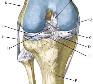
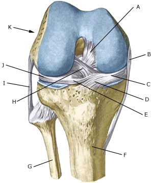
The bones in the knee joint comprise the thigh bone (femur), shin bone (tibia) and the knee cap (patella). There is furthermore a small joint between the shin bone and the calf bone (fibula). The knee joint is strengthened by a joint capsule which is in turn strengthened on the sides by an outer and an inner collateral ligament (ligamentum collaterale laterale (LCL) and ligamentum collaterale mediale (MCL)). Inside the knee there are two ligaments, the anterior and posterior cruciate ligaments (ligamentum cruciatum anterius (ACL) and ligamentum cruciatum posterius (PCL)).


  1. Ligamentum cruciatum posterius
  2. Ligamentum collaterale mediale/tibiale
  3. Meniscus medialis
  4. Insertio anterior menisci medialis
  5. Ligamentum transversum genus
  6. Tibiae
  7. Fibulae
  8. Ligamentum cruciatum anterius
  9. Ligamentum collaterale laterale/fibulare
  10. Meniscus lateralis
  11. Femur

KNEE JOINT FROM THE FRONT

Cause: Rupture of the anterior cruciate ligament usually occurs during running with fast changes of direction or as the result of a fall while skiing. Most injuries occur without contact with an opponent. Cruciate ligament injuries in adolescents are being diagnosed with increasingly regularity.

Symptoms: Usually a snap can be heard or felt and continued sports activity must be aborted. The knee swells within the first few hours, after which the knee can not bend completely. You can subsequently often sense that the leg gives way (knee failure).

Acute treatment: Click here.

Examination: If a partial or complete rupture of the cruciate ligament is suspected, you should seek medical attention (casualty ward) immediately, to obtain a diagnosis. The doctor can perform various tests on the knee (front drawer looseness, Lachmann Pivot shift) to examine the stability of the knee. It should be noted that the looseness in the knee can often only be demonstrated after two weeks. The fluid in the knee can be drained. Blood in the knee provides a very strong suspicion of a rupture of the anterior cruciate ligament. The examination is often more difficult in adolescents. The bleeding that occurs after a rupture of the anterior cruciate ligament can usually be seen in an ultrasound scan (Ultrasonic image) (article) or MR-scan. Arthroscopy (a telescopic examination of the knee) is the best suited examination if there is any doubt in the diagnosis.

Treatment: Caution will normally be exercised regarding operating on children in the pre-teenage age group, however, teenagers nearing a fully grown state will usually be advised surgery with the insertion of a new cruciate ligament (article-1) (article-2) (article-3).

Rehabilitation of children and adolescents: INSTRUCTION

An intensive rehabilitation period of at least six months is to be expected. It is important that the knee is stretched completely at least twice a day. The surgeon should be consulted if problems occur with stretching the knee completely

Bandage: Hinge bandages (Don-Joy) can be utilised the first few weeks. Tape treatment of cruciate ligament ruptures in the knee has no sure effect.

Complications: In case of lack of progress you should consider various complications:

In cases where the knee cap tendon is used as a new cruciate ligament an inflammation is often seen where the knee cap tendon has been removed (article).
In children and adolescents there is a risk of affecting the development of the leg operated upon, as it is necessary to pierce the growth zone to insert the artificial cruciate ligament (article).


Special:
Since there is a risk that the injury can cause permanent disability, the injury should be reported to your insurance company.

Rupture of the posterior cruciate ligament

Diagnosis: RUPTURE OF THE POSTERIOR CRUCIATE LIGAMENT
(RUPTURA LIGAMENTUM CRUCIATUM POSTERIUS)


Anatomy:
The bones in the knee joint comprise the thigh bone (femur), shin bone (tibia) and the knee cap (patella). There is furthermore a small joint between the shin bone and the calf bone (fibula). The knee joint is strengthened by a joint capsule which is in turn strengthened on the sides by an outer and an inner collateral ligament (ligamentum collaterale laterale (LCL) and ligamentum collaterale mediale (MCL)). Inside the knee there are two ligaments, the anterior and posterior cruciate ligaments (ligamentum cruciatum anterius (ACL) and ligamentum cruciatum posterius (PCL)).


  1. Ligamentum cruciatum posterius (
  2. Ligamentum collaterale mediale/tibiale
  3. Meniscus medialis
  4. Insertio anterior menisci medialis
  5. Ligamentum transversum genus
  6. Tibiae
  7. Fibulae
  8. Ligamentum cruciatum anterius
  9. Ligamentum collaterale laterale/fibulare
  10. Meniscus lateralis
  11. Femur

KNEE JOINT FROM THE FRONT

Cause: Rupture of the posterior cruciate ligament usually occurs following a blow or kick direct on the front of the shin bone just below the knee.
(Photo)

Symptoms: Usually a snap can be heard or felt and continued sports activity must be aborted. The knee can swell within the first few hours, after which the knee can not bend completely. You can subsequently often sense that the leg gives way (knee failure).

Acute treatment: Click here.

Examination: If a partial or complete rupture of the cruciate ligament is suspected, you should seek medical attention (casualty ward) immediately, to obtain a diagnosis. The doctor can perform various tests on the knee (rear drawer looseness) to examine the stability of the knee. It should be noted that the looseness in the knee can often only be demonstrated after two weeks. The examination is often more difficult in children and adolescents. It is often necessary to supplement the examination with a MR-scan, ultrasound scan (Ultrasonic image) (article), or arthroscopy to make the diagnosis with certainty.

Treatment: Treatment of a rupture of the posterior cruciate ligament usually comprises relief and rehabilitation. It is only in cases of pronounced looseness, or if the rupture is combined with other ligament ruptures, that surgery is recommended (article).

Rehabilitation of children and adolescents: INSTRUCTION

A rehabilitation period of at least six months is to be expected.

Bandage: Hinge bandages (Don-Joy) can be utilised the first few weeks. Tape treatment of cruciate ligament ruptures in the knee has no sure effect.

Complications: If smooth progress is not achieved, it should be considered whether the diagnosis is correct or whether complications have arisen. Amongst others the following should be considered:

Special: Since there is a risk that the injury can cause permanent disability, the injury should be reported to your insurance company.

Cartilage damage in the joint

Diagnosis: CARTILAGE DAMAGE IN THE JOINT
(OSTEOCHONDRALE LÆSIONER, OSTEOCHONDRITIS DISSICANS)


Anatomy:
The bones in the knee joint comprise the thigh bone (femur), shin-bone (tibia) and the knee cap (patella). The articular surfaces of the thigh bone, shin-bone and knee cap are lined with a cartilage covering of a few millimetre’s thickness which serves to reduce the load or strain on the joint surfaces.

Cause: Twists in the knee joint causing the thigh bone and shin bone to collide can cause damage to the cartilage in the knee. In some cases a piece of cartilage can be shed which can wander in the joint (joint-mouse) and become jammed. The synovial membrane can become inflamed, and increase the production of synovial fluid. In other cases, and for unknown reasons, a piece of bone in the knee can loosen (osteochondritis dissicans).

Symptoms: Pain in the joint when under load or strain. Often, periodic swelling of the joint (synovitis).

Examination: Normal medical examination is often not sufficient. To make the diagnosis correctly it is therefore necessary to perform an arthroscopic examination (telescopic examination of the joint), (Photo) or an MR-scan (article-1) (article-2) (article-3). Ultrasound scanning will be able to reveal many, but not all, cartilage damage (Ultrasonic image), which is why an MR-scan is recommended.

Treatment: It is important to make the diagnosis very early if a piece of bone has become loose (osteochondritis dissicans), as the sooner treatment begins, the better the result achieved (article). Treatment can comprise relief or surgery dependant upon the extent of the condition, and whether a piece of bone has possibly become loose (article-1) (article-2). For cartilage damage caused by a twist, the treatment comprises relief from the painful activities until the pain is no longer experienced, after which gradual re-training can be commenced. There is no treatment that can restore the damaged cartilage, which has itself poor restorative ability. Different procedures to enhance the healing can be attempted using arthroscopy, however, the results are generally unsatisfactory. Results from experimental cartilage transplants are still not successful enough to warrant introduction as a routine treatment in the near future. Joint-mouse that provokes the symptoms must be surgically removed. Injection of corticosteroid in the knee joint, performed under ultrasound guidance, can be attempted to combat prolonged and pronounced cases of fluid accumulation in the knee joint (synovitis).

Rehabilitation of children and adolescents: Rehabilitation is completely dependent upon the type of cartilage damage (size and position in the joint) and treatment (conservative or surgical). It is generally attempted to strengthen the musculature around the knee joint.
See: Rehabilitation of children and adolescents in general.

Complications: Large cartilage damage which is positioned on the weight-bearing parts of the joint represents one of the most serious sports injuries, and often results in an end to the sporting career.

Special: As there is a risk that the injury can cause permanent disability, all cases should be reported to your insurance company.

Cartilage damage on the knee cap

Diagnosis: CARTILAGE DAMAGE ON THE KNEE CAP
(CHONDROMALLACIA PATELLAE)


Anatomy:
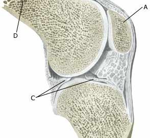
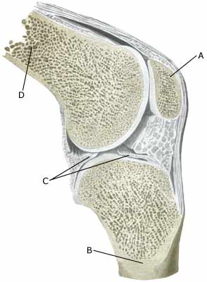
The bones in the knee joint comprise the thigh bone (femur), shin-bone (tibia) and the knee cap (patella). The articular surfaces of the thigh bone, shin-bone and knee cap are lined with a cartilage covering of a few millimetre’s thickness which serves to reduce the load or strain on the joint surfaces.

  1. Patella
  2. Tibiae
  3. Meniscus lateralis
  4. Femur

KNEE JOINT

Cause: Repeated and uniform loads can cause damage to the cartilage, and subsequently the bone beneath the cartilage (degenerative arthritis). Degenerative arthritis changes (osteoarthrosis) on the rear of the kneecap occur often after a fall on the knee and with many smaller over-loads, however, in many cases the cause is unknown. A weak thigh muscle and increased outward turning of the foot (pronation) have been suspected of being a contributory factor to the disease. The frequency of chondromallacia is dwindling, most probably due to the fact that physicians have become better at making other diagnoses (article).

Symptoms: Pain in the joint when under load or strain, especially with bent knee (for example stairway steps). A sensation of stiffness in the knee after a prolonged period of sitting. Swelling of the joint (synovitis) in occasional cases.

Examination: Normal medical examination is often sufficient. A characteristic of the condition is that the pain occurs when the knee cap presses against the thigh bone, producing a rough, grating sensation. If there are any doubts regarding the diagnosis the examination can be supplemented by X-rays, arthroscopic examination or an MR-scan. (article).

Treatment: Treatment comprises relief from the painful activities until the pain is no longer experienced, after which gradual re-training can be commenced primarily aimed at strengthening the thigh muscles. There is no treatment that can restore the damaged cartilage on the knee cap. For children, treatment of chondromalacia (a softening of the articular cartilage) is non-operative, as the condition has a good prognosis in the younger age groups (article). Several different surgical treatments have been attempted with unsatisfactory results (article).

Rehabilitation of children and adolescents: INSTRUCTION

Bandage: Some patients have felt that the discomfort from cartilage damage behind the knee cap has been helped by applying a knee bandage that holds the knee cap slightly to the side. Alternatively, knee cap stabilising tape can be used, (tape-description).

Complications: If smooth progress is not achieved it should be considered whether the diagnosis is correct, which will often require supplementary examination (X-ray, ultrasound scanning, MR scanning or arthroscopy). The following should especially be considered:

Eye injuries

Diagnosis: EYE INJURIES
(CONTUSIO OCULI)


Anatomy:
The eye is protected by the eyelid. An eye nerve runs from the back of each eye to the brain. The visual centre of the brain is located in the rear part of the brain.

Cause: Visual disturbance following a blow to the eye can be due to an injury to the eyeball, optic nerve or the brain.

Symptoms: Visual disturbance in the form of double vision, blurred vision, loss of visual field, newly arisen spots in front of the eyes, pain, bleeding on the outer part of the eyeball (both in the white (conjunctiva) and the anterior chamber of the eye at the pupil (hyphaema)), change in the shape of the pupil as well as stinging and irritation sensations in the eye (article-1),(article-2).

Examination: In all cases where the visual disturbance does not slowly disappear, with bleeding in the anterior chamber (hyphaema), and with loss of visual field, medical examination should be performed (possibly by specialist eye doctor) at the earliest opportunity.

Treatment: Treatment usually consists of rest and relief. Some serious eye injuries can require medicinal (rarely surgical) treatment.

Rehabilitation: Normal sports activity can usually be resumed as soon as the symptoms have disappeared.
Also read Rehabilitation of children and adolescents in general.

Prevention: More widespread use of helmets in different sports will unquestionably reduce the number of eye injuries.

Dental injuries

Diagnosis: DENTAL INJURIES


Anatomy:
Milk teeth are replaced in childhood, where the permanent teeth appear. The front teeth are usually replaced in the 6-8 year age group.

Cause: Direct blows to the teeth can cause the teeth to fracture, fall out or cause damage to the blood supply to the tooth bringing about permanent damage.

Symptoms: Loose teeth, bleeding from the gums, pain in the tooth.

Examination: Examination by a dentist should be performed in all cases where the tooth is knocked out, loose or crooked. The results of the treatment are directly dependent upon how quickly you can be examined.

Treatment: If the tooth is knocked out you should try to put it in place again or keep it in a moist environment, most favourably in salt water (one teaspoon cooking salt in one litre water) or second best in the mouth under the tongue (not children or unconscious persons) or in a handkerchief made moist with saliva to avoid drying out. You should seek acute dental assistance. The dentist can attempt to replace the tooth so that it can re-attach itself. The chances of good results are reduced for each hour which elapses before reaching the dentist.

Rehabilitation: Normal sports activity can be resumed within a short space of time.
Also read Rehabilitation of children and adolescents in general.

Special: Preventive mouth guards significantly reduce the risk of dental injury. It is recommended to utilise mouth guards in a wide variety of sports (contact sports). Resumption of contact sports following a dental injury requiring treatment should be delayed until the tooth has attached itself again in order to avoid possible blows to the tooth in the re-attachment phase (article-1), (article-2). All dental injuries should be reported to your insurance company.

Nosebleed

Diagnosis: NOSEBLEED
(EPISTAXIS)


Anatomy:
It is only the top of the bridge of the nose, close to the skull, that consists of bone (nasal bone). The remainder of the bridge of the nose consists of cartilage (nasal septum).

Cause: A nosebleed usually occurs following a direct blow to the nose.

Symptoms: Bleeding from the nose. In some cases the blow starting the nosebleed can cause other injuries, (fracture of the nasal bone or concussion of the brain).

Examination: A nosebleed does not usually require medical attention. You should, however, ensure that the nasal septum is correctly positioned. If the blow has been particularly hard and there is general malaise or pronounced tenderness, the patient should be attended to by a doctor.

Treatment: Almost all nosebleeds will stop if the whole of the nose that is comprised of cartilage is squeezed for 5 minutes (time should be taken). It is in other words not sufficient merely to press the lower part of the nostrils together. It is recommended to stand or sit under the treatment. Ice can be placed over the bridge of the nose. The bleeding can be made to stop by packing the nose with cotton wool or gauze. An ear, nose and throat specialist should be consulted if the bleeding occurs repeatedly without a blow, as some cases can be treated by cauterising or burning a blood vessel in the nose. Only very rare cases require hospitalization (article).

Rehabilitation of children and adolescents: The sports activity can be resumed as soon as the bleeding has ceased if no other symptoms are in evidence.

Concussion of the brain

Diagnosis: CONCUSSION OF THE BRAIN
(COMMOTIO CEREBRI)


Anatomy:
The brain is surrounded by the membrane of the brain (meninx) and protected by the cranial bones.

Cause: With a violent blow or shaking of the head, the brain can collide against the cranial bones with such a force that bleeding can occur, or fluids can seep, on to or in to the surface of the brain.

Symptoms: Headache, general uneasiness, nausea, visual disturbance, drowsiness, increasing remoteness, unconsciousness, convulsions and in worst case, death, (article). In the rare cases where a fatality occurs in sport due to a blow to the head, it is often caused by incurring two head injuries in the same match.

Examination: All athletes who receive a blow to the head and subsequently complain of uneasiness, visual disturbance or haziness should immediately cease further sport and undergo medical examination. All head injuries must be taken very seriously! (article).

Treatment: Rest and relief until the symptoms have abated (article). It is naturally highly inappropriate, and can be extremely hazardous, to take head ache pills in order to continue sports activity.

Rehabilitation of children and adolescents: Rest and relief until the symptoms have abated. Training can subsequently be cautiously resumed, but should be stopped immediately if symptoms are experienced again (for example head ache).
See: Rehabilitation of children and adolescents in general.

Special: More widespread use of helmets in different sports will unquestionably reduce the number of concussions and after effects thereof. It is imperative that athletes with head injuries which have brought about groggy moments are removed from the sports activity and not permitted to resume until the symptoms have gone during the following days (article 1), (article 2).

Fracture of the nasal bone

Diagnosis: FRACTURE OF THE NASAL BONE
(FRACTURA NASI)


Anatomy:
It is only the upper part of the bridge of the nose, close to the cranium, which actually comprises bone (nasal bone). The remainder of the bridge of the nose is made of cartilage (nasal septum).

Cause: Direct blows to the nose can cause a fracture of the nasal bone. A dislocation of the nasal septum can also occur.

Symptoms: Pain and swelling of the bridge of the nose. In the event of fracture the upper part of the nasal bone will be tender when applying pressure. If there is an accompanying head-ache, or a general feeling of being unwell, the possibility of concussion must be considered. Rare cases can cause bleeding in the nasal septum, bringing about swelling and causing the nose to be blocked (requires acute medical treatment).

Acute treatment: Click here.

Examination: All athletes who receive a blow to the head and subsequently complain of uneasiness, visual disturbance or haziness should immediately cease further sport and undergo medical examination. All head injuries must be taken very seriously! If the pain is purely localised to the nose, and there are free airways through both nostrils, a medical examination should be performed if the nose still appears crooked after three days. Urgent medical attention should be sought if the nasal septum swells and blocks one or both nostrils. X-ray examination of the nose is almost never recommended. The patient will be referred to a specialist ear, nose & throat doctor if there are any suspicions of a fracture of the nasal bone.

Treatment: The fracture will be re-placed and fixed if the fracture has caused the nose to be crooked. Fractures of the nasal bone where the nose does not become crooked are treated with rest and relief until the pain abates. Contact sport can usually be resumed after approximately four weeks.

Rehabilitation of children and adolescents: Rest and relief until the symptoms have abated. Training can subsequently be cautiously resumed, but should be stopped immediately if concussion symptoms are experienced again (for example head-ache). See Rehabilitation of children and adolescents in general.

Bandage: Special facial bandages can be manufactured for use with a fracture of the nasal bone which will allow a speedier resumption of sports activity (article-1), (article-2).

Tendinitis at the ischiatic bone

Untitled Document

Diagnosis: TENDINITIS AT THE ISCHIATIC BONE
(APOPHYSITIS TUBER ISCHIADICUM


Anatomy:
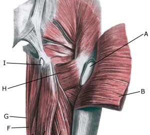
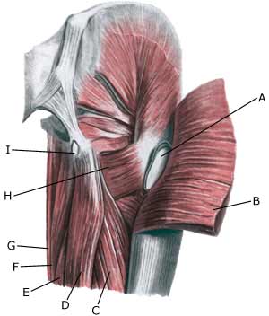
The large posterior thigh muscles (hamstring muscles) have a common muscle tendon fastening on the ischiatic bone (tuber ischiadicum), where a growth zone is also located in children and adolescents. The posterior thigh muscles flex the knee and stretch the hip.

  1. Bursa trochanterica m. glutei maximi
  2. M. gluteus maximus
  3. M. biceps femoris (caput longum)
  4. M. semitendinosus
  5. M. semimembranosus
  6. M. adductor magnus
  7. M. gracilis
  8. M. quadratus femoris
  9. Bursa ischiadica m. glutei maximi

(Photo)

RIGHT GLUTEAL MUSCLES
FROM THE REAR

Cause: Inflammation of the tendon fastenings (tendinitis) at the ischiatic bone (tuber ischiadicum) occurs following repeated uniform (over)loads (e.g. running, sprinting) causing microscopic ruptures in the tendon, and especially at the tendon fastening in the growth zone. Inflammation is a warning that the training performed is too strenuous for the muscles in question. In some cases, a single strenuous load can cause a piece of the ischiatic bone to be torn off in the growth zone (article).

Symptoms: Pain in the growth zone in the ischiatic bone can occasionally radiate down into the rear of the thigh. The pain is aggravated when applying pressure on the bone (e.g. sitting position), stretching and activating the posterior thigh muscles (flexing the knee against resistance).

Examination: In slight cases with only minimal tenderness and no discomfort with walking, medical examination is not necessarily required. The extent of the tenderness is, however, not always a mark of the degree of the injury. In cases of more pronounced pain or tenderness, medical examination is required to ensure the correct diagnosis and treatment. The diagnosis is usually made on the basis of a normal medical examination, however, if there is any doubt concerning the diagnosis, this can be confirmed by ultrasound scanning or MR scanning (article). X-ray can be considered if sudden powerful pain is experienced, as this could indicate suspicions of a bone tear.

Treatment: The treatment usually comprises relief, stretching and rehabilitation (article). It can be necessary to re-join the piece of the ischiatic bone which has torn off in the growth zone under surgery in some cases, however, many cases can be successfully treated with relief (article).

Rehabilitation of children and adolescents: INSTRUCTION

Complications: If the treatment does not progress according to plan, it should be considered if the diagnosis is correct or whether complications have arisen. The following should in particular be considered:

Special: Adolescents nearing the end of their teenage years should refer to the adult SportNetDoc under tendonitis at the ischiatic bone.