Alle indlæg af urtehave_com

KONDITION

STEP3

Training ladder for:
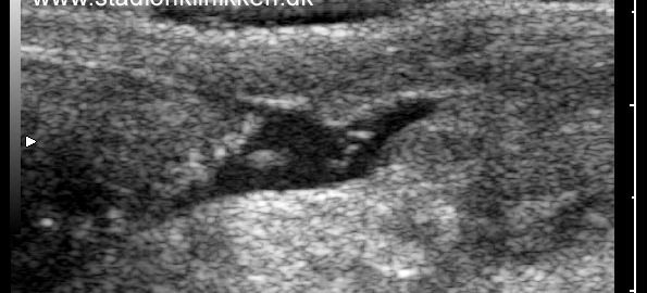
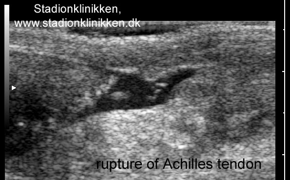
RUPTURE OF THE ACHILLES TENDON
(RUPTURA TRAUMATICA TENDINIS ACHILLES)

STEP 3

KONDITION
Unlimited: Cycling. Swimming. Light jogging on a smooth surface.

UDSPÆNDING
(5 min)

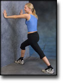
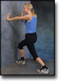
Stand with the injured leg stretched backwards with the toes facing front. Slowly bend the knee so that the calf muscles become increasingly stretched. Hold the position for 20 seconds and relax for 20 seconds before repeating.

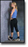
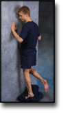
Stand with the injured leg outstretched with the ankle joint bent up against the wall. Press your abdomen against the wall so that the calf muscles become increasingly stretched. Hold the position for 20 seconds and relax for 20 seconds before repeating.

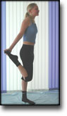
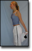
Stand on the good leg while gaining support on the wall with the hand on the same side. Bend the knee of the injured leg and draw the heel towards the buttocks. Take hold of the big toe side of the foot and draw slowly upwards so that the ankle joint is stretched to the maximum and drawn slightly outwards so that increased stretching is experienced on the inside of the shin bone. Hold the position for 20 seconds and relax for 20 seconds before repeating.

Stand on the good leg while gaining support from the wall with the opposite hand. Bend the knee of the injured leg and draw the heel towards the buttocks. Take hold of the little toe side of the foot and draw slowly upwards so that the ankle joint is stretched to the maximum and drawn over the good leg so that increased stretching is experienced on the outer side of the shin bone. Hold the position for 20 seconds and relax for 20 seconds before repeating.

KOORDINATION
(5 min)

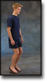
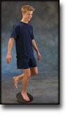
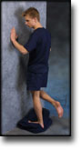
Seesaw. Balance on two legs, possibly using a hand as support against the wall, balancing subsequently on one leg without support. Look straight ahead and keep knees bent.

STYRKE
(50 min)

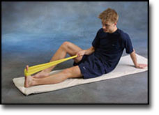
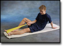
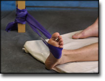
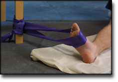
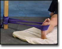
Sit on the floor. Tip the foot up and down with elastic under the forefoot so that the elastic becomes taut.

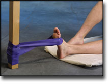
Sit on the floor. Tip the foot up and down with elastic on top of the forefoot so that the elastic becomes taut.

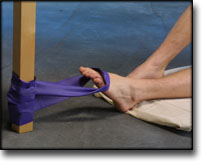
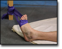
Sit on the floor. Tip the foot from side to side with elastic on the outer side of the foot, without moving the knee.

Sit on the floor. Tip the foot from side to side with elastic on the inner side of the foot, without moving the knee.

Stand on a soft surface. Rise slowly up on tiptoe and go down again.

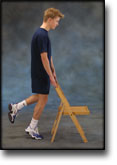
Stand behind a chair. Rise slowly up on tiptoe and go down again.

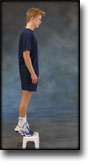
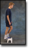
Stand with both forefeet on a stool or doorstep with the heel out over the edge. Slowly rise up on to your toes with your weight on the healthy leg. Go slowly down on the injured leg as far as you can go. Use the healthy leg to rise up on to your toes again. The exercise should be performed with stretched, as well as bent knee. Wearing a rucksack and gradually increasing the ballast in the rucksack can increase the load.

Stretching is carried out in the following way: stretch the muscle group for 3-5 seconds. Relax for 3-5 seconds. The muscle group should subsequently be stretched for 20 seconds. The muscle is allowed to be tender, but must not hurt. Relax for 20 seconds, after which the procedure can be repeated. The time consumed for stretching, coordination and strength training can be altered depending on the training opportunities available and individual requirements.

KONDITION

STEP2

Training ladder for:
RUPTURE OF THE ACHILLES TENDON
(RUPTURA TRAUMATICA TENDINIS ACHILLES)

STEP 2

KONDITION
Unlimited: Cycling. Swimming. Running in deep water.

UDSPÆNDING
(Ingen øvelser)

KOORDINATION
(5 min)

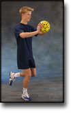
Stand on one leg. Play the ball up against the wall.

STYRKE
(55 min)

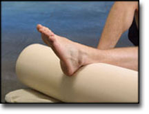
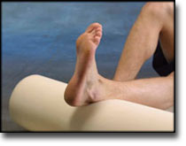
Lay on the floor with a cushion under the calf. Tip the foot up and down without putting any resistance on the foot.

Sit on the floor. Tip the foot up and down with elastic on top of the forefoot so that the elastic becomes taut.

Sit on the floor. Tip the foot from side to side with elastic on the outer side of the foot, without moving the knee.

Sit on the floor. Tip the foot from side to side with elastic on the inner side of the foot, without moving the knee.

Stretching is carried out in the following way: stretch the muscle group for 3-5 seconds. Relax for 3-5 seconds. The muscle group should subsequently be stretched for 20 seconds. The muscle is allowed to be tender, but must not hurt. Relax for 20 seconds, after which the procedure can be repeated. The time consumed for stretching, coordination and strength training can be altered depending on the training opportunities available and individual requirements.

KONDITION

STEP1

Training ladder for:
RUPTURE OF THE ACHILLES TENDON
(RUPTURA TRAUMATICA TENDINIS ACHILLES)

STEP 1

The indications of time after stretching, coordination training and strength training show the division of time for the respective type of training when training for a period of one hour. The time indications are therefore not a definition of the daily training needs, as the daily training is determined on an individual basis.

KONDITION
Unlimited: Cycling. Swimming.

UDSPÆNDING
(Ingen øvelser)

KOORDINATION
(5 min)

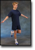
Stand on one leg on the floor or a mattress. Look straight ahead and keep the knee slightly bent.

STYRKE
(55 min)

Lay on the floor with a cushion under the calf. Tip the foot up and down without putting any resistance on the foot.

Sit on the floor. Tip the foot from side to side with elastic on the outer side of the foot, without moving the knee.

Sit on the floor. Tip the foot from side to side with elastic on the inner side of the foot, without moving the knee.

Stretching is carried out in the following way: stretch the muscle group for 3-5 seconds. Relax for 3-5 seconds. The muscle group should subsequently be stretched for 20 seconds. The muscle is allowed to be tender, but must not hurt. Relax for 20 seconds, after which the procedure can be repeated. The time consumed for stretching, coordination and strength training can be altered depending on the training opportunities available and individual requirements.

treatment-article3

An update on repair of Achilles tendon rupture. Acute and delayed.

Kuwada GT. J Am Podiatr Med Assoc 1999 Jun;89(6):302-6.

Current surgical treatments for Achilles tendon rupture are thoroughly discussed. New repair techniques, such as the use of soft-tissue anchors, are reviewed, as is the use of synthetic mesh to augment the surgical repair. A classification system devised by the author is presented to make it easier to select the appropriate surgical procedure or combination of procedures in delayed rupture repair. Postoperative physical therapy is paramount in the return to preinjury level of activity for these patients.

treatment-article2

Diagnosis and treatment of acute ruptures of the Achilles tendon.

Current concepts review. Popovic N, Lemaire R. Acta Orthop Belg 1999 Dec;65(4):458-71.

Subcutaneous rupture of the Achilles tendon seems to have become more common in recent years. This results from a combination of more awareness in the medical field and greater participation in physical activities by the general population. The causes of Achilles tendon rupture are multifactorial and still unclear. The diagnosis can be made based on physical examination; special diagnostic studies are rarely necessary. The literature on ruptures of the Achilles tendon and associated treatment has expanded over the past decade. The lack of a universal, consistent protocol for subjective and objective evaluation following treatment of Achilles tendon rupture has prevented any comparison of results. There is still controversy concerning the best treatment. From a literature review, it appears that a satisfactory outcome may be achieved with either nonoperative or operative treatment but surgical repair appears to provide better functional capacity. Lower rerupture rates and slightly improved strength and functional ability may be expected with surgical treatment; however, the rate of minor complications is higher than with nonoperative treatment. Reports in the literature indicate that in active, young, very demanding individuals, surgical repair should be considered, with nonsurgical treatment reserved for elderly or sedentary patients. There is no single, uniformly accepted surgical technique for Achilles tendon repair. Most acute ruptures have been treated successfully with simple end-to-end suture, although various augmentation procedures have been combined with simple suture with satisfactory outcomes. To minimize the complications typically associated with open surgery, percutaneous techniques to repair the ruptured Achilles tendon have been advocated, and the results are reported to be promising, although not without failures and complications. Several recent studies have reported functional benefits of early postoperative tendon mobilization in well-motivated patients, since treatment results are determined not only by the method of repair but also, and perhaps more importantly, by the early postoperative functional rehabilitation.

treatment-article1

Tendo Achillis rupture; surgical repair is a safe option.

Mellor SJ, Patterson MH. Injury 2000 Sep;31(7):489-91.

We reviewed the results of 67 tendo Achillis repairs performed at one District General Hospital over a 5-year period, using details from a comprehensive database. At a mean follow-up of 2.5 years, the re-rupture rate was 3% (2 out of 67). The infection rate was 1.5% (1 out of 67). A review of the relevant literature has revealed that quoted infection rates following tendo Achillis repair vary considerably, and are as high as 21%. Several authors have advised against repair based on those data. In the light of our results, we can recommend surgical repair of tendo Achillis rupture as a safe option.

Rupture of the Soleus muscle

Diagnosis: RUPTURE OF THE SOLEUS MUSCLE
(Ruptura M soleus)


Anatomy:
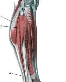
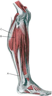
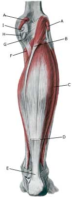
The calf muscle (M Gastrocnemicus) is comprised of two muscle heads which gather in a wide tendinous ligament and continue in to the Achilles tendon. Another of the larger calf muscles (M Soleus) is attached to the front side of the Achilles tendon and thus forms a part of the Achilles tendon. The Achilles is attached to the heel bone (calcaneus).

  1. Tendo calcaneus (Achillis)
  2. M. soleus
  3. M. gastrocnemius

LOWER LEG

  1. M. gastrocnemius
  2. M. plantaris
  3. M. soleus
  4. Tendo m. gastrocnemii
  5. Tendo calcaneus (Achillis)
  6. M. popliteus
  7. Bursa m. semimembranosi
  8. M. semimembranosus
  9. Bursa subtendinea m. gastrocnemii medialis

KNEE FROM THE REAR

Cause: Full or partial rupture of the soleus muscle usually occurs when the calf muscle becomes stretched while it is contracting (eccentric contraction). Partial ruptures represent the majority of the ruptures. The rupture occurs in many instances at the point of attachment of the soleus muscle to the Achilles tendon, which will often trigger an inflammation of the Achilles tendon as a result of the soleus rupture.

Symptoms: Pain when activating the calf muscle (running and jumping), when applying pressure on the Achilles tendon approx. 4 cm. above the anchor point on the heel bone or higher up in the calf muscle, and when stretching the tendon. Walking on tip-toe will aggravate the pain.

Acute treatment: Click here.

Examination: In all cases when there is a sense of a “crack”, or sudden shooting pains in the Achilles tendon, medical attention should be sought as soon as possible. Ultrasound scanning or MRI examination is used to advantage when making the diagnosis, as even full ruptures can easily be overlooked by normal clinical examination.

Treatment: Treatment of the rupture can comprise rest, stretching and training.

Complications: If there is not a steady improvement in the condition an ultrasound scan should be performed to exclude:

inflammation of the bursa

Diagnosis: INFLAMMATION OF THE BURSA
(Bursitis)


Anatomy:
There are numerous bursas around the foot for the purpose of reducing the pressure on the muscles, tendons and ligaments which lie close to bone projections. The bursas at the achilles tendon are those which most often give rise to symptoms.

Cause: The bursas can become inflamed, produce fluid, swell and become painful with repeated over-load or due to blows.

Symptoms: Pain when applying pressure to the bursa, which sometimes, but far from always, can give the impression of being swollen.

Acute treatment: Click here.

Examination: Medical examination is usually not required in light cases with only minimal tenderness. With more pronounced pain, or lack of improvement, medical examination should always be performed for confirm the diagnosis and commencement of treatment if required. The diagnosis is best made using ultrasound examination.

Treatment: Treatment is primarily concentrated on providing rest. If the provoking factor is known (i.e. tight shoes), this should naturally be corrected. Treatment can be supplemented with rheumatic medicine (NSAID) or injection of corticosteroid in the bursa preceded by draining, which can be best performed if ultrasound-guided.

Rehabilitation: Treatment is completely dependent upon which bursa is inflamed, but the sports activity can be cautiously resumed when the pain has diminished, especially if the provoking factor has been identified and removed.
Also read rehabilitation, general.

Bandage: In some cases a ring of felt (for example) can be taped around the tender bursa which will reduce the pressure from shoes. It is naturally important that the hole in the ring is positioned directly above the bursa.

Complications: If there is not a steady improvement in the condition consideration must be given as to whether the diagnosis is correct, or if complications have arisen:

In rare cases, the bursa can be infected with bacteria. This is a serious condition if the bursa becomes red, warm and increasingly swollen and tender. This condition requires immediate examination and treatment.

KONDITION

STEP4

TRAINING LADDER FOR CHILDREN AND ADOLESCENTS:
FOR INFLAMMATION OF THE GROWTH ZONE OF THE HEEL BONE
(APOPHYSITIS CALCANEI, MB SEVER)

STEP 4

The majority of young patients can manage with relief until the pain subsides, following which the sports activity can be slowly resumed. The following rehabilitation program will cover the needs for the vast majority of children with inflammation of the growth zone of the heel bone. Older teenagers involved in sports at a high level can advantageously use the rehabilitation program for adults.
KONDITION
Unlimited: Cycling. Swimming. Running with increasing distance on a soft surface.

UDSPÆNDING
(5 min)

Stand with the injured leg stretched backwards with the toes facing front. Slowly bend the knee so that the calf muscles become increasingly stretched. Hold the position for 20 seconds and relax for 20 seconds before repeating.

Stand with the injured leg outstretched with the ankle joint bent up against the wall. Press your abdomen against the wall so that the calf muscles become increasingly stretched. Hold the position for 20 seconds and relax for 20 seconds before repeating.

KOORDINATION
(5 min)

Seesaw. Balance on two legs, possibly using a hand as support against the wall, balancing subsequently on one leg without support. Look straight ahead and keep knees bent.

STYRKE
(10 min)

Sit on the floor. Tip the foot up and down with elastic under the forefoot so that the elastic becomes taut.

Sit on the floor. Tip the foot up and down with elastic on top of the forefoot so that the elastic becomes taut.

Sit on the floor. Tip the foot from side to side with elastic on the outer side of the foot, without moving the knee.

Sit on the floor. Tip the foot from side to side with elastic on the inner side of the foot, without moving the knee.

Stand with both forefeet on a stool or doorstep with the heel out over the edge. Slowly rise up on to your toes with your weight on the healthy leg. Go slowly down on the injured leg as far as you can go. Use the healthy leg to rise up on to your toes again. The exercise should be performed with stretched, as well as bent knee. Wearing a rucksack and gradually increasing the ballast in the rucksack can increase the load.

Stretching is carried out in the following way: stretch the muscle group for 3-5 seconds. Relax for 3-5 seconds. The muscle group should subsequently be stretched for 20 seconds. The muscle is allowed to be tender, but must not hurt. Relax for 20 seconds, after which the procedure can be repeated.

The time consumed for stretching, coordination and strength training can be altered depending on the training opportunities available and individual requirements.