Kategoriarkiv: Hip

Inflammation of the pubic bone joint (osteitis pubis)

INFLAMMATION OF THE PUBIC JOINT

Diagnosis: INFLAMMATION OF THE PUBIC JOINT
(Osteitis pubis)


Anatomy:
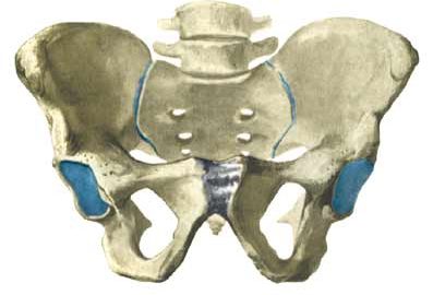
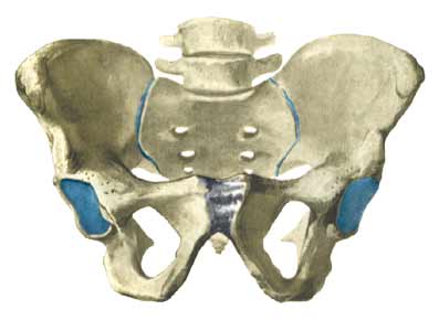
The pelvis consists of two halves joined at the back by the sacral bone (os sacrum) and at the front by the pubic joint (symphysis). The abdominal muscle (M rectus abdominis) if fastened on the pubic bone and the adductor muscles of the thigh (amongst others the M adductor longus) are fastened in the groin just below the pubic bone.

  1. Symphysis pubica

PUBIC SYMPHYSIS

Cause: occurs in the joint, whereby the joint can become loose so that the two halves of the pelvis can move slightly in relation to one another due to heavy loads. (In many areas the symptoms are the same as those seen in pregnant women with pelvic loosening). The inflammation is a warning that the training is too strenuous, and that if the load is not reduced a chronic condition with a significantly longer rehabilitation period may follow. Inflammation of the pubic joint is often preceded by long-term inflammation of the abdominal muscle fastening on the pubic bone and inflammation of the adductor muscle of the thigh. Inflammation of the pubic joint (osteitis pubis) is especially seen in sports characterised by activity with sprinting with sudden changes of direction (soccer, ice hockey).

Symptoms: Pain around the pubic bone, the abdominal muscle fastening and in the groin. Aggravated with asymmetric loads (jumping on one leg).

Acute treatment: Click here.

Examination: Inflammation of the pubic bone joint is always an athletically serious condition that always requires medical examination. A normal medical examination is usually sufficient in order to make the diagnosis. Tenderness will often be present on the pubic bone joint (direct and indirect). The examination can be supplemented with X-ray, scintigraphy, MRI- and ultrasound scan. The diagnosis of groin pain can be particularly difficult (article)

Treatment: Inflammation of the pubic bone joint is often the end stage of a poorly treated chronic overloading of the muscle fastenings on the pubic bone and the groin. In severe cases the rehabilitation period can be expected to last a year (and in the worst case render a return to sports an impossibility). It is therefore of the utmost importance that the treatment be started as soon as possible, without continuing the condition inducing sports activity. The treatment primarily consists of relief and subsequently slowly increasing training of the musculature around the pubic bone joint (stomach, groin), pelvis and loin. In case of lack of progress with rehabilitation, a medical treatment can be considered in the form of rheumatic medicine (NSAID) or the injection of corticosteroid around the inflamed tendon fastening or in the pubic bone joint (article). The injection can advantageously be ultrasound guided. Since the injection of corticosteroid always is part of a long-term rehabilitation of a very serious, chronic injury, it is decisively necessary, that the rehabilitation course stretches over several months to reduce the risk of relapse. A rehabilitation period of ½ – 1 year before maximum load in the form of maximal running with directional change is allowed is not unusual. Some have tried operating for this condition, which usually returns to normal when the load (sports activity) ceases (article). Particularly careful consideration should be made before accepting a surgical offer with the risks this entails, for a condition that does not threaten mobility nor health (article).

Complications: If progress is not smooth the correctness of the diagnosis should be considered. In particular, the following should be considered:

Special: Shock absorbing shoes or inlays will reduce the load in the groin. In case of lack of progress or relapse after successful rehabilitation, a running style analysis can be considered to evaluate whether correction of the running style should be recommended.

Inflammation of the abdominal muscle fastening on the pubic bone (tendinitis m rectus abdominis)

INFLAMMATION OF THE ABDOMINAL MUSCLE AT THE POINT OF ATTACHMENT ON THE PUBIC BONE

Diagnosis: INFLAMMATION OF THE ABDOMINAL MUSCLE AT THE POINT OF ATTACHMENT ON THE PUBIC BONE
(Tendinitis M rectus abdominis)


Anatomy:
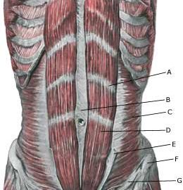
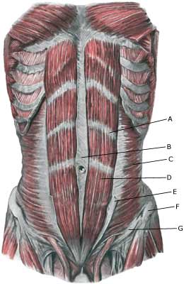
The pelvis consists of two halves, joined at the back by the sacral bone (os sacrum) and at the front by the pubic joint (symphysis). The abdominal muscle (M rectus abdominis) is fastened on the pubic bone and helps bend the back (so that the head is brought towards the knee). The adductor muscles in the thigh (amongst others the M adductor longus) are fastened in the groin just below the pubic bone.

  1. Intersectio tendinea
  2. Linea alba
  3. M. transversus abdominis
  4. M. rectus abdominis
  5. Vagina m. recti abdominis (lamina anterior)
  6. Spina iliaca anterior superior
  7. Lig. inguinale

STOMACH MUSCLES

Cause: Inflammation of the tendon (tendinitis) occurs upon repeated uniform (over)loads. Microscopic ruptures can thus occur in the tendon and in particular at the fastening on the pubic bone, causing an inflammation. Tendinitis is a warning that the exercise load is too strenuous for the muscle tendon in question, and that if the load is not reduced a rupture can occur, with a significantly longer subsequent rehabilitation period. In some cases the inflammation can spread to the pubic joint (osteitis pubis).

Symptoms: Pain around the abdominal muscle fastening on the pubic bone, worsening when applying pressure, stretching and activation of the abdominal muscles (sit-ups, stomach crunches).

Acute treatment: Click here.

Examination: In light cases, medical examination is not necessarily required. Severe cases or cases not improved by treatment, should be evaluated by a doctor to make a precise diagnosis. A normal medical examination is usually sufficient in order to make the diagnosis. If upon examination there is pain when applying pressure on the muscle fastening in the groin, worsening at the same location upon stretching and activation (sit-ups), there is hardly any doubt concerning the diagnosis even though the diagnosis of groin pain can be particularly difficult (article). However, if there is any doubt concerning the diagnosis an ultrasound scan can be performed. Ultrasound examination is well suited to evaluate the muscles in the area.

Treatment: The treatment usually consists of relief and cautious rehabilitation. In case of lack of progress with rehabilitation, medical treatment can be considered in the form of rheumatic medicine (NSAID) or the injection of corticosteroid along the inflamed part of the muscle fastening on the pubic bone. Surgery is rarely necessary (and the results are not convincing).

Complications: If satisfactory progress is not achieved, it should be considered whether the diagnosis is correct or whether complications have arisen. In particular the following should be considered:

Special: Shock absorbing shoes or inlays will reduce the load on the muscle fastenings. In case of lack of progress or relapse after successful rehabilitation, a running style analysis can be considered to evaluate whether correction of the running style should be recommended.

Inflammation of adductor muscle of the thigh (tendonitis m adduktor longus)

INFLAMMATION OF THE ADDUCTOR OF THE THIGH

Diagnosis: INFLAMMATION OF THE ADDUCTOR OF THE THIGH
(Tendinitis M adduktor longus)


Anatomy:
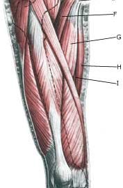
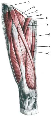
The adductors in the thigh (M adductor longus, M adductor brevis and M adductor magnus along with M gracilis and M pectineus) are all fastened in the groin on the pubic bone.

  1. Spina iliaca anterior superior
  2. M. iliopsoas
  3. Lig. inguinale
  4. Lig. lacunare
  5. Tuberculum pubicum
  6. M. pectineus
  7. M. adductor longus
  8. M. gracilis
  9. M. adductor magnus

THIGH FROM THE FRONT


(Photo)

Cause: Inflammation of the tendon (tendinitis) occurs upon repeated uniform (over)loads. Microscopic ruptures can thus occur in the tendon and in particular at the point of attachment, which causes inflammation. Inflammation of the tendon is a warning that the activity is too strenuous on the particular muscle tendon, and that if the load is not reduced a rupture may occur with a significantly longer subsequent rehabilitation period. The adductor longus muscle, which feels like a firm string in the groin, is one of the adductor muscles, that are most frequently damaged. The muscle is especially susceptible to injury in sports characterized by activity with sprinting with sudden directional changes, a lot of weight training and modest agility training (soccer, hockey), while it is very rarely seen in sports where there is typically great agility (gymnastics). In some cases the inflammation can spread to the fastening of the abdominal muscle on the pubic bone and to the pubic bone joint (symphysis).

Symptoms: Pain when applying pressure along the tendon, worsening upon stretching and activation of the muscle tendon (closing of stretched legs against resistance).

Acute treatment: Click here.

Examination: In light cases medical examination in not necessarily required. Severe cases or cases not improved by treatment should be evaluated by a doctor so that a precise diagnosis can be made. A normal medical examination is usually sufficient in order to make the diagnosis. If in the medical examination there is pain when applying pressure on the muscle attachment point in the groin, and aggravation at the same location upon stretching and activation of the adductor, there is hardly any doubt about the diagnosis. The diagnosis of groin pain can be particularly difficult (article) However, if there is any doubt concerning the diagnosis an ultrasound scan can be performed. Ultrasound is well suited to evaluate the muscles in the area.

Treatment: Since tendinitis of the adductor (M adductor longus) in the thigh is one of the sports injuries with the highest risk of becoming chronic, it is imperative that treatment starts as soon as the first symptoms are felt (and not after months of increasing discomfort). The treatment involves relief, stretching and slowly increasing load within the pain threshold (article). Ice treatment should be used after training if tenderness is felt in the groin. If there is a lack of progress with relief and rehabilitation, medical treatment in the form of rheumatic medicine (NSAID) or the injection of corticosteroid around the inflamed part of the tendon may be considered. The injection of corticosteroid is always part of long-term rehabilitation of a very serious chronic injury. It is therefore necessary that the rehabilitation period after the injection treatment stretches over several months to reduce the risk of relapse and ruptures. The tendon can naturally not sustain maximal load after a long-term injury period and only a short-term rehabilitation period. It is not unusual for rehabilitation to stretch over six months, before maximal load in the form of running with directional change is permitted. In cases where there is lack of progress after rehabilitation and conservative treatment, operative treatment can be attempted. The long-term results of operations are often disappointing.

Complications: If satisfactory progress is not achieved, it should be considered whether the diagnosis is correct or whether complications have arisen. In particular the following should be considered:

Special: Shock absorbing shoes or indlays will reduce the load in the groin. In case of lack of progress or relapse after successful rehabilitation, a running style analysis can be considered to evaluate whether correction of the running style should be recommended.

Stress fracture of the femoral neck

STRESS FRACTURE IN THE FEMORAL NECK

Diagnosis: STRESS FRACTURE IN THE FEMORAL NECK
(Stress fraktur)


Anatomy:
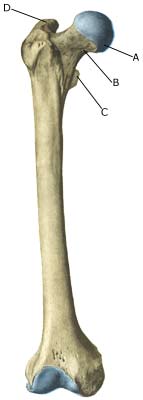
The femur and the hip bone form the hip joint.

  1. Caput femoris
  2. Collum femoris
  3. Trochanter minor
  4. Trochanter major

PELVIS AND THIGH BONE FROM THE FRONT


Cause: Repeated loads, especially when walking or running can cause cracks (stress fractures) in the femoral neck (collum femoris) (article-1) (article-2).

Symptoms: Pain in the hip when applying pressure (direct and indirect tenderness) and when under load (walking, running).

Examination: X-ray. Since many stress fractures cannot be seen early in the course of events, X-ray examination can be repeated after a few weeks. Scintigraphy, CT- and MRI and ultrasound scan can often diagnose stress fractures far earlier than X-rays (Ultrasonic image)
It is imperative for the result of the treatment that the diagnosis is made as early as possible (article).

Treatment: Relief. In some cases surgery is necessary (article).

Rehabilitation: The rehabilitation is completely dependent on the type of fracture and treatment (conservative or operative).
Also read rehabilitation, general.

Complications: If progress is not smooth, you should be re-examined to ensure that the fracture heals according to plan. In some cases a false joint can be formed (pseudoarthrosis), which requires surgical treatment.

Special: Shock absorbing shoes or inlays will reduce the load.

Rupture of the superficial hip flexor (ruptura musculus rectus femoris)

BRISTNING AF DEN OVERFLADISKE HOFTEBØJER

Diagnosis: BRUPTURE OF THE SUPERFICIAL HIP FLEXOR
(Ruptura musculus rectus femoris)


Anatomy:
The superficial hip flexor (the forward straight thigh muscle, musculus quadriceps femoris) originate from the front edge of the hip (processus spinosus anterior inferior) and from the upper edge hip joint socket (acetabulum). The muscle is joined by three of the other thigh muscles and is attached in a common joint muscle tendon (quadriceps) on the upper edge of the kneecap (patella). The function of the superficial hip flexor is to stretch the knee and bend in the hip.

  1. Spina iliaca anterior superior
  2. M. iliopsoas
  3. Lig. inguinale
  4. Lig. lacunare
  5. Tuberculum pubicum
  6. M. pectineus
  7. M. adductor longus
  8. M. gracilis
  9. M. adductor magnus

THIGH FROM THE FRONT

Cause: When a muscle is subjected to loads (repeated smaller loads or one very powerful load), beyond the strength of the muscle (jumping, kicking), a rupture occurs. The rupture can be microscopic and due to repeated loads in continuing sports activity, many small loads can trigger a chronic inflammation or a rupture. The vast majority of cases are partial muscle ruptures.

Symptoms: In light cases a local tenderness is felt after the load (“muscle strain”, “imminent pulled muscle”, “tendinitis”). The symptoms can often decrease after a thorough warm-up, only to return when the sports activity has ceased. In severe cases a sudden shooting pain is felt in the muscle (“partial muscle rupture”, “pulled muscle”) and in the worst case a sudden snap is felt rendering the muscle unusable (“total muscle rupture”). With muscle injuries the following three symptoms are characteristic: pain when applying pressure, stretching and activation against resistance. In total ruptures a defect in the muscle can often be seen and felt, and a swelling is felt above or below the rupture (the contracted muscular belly and the bleeding).

Acute treatment: Click here.

Examination: In light cases with only minimal tenderness and no discomfort with ordinary walking, medical examination is not necessarily required. The extent of the tenderness is, however, not always a mark of the degree of the injury. In cases of more pronounced pain or tenderness, medical examination is required to ensure the correct diagnosis and treatment. A normal medical examination is usually sufficient in order to make the diagnosis, however, if there is any doubt concerning the diagnosis an ultrasound scan can be performed (Ultrasonic image). X-ray examination is recommended when it is suspected that the bone on which the muscle tendon fastens has been torn off.

Treatment: The treatment usually comprises relief and rehabilitation. Only in very rare cases is surgery indicated (e.g. total ruptures in the muscle tendon, close to the fastening). Even large ruptures in the femoral muscle can usually be rehabilitated without resulting in functional harm (but often cosmetic damage, with an irregular femoral muscle). If the condition concerns tendinitis where there has been no sensation of a “snap” in the muscle, and smooth improvement has not been achieved after relief and gradually increasing rehabilitation, treatment can be supplemented with rheumatic medicine (NSAID) oand possibly injection of corticosteroid in the area surrounding the inflamed part of the muscle attachment point. If it concerns ruptures, (“total or partial muscle rupture”) the injection of corticosteroid is not indicated.

Complications: If the treatment does not progress according to plan, it should be considered whether the diagnosis is correct or whether complication have arisen. In particular the following should be considered:

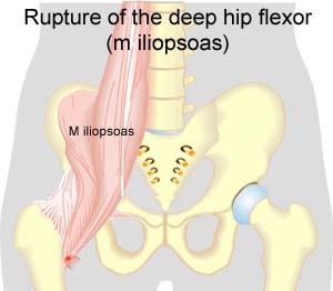
Rupture of the deep hip flexor (M iliopsoas)

RUPTURE OF THE DEEP HIP FLEXOR

Diagnosis: RUPTURE OF THE DEEP HIP FLEXOR
(Ruptura musculus iliopsoas)


Anatomy:
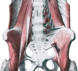
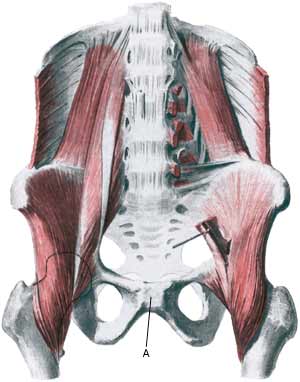
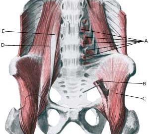
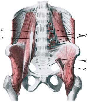
The deep hip flexor (M iliopsoas) consists of two muscles. The Psoas muscle originate from the lumbar vertebra and the Iliacus muscle from the inside of the hip bone. The two muscles fuse and are both fastened on the inside of the femur (trochanter minor). The iliopsoas is the strongest flexor muscle of the hip joint.

  1. Origines m. psoatis
  2. M. psoas major
  3. M. iliacus
  4. M. psoas major
  5. M. psoas minor

PELVIS FROM THE FRONT

Cause: When a muscle is subjected to a load beyond the strength of the muscle (jumping, kicking), a rupture occurs. The vast majority of ruptures are partial muscle ruptures.

Symptoms: In light cases a local tenderness is felt after the load (“sprained muscle”, “imminent pulled muscle”) e.g. kicking a ball with the instep. In severe cases a sudden shooting pain is felt in the muscle (“partial muscle rupture”, “pulled muscle”) and in the worst case a violent snap is felt, rendering the muscle unusable for e.g. walking up stairs (“total muscle rupture”). With muscle injuries the following three symptoms are characteristic: pain upon pressure, stretching and activation against resistance. In some cases the bleeding can be so great that it entraps the nerve to the bone (nervus femoralis) with increasing pains, reduction of power and symptoms into the leg (article).

Acute treatment: Click here.

Examination: In light cases with only minimal tenderness and no discomfort when walking normally (“sprained muscle”, “imminent pulled muscle”), medical examination is not necessarily required. The severity of the tenderness is, however, not always a measure of the extent of the injury. In case of more pronounced tenderness or pain medical examination is required to ensure the correct diagnosis and treatment. A normal medical examination is usually sufficient in order to make the diagnosis, however, if there is any doubt concerning the diagnosis an ultrasound scan can be performed. It is known that the larger the bleeding is assessed in the ultrasound scan, the longer the rehabilitation period (Ultrasonic image), (article).

Treatment: The treatment usually consists of relief and careful rehabilitation (article). Only in very rare cases is surgery indicated (e.g. in case of total ruptures or very heavy bleeding).

Complications: In case of lack of progress it should be considered if the diagnosis is correct or whether complications have arisen. In particular the following should be considered:

treatment-article

SportNetDoc

Surgical repair of chronic complete hamstring tendon rupture in the adult patient.

Cross MJ, Vandersluis R, Wood D, Banff M. Am J Sports Med 1998 Nov-Dec;26(6):785-8.

Complete rupture of the hamstring tendons in the adult is a rare injury. This report discusses complete rupture of the hamstring tendons in nine patients treated by late operative repair. All patients were referred from outside centers for a second opinion after failed nonoperative treatment. The diagnosis was made quite easily on clinical grounds and was confirmed at surgery. Surgical treatment in all cases consisted of reattachment of the hamstring tendons to the origin on the ischium, and in all cases it was necessary to perform neurolysis of the sciatic nerve. Good results were achieved in all cases, at follow-up all patients were satisfied with the surgery.

Slipped disc

SLIPPED DISC

Diagnosis: SLIPPED DISC
(PROLAPSUS DISCI INTERVERTEBRALIS LUMBALIS)


Anatomy:
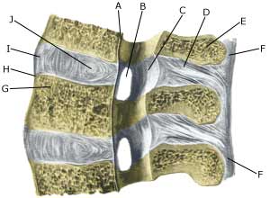
The back is constructed of box-like vertebrae which are held in place partly with the help of the shape of the bones, partly by ligaments and partly by the large and small back and stomach muscles. Cartilage discs (disci) are located between the vertebrae which act as shock absorbers. The discs lie close to the spinal canal from where nerves depart to the legs.

  1. Ligamentum longitudinale posterius
  2. Foramen intervertebrale
  3. Ligamentum flavum
  4. Ligamentum interspinale
  5. Processus spinosus
  6. Ligamentum supraspinale
  7. Corpus vertebrae
  8. Ligamentum longitudinale anterius
  9. Discus intervertebralis
  10. Nucleus pulposus

BACK VERTEBRAE

(Photo)

Cause: If the back is subjected to a load which exceeds its capabilities, a crack in one of the discs may occur so that the liquid content in the centre of the disc (nucleus pulposus) can be squeezed out and apply pressure on a nerve root.

Symptoms: Pain and stiffness in the lower back (lumbago), radiating to one of the legs. There can be sensory interference and reduction in strength of the leg. Symptoms are often aggravated by coughing. In rare cases the nerves can be so severely compressed that problems can arise in control of bladder and bowels, requiring acute surgical treatment.

Examination: If a slipped disc is suspected medical attention should be sought at once to establish the diagnosis and which treatment should be initiated.

Treatment: If examination reveals signs of a slipped disc without alarming symptoms (problems in control of bladder and bowels or substantial deterioration of muscle/paralysis), treatment will primarily be directed at altering the imbalance between the load the back is subjected to, opposed to the level the back is trained to manage. It is therefore recommended that you are instructed (possibly by a physiotherapist) in the appropriate way to put a strain on the back, and which loads and movements should be avoided (“ergonomic guidance”). A few days’ rest and relief may be needed to subdue the pain, after which steadily increasing training should be started with back and stomach stabilising and strengthening exercises. If painkillers are required, paracetamol can be recommended, possibly combined with rheumatic medicine (NSAID). Chronic back pain may suggest stronger painkillers, however, stronger medicine should be used with extreme caution as it can quickly lose its effect and there is a risk of increased dependence on the medicine. By far the majority of slipped discs can be managed through correct training (article 1). In cases where the above treatment does not produce progress in the condition, a CT or MRI scan will be considered with a view to possible operation. CT and MRI scan and operation is therefore first considered if the rehabilitation programme does not succeed (article 2). In cases with alarming symptoms (problems in control of bladder and bowels or substantial deterioration of muscle/paralysis) acute hospitalisation is recommended for evaluation of the need for acute surgery.

Special: Training should be performed on a “lifelong” basis to reduce the risk of relapse after a successful rehabilitation. Smoking causes increased risk of lumbago by reducing the flow of blood to the cartilage discs (disci), implying that daily small injuries do not heal so well. Stopping smoking is therefore an important part of the treatment. Shock absorbing shoes or insoles will reduce the load on the back.

Lumbago

LUMBAGO

Diagnosis: LUMBAGO
(Insufficientia dorsi)


Anatomy:
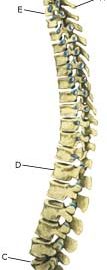
The back is constructed of box-like vertebrae which are held in place partly with the help of the shape of the bones, partly by ligaments and partly by the large and small back and stomach muscles.

  1. Vertebra prominens
  2. Vertebra coccygea I
  3. Promontorium
  4. L I
  5. Th I
  6. Axis

SIDE VIEW OF BACK

Cause: If the back is subjected to a load which exceeds its capabilities, over-load conditioned pain will be triggered from the muscles, tendons, ligaments, and possibly from the cartilage discs (disci) and bones. Pain in the lower back is one of the most frequent sports injuries (comprises approx. 10% of injuries in a top flight football club).

Symptoms: Pain and stiffness in the lower back (lumbago), occasionally radiating to the leg (“sciatica”).

Examination: Slight back discomfort does not necessarily require medical examination, however, all cases with strong or repeated back pain should be examined. The doctor will be able to evaluate whether further examination is required, i.e. x-ray, CT or MRI scan.

Treatment: Treatment will primarily be directed at altering the imbalance between the load the back is subjected to, opposed to the level the back is trained to manage. It is therefore recommended that you are instructed (possibly by a physiotherapist) in the appropriate way to put a strain on the back, and which loads and movements should be avoided (“ergonomic guidance”). A few days’ rest and relief may be needed to subdue the pain, after which steadily increasing training should be started with back and stomach stabilising and strengthening exercises. If painkillers are required, paracetamol can be recommended, possibly combined with rheumatic medicine (NSAID). Chronic back pain may suggest stronger painkillers, however, stronger medicine should be used with extreme caution as it can quickly lose its effect and there is a risk of increased dependence on the medicine. With acute lumbago without signs of a slipped disc, assistance can be sought from manipulative treatment by a doctor, physiotherapist or chiropractor.

Complications: If the pain does not decline under the treatment, clinical (re)examination by a doctor should be performed. Special consideration should be given to:

However, many other causes of lumbago are found, of which some will require further examination. In the majority of cases, the treatment will be identical. It will not be possible for some to be completely free of discomfort (i.e. with heavy degenerative arthritis in the lumbar region), and the aim of the treatment will often be to reduce the frequency and degree of pain.

Special: Training should be performed on a “lifelong” basis to reduce the risk of relapse after a successful rehabilitation. Smoking causes increased risk of lumbago by reducing the flow of blood to the cartilage discs (disci), implying that daily small injuries do not heal so well. Stopping smoking is therefore an important part of the treatment. Shock absorbing shoes or insoles will reduce the load on the back.

Fluid accumulation in the hip joint

FLUID ACCUMULATION IN THE JOINT

Diagnosis: FLUID ACCUMULATION IN THE JOINT
(Synovitis / coxitis)


Anatomy:
The hip joint consists of the hip socket (acetabulum) and the femoral head (caput femoris). The articular surfaces are coated with a cartilage layer a few mm thick, which reduces the load on the articular surfaces.

Cause: Many repeated loads or one violent load can cause an inflammation of the synovial membrane (synovitis), fluid formation, swelling, restriction of movement and pain in the hip joint. The condition is relatively often seen in children (Ultrasonic image).

Symptoms: Pain in the joint upon movement with load. Often there will be movement restriction with rotation of the hip joint.

Examination: It will often be necessary to supplement the ordinary clinical examination with an ultrasound scan, where the fluid in the hip joint can easily be seen (Ultrasonic image).

Treatment: The treatment primarily comprises relief from the pain inducing activity until the swelling has abated. Rehabilitation, within the pain threshold, can subsequently be commenced. In cases of lack of progress with relief the treatment can be supplemented with a medical treatment in the form of rheumatic medicine (NSAID) or by drainage of the joint fluid and the injection of corticosteroid, which should be done with ultrasound guidance.

Complications: In particular you should consider a bacterial infection in the joint (pyarthron), which is determined by ultrasound guided draining of the joint fluid, the child hip diseases (epifysiolysis capitis femoris and Calvé-Legg-Perthes disease), where the joint head on the femur slips or collapses (X-ray examination will determine the diagnosis), arthritis or:

Special: There are two childrens’ hip diseases that should always be considered in children with hip pain.

  • SLIPPING OF THE FEMORAL HEAD IN THE GROWTH ZONE (EPIFYSIOLYSIS CAPITIS FEMORIS), which mainly affects boys age 11-16 years (article). There will often be limping, groin pain, but sometimes the pain is sensed in the knee. It is important in order to obtain a good result from the treatment to be examined by a doctor and have the diagnosis made as soon as possible (with X-rays).

  • CALVÉ-LEGG-PERTHES DISEASE is a disease which mainly affects boys age 3-11 years (article). The bone core in the femoral head is dissolved and flattened. There will often be limping, tiredness and pain in the groin, but once in a while the pain is sensed in the knee instead. It is important in order for a good result of the treatment to be examined by a doctor and have the diagnosis made as soon as possible (by means of X-rays or ultrasound).