Alle indlæg af urtehave_com

Inflammation of the bursa

INFLAMMATION OF THE BURSA

Diagnosis: INFLAMMATION OF THE BURSA
(Bursitis)


Anatomy:
On the forearm there are numerous bursas reducing the pressure on muscles and tendons, where these lie close to the bone.

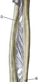
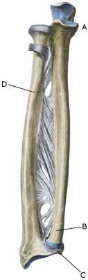
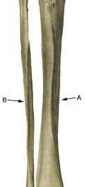
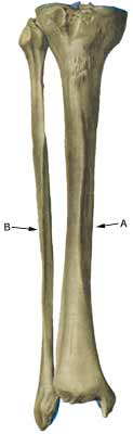
Cause: In case of repeated loads or blows the bursas can become inflamed, produce fluid, swell and become painful. One of the most frequent bursitis forms on the forearm, is inflammation of the bursa located between the biceps tendon and the fastening on the radius (tuberositas radii) (article).

Symptoms: Pain upon applying pressure on the bursa, which sometimes (but far from always) may feel swollen. Aggravated upon activation of the muscle located immediately above the bursa.

Acute treatment: Click here.

Examination: In slight cases with only minimal tenderness, medical examination is not necessarily required. In cases of more pronounced pain or lack of progress, a medical examination should be carried out to ensure a correct diagnosis and treatment. The diagnosis is usually made on the basis of a normal medical examination, however, if there is any doubt surrounding the diagnosis, it can easily and quickly be confirmed under an ultrasound scan.

Treatment: The treatment primarily consists of relief, and removal of the provoking cause (if identified). The treatment can be supplemented with rheumatic medicine (NSAID) or the injection of corticosteroid in the bursa preceded by draining of the bursa, which can advantageously be done under ultrasound guidance.

Rehabilitation: The treatment is completely dependant on which bursa is inflamed, but sports activity can usually be cautiously resumed once pain has decreased, particularly if it has been possible to remove the provoking cause.
Also read rehabilitation, general.

Complications: If progress is not smooth, it should be considered if the diagnosis is correct or whether complications have arisen. In rare cases the bursa can become infected with bacteria. This is a serious condition where the bursa becomes red, warm and increasingly swollen and tender, and requires immediate medical examination and treatment.

If there is no progress with relief, medical treatment (rheumatic medicine (NSAID) and the ultrasound guided injection of corticosteroid), surgical removal of the bursa may be attempted.

Chronic compartment syndrome

CHRONIC COMPARTMENT SYNDROME

Diagnosis: CHRONIC COMPARTMENT SYNDROME


Anatomy:
The forearm muscles are divided into three groups (muscle compartments) of powerful, partially unyielding, muscle membranes (fascias); a front, an outer and a rear muscle compartment. Each muscle group has its own blood and nerve supply.

Cause: The muscles can increase so quickly following intensive training of the forearm that the muscle membranes surrounding the muscles cannot keep up, causing the pressure in the muscle compartment to increase. The pressure can in some cases increase so greatly that impingement of blood vessels and nerves can occur. In other cases, chronic muscle compartment syndrome can arise due to development of scar tissue in the muscle (following previous muscle ruptures).

Symptoms: With chronic compartment syndrome there is slowly insetting pain in the muscles after a few minutes activity. There is a sensation that the muscle is “tightened” and becomes hard, which is accompanied by discomfort. If the activity is stopped the discomfort diminishes, but returns after a short period of resuming the sports activity once again. There is often sensory disturbance in the fingers.

Examination: The diagnosis is made on the basis of the characteristic history and possibly with a pressure measurement in the muscle compartment.

Treatment: With chronic muscle compartment syndrome the treatment primarily comprises relief and slowly increasing training intensity and possibly rheumatic medicine (NSAID). If there is scar tissue in the muscle, ultrasound guided injection of corticosteroid around the scar tissue formation can be attempted. In cases where there is a lack of progress a surgical splitting of the muscle membranes can be performed, which is usually a minor procedure with good results (article).

Rehabilitation: INSTRUCTION

Rehabilitation: Rehabilitation is dependant upon which muscle group has been affected. Once the pain has diminished, the sports activity can generally be slowly resumed according to the principles mentioned under rehabilitation, general.

Complications: In cases of lack of progress with relief and slow rehabilitation, an ultrasound scan should be performed before possible surgery to rule out complications from an earlier muscle rupture.

Acute compartment syndrome

ACUTE COMPARTMENT SYNDROME

Diagnosis: ACUTE COMPARTMENT SYNDROME


Anatomy:
The forearm muscles are divided into three groups (muscle compartments) of powerful, partially unyielding, muscle membranes (fascias); a front, an outer and a rear muscle compartment. Each muscle group has its own blood and nerve supply.

Cause: The pressure in a muscle compartment can rise so fast (due to bleeding or fluid extraction) that the muscle membranes cannot keep up. The pressure in the muscle compartment can therefore increase so greatly that impingement of blood vessels and nerves can occur (article).

Symptoms: With the acute muscle compartment syndrome there is increasing pain, which is often more powerful than expected from the primary evaluation of the extent of the injury. At the same time sensory disturbances can occur in the fingers.

Acute treatment: Click here.

Examination: The diagnosis is made on the basis of the characteristic history, and by a pressure measurement in the muscle compartment.

Treatment: With acute muscle compartment syndrome the treatment in severe cases comprises acute splitting of the muscle membrane. It is imperative for the continuing function of the muscle that this operation be acute, which is, of course, only possible if the athlete seeks acute medical attention (article-1) (article-2).

Rehabilitation: The rehabilitation is completely dependant on which muscle groups are affected, the provoking cause (blow to the muscle, muscle rupture or over-training) and which treatment that has been performed (relief, surgical splitting). Once pain has decreased, the sports activity can generally be slowly resumed according to the principles as mentioned under rehabilitation, general.

Complications: Muscles and nerves can suffer permanent damage if the treatment is not started as soon as possible.

Special: Since there is a risk of permanent disability, the injury should be reported to your insurance company.

Bone fracture

BONE FRACTURE

Diagnosis: BONE FRACTURE
(Fractura antebrachii)


Anatomy:
The forearm bones consist of the ulna and the radius.

  1. Elbow
  2. Ulna
  3. Wrist
  4. Radius

FOREARM BONES FROM THE FRONT

Cause: A bone fracture can occur in cases of a direct blow or fall on the arm, and can occur anywhere on the bone. It is often seen in children that the bone merely “bends” (green-stick fracture).

Symptoms: Sudden pain and pain induced constriction of movement of the arm after a fall or blow. An angling of the forearm can occasionally be seen. In rare case, acute compartment syndrome can develop.

Acute treatment: Click here.

Examination: Sudden, powerful pains in the arm with constriction of movement after a fall, should always lead to acute medical examination. The fracture is usually visible on x-rays, and on the basis of the type of fracture, the correct treatment can be determined.

Treatment: In cases of considerable dislocation or angling of the bones, the fracture will be reset under an anaesthetic, followed by bandaging for a few weeks. In certain types of fractures, an operative fixation can be necessary.

Rehabilitation: When pain has decreased fitness training in the form of cycling may be started along with rehabilitation as specified under rehabilitation, general. The rehabilitation period is completely dependent upon the type of fracture and the treatment administered.

Bandage: Special plastic bandages can be made for use following a fracture of the forearm when sports are resumed.

Complications: In the vast majority of cases the fracture heals without complications, although in some cases a poor healing occurs which can affect the blood vessel and the nerve supply to the arm, resulting in chronic compartment syndrome. If satisfactory progress is not achieved, you should therefore consult your doctor.

Inflammation of the outer shin bone


Diagnosis: INFLAMMATION OF THE OUTER SHIN BONE
(LATERAL TIBIAL PERIOSTITIS)


Anatomy:
The calf muscles are divided in three muscle groups (compartments) by powerful muscle membranes (fascies). A forward, an exterior and a rear muscle group (which is divided in a superficial and a deep part). The large rear muscle groups’ muscle membrane is fastened on the inner edge of the shin bone, while the forward muscle group is fastened on the external edge of the shin bone (photo).

Cause: In cases of repeated uniform loads, a stress related inflammation, can occur where the muscle membrane fastens on the edge of the shin bone. The condition is most often seen in athletes who jump a lot and with frequent change of running surface or shoes.

Symptoms: Pain upon applying pressure, particularly on the exterior edge of the shin bone and upon straining, and is aggravated when the foot is bent upwards against resistance (extension). Occasionally an irregular bone edge can be felt on the shin bone.

Acute treatment:

Examination: In light cases medical examination is not necessarily required. In cases with more pronounced pain or lack of progress despite relief, a medical examination is recommended to ensure the diagnosis and rule out amongst other things a stress fracture. A medical examination is usually sufficient in order to make the diagnosis. It can be necessary to supplement with X-ray, Scintigraphy, ultrasonography or MRI (article).

Treatment: The treatment involves relief, stretching and slow rehabilitation. It is imperative that there are good shock absorbing soles in the shoes.

Rehabilitation of children and adolescents: INSTRUCTION

Complications: If the course does not progress smoothly, you should be medically re-examined to ensure that the diagnosis is correct, and that complications have not arisen in the form of stress fracture.

Special: Shock absorbing shoes or inlays will reduce the load.


Inflammation of the inner shin bone

Diagnosis: INFLAMMATION OF THE INNER SHIN BONE
(MEDIAL TIBIAL PERIOSTITIS, “SHIN SPLINT”)


Anatomy:
The calf muscles are divided in three muscle groups (compartments) by powerful muscle membranes (fascies). A forward, an exterior and a rear muscle group (which is divided into a superficial and a deep part). The large rear muscle groups’ muscle membrane is fastened on the inner edge of the shin bone (tibia), while the forward muscle group is fastened on the external edge of the shin bone (photo).

Cause: In cases of repeated uniform loads, a stress related inflammation, can occur where the muscle membrane fastens on the edge of the shin bone. The condition is most often seen in athletes with a tendency to rotate the foot outwards (hyperpronation) or with a high foot arch. Frequent change of running surface or shoes increases the risk.

Symptoms: Pain on the inner edge of the shin bone, aggravated upon applying pressure, load (running) and stretching in the foot joint against resistance (flexion). The pain is often localized to the lower part of the shin bone. Sometimes an irregular bone edge can be felt on the shin bone.

Acute treatment:

Examination: In light cases medical examination is not necessarily required. In cases with more pronounced pain or lack of progress despite relief, medical examination is recommended to ensure the diagnosis and rule out amongst other things a stress fracture. A medical examination is usually sufficient in order to make the diagnosis. In some cases it may be necessary to supplement with X-rays, scintigraphy or ultrasound scanning.

Treatment: The treatment comprises relief, stretching and slow rehabilitation. It is imperative that there are good shock absorbing soles in the shoes.

Rehabilitation of children and adolescents: INSTRUCTION

Complications: If the course does not progress smoothly, you should be medically re-examined to ensure that the diagnosis is correct, and that complication have not arisen in the form of stress fracture.

Special: Shock absorbing shoes or inlays will reduce the load.

Bone fracture

Diagnosis: BONE FRACTURE OF THE SHIN BONE
(FRACTURE)


Anatomy:
The bones in the lower leg comprise the shin bone (tibia) and the calf bone (fibula).

  1. Tibia
  2. Fibula

SHIN BONE FROM THE FRONT

Cause: Violent loads in the form of a blow or twist can cause a fracture on the shin-bone as well as the calf bone (article). Fracture of the fibula is often seen after a kick on the outer side of the shin bone.

Symptoms: Pain upon applying pressure (direct and indirect tenderness) and when under load (walking and running).

Acute treatment: Click here.

Examination: X-ray.

Treatment: The treatment comprises relief and bandaging. In some cases surgery is necessary depending on the type of fracture and any displacement of the fracture-surfaces (article).

Rehabilitation of children and adolescents: Which loads and rehabilitation that can be permitted is completely dependent upon the severity and the treatment of the fracture. It is therefore important that the rehabilitation is performed in close cooperation with the doctors controlling the treatment. Approximately six months’ rehabilitation must be expected before shin bone fractures allow resumption of maximum load, and approximately 3 months’ rehabilitation before calf bone fractures allow maximum load.

Plastic bandage: Individual plastic bandages can be manufactured for use during sports activity after bone fractures. Individual plastic bandages are particularly well-suited after a fracture of the fibula.

Complications: If progress is not smooth you should be medically re-examined to ensure that the fracture is healing according to plan. In some cases a false joint can be formed (pseudoarthrosis) (article), requiring surgical treatment.

Scheuermans decease

Diagnosis: SCHEUERMANN’S DISEASE


Anatomy:
The back is constructed of box-like vertebrae which are held in place partly with the help of the shape of the bones, partly by ligaments and partly by the large and small back and stomach muscles.


  1. Vertebra prominens
  2. Vertebra coccygea I
  3. Promontorium
  4. L I
  5. Th I
  6. Axis

THE BACK SEEN FROM THE SIDE

Cause: Scheuermann’s disease occurring in approximately 4% of the population (article). A curvature of the back occurs (bending over forwards) due to the vertebrae becoming wedge shaped. There are also characteristic x-ray finds. The cause of the condition is unknown, but evidence tends to suggest that the condition is hereditary (article).

Symptoms: Back curvature localised high in the back (thoracal Scheuermann) gives often only few, if any, symptoms. Back curvature localised in the lower back (thoracolumbal or lumbal Scheuermann) does entail back pain for the majority (article).

Examination: The diagnosis is usually made following a medical examination supplemented with an x-ray (at least 3 adjacent vertebrae with at least 5 degrees wedge form, Schmorlske impressions, flattening of discs, irregular end plates) (article). The crooked back is often mistaken in the beginning for “bad posture”. In some cases, CT or MRI scanning is recommended.

Treatment: The vast majority of cases can be treated with training, attempting to maintain the mobility of the back, counteract the curvature tendency and strengthen the stomach and back muscles. A corset can in some cases be used until the young person is fully grown. An operation can be performed only in very rare cases. The condition has a good prognosis (article), and even after an operation it is still possible to take part in many different forms of sport (article).

Rehabilitation of children and adolescents: INSTRUCTION

Complications: In some cases a crooked back can have other causes (infection, nerve disease, inborn bone change, rheumatic illness, bone disease, metabolic disorder).

Crooked back

Diagnosis: CROOKED BACK
(SCOLIOSIS)


Anatomy:
The back is constructed of box-like vertebrae which are held in place partly with the help of the shape of the bones, partly by ligaments and partly by the large and small back and stomach muscles.


  1. Vertebra prominens
  2. Vertebra coccygea I
  3. Promontorium
  4. L I
  5. Th I
  6. Axis

THE BACK SEEN FROM THE SIDE

Cause: The cause of scoliosis in children and adolescents is unknown in the majority of cases (idiopathic). Scoliosis is most commonly seen during the growing years, and is more often seen in girls than boys (Photo).

Symptoms: Scoliosis does not necessarily cause pain or other symptoms.

Examination: It is important that the diagnosis is made as soon as possible as better results are achieved if the treatment is commenced as soon as it is necessary. One should therefore always be aware of the early signs of scoliosis (uneven shoulder or hip level, one or both shoulder blades being prominent, slanting waist). A normal medical examination will usually be sufficient to make the diagnosis. X-rays will reveal the degree of severity of the scoliosis. In some cases, CT or MRI scanning is recommended (article).

Treatment: Treatment is dependant upon the degree of severity. The majority of cases will normally be able to be controlled without treatment (article). It will normally be possible to take part in sports activities without any problems (article). Strength training and stretching of the stomach and back muscles is recommended. Supportive bandaging can be used if the scoliosis becomes worse (> 25-30 degrees) and the young person is still growing. It is normally possible to take part in sport at the usual level despite the bandaging (article). An operation may become necessary if the scoliosis becomes pronounced (> 40-50 degrees), and even earlier in some special cases. Certain forms of sport can be resumed 6-9 months after the operation (article).

Rehabilitation of children and adolescents: INSTRUCTION

Complications: In some cases the presence of scoliosis can have other causes (infection, nerve disease, inborn bone change, rheumatic illness, bone disease, metabolic disorder).

Fracture of the vertebral arch

Diagnosis: FRACTURE OF THE VERTEBRAL ARCH
(SPONDYLOLYSIS)


Anatomy:
The back is constructed of box-like vertebrae which are held in place partly with the help of the shape of the bones, partly by ligaments and partly by the large and small back and stomach muscles (sketch).

Cause: There are several causes for looseness of the lumbar vertebrae (spondylolyse): inborn (dysplastic), fracture of the vertebral arch and degenerative. Fracture of the vertebral arch is the most frequent cause amongst adolescents and athletes, and is seen in 5% of the normal population. It is most often seen in athletes who repeatedly bend the back backwards (high-jumpers, gymnasts, pole vaulters, footballers) (article-1) (article-2). In some cases, the looseness can cause one of the vertebrae to slide forwards and apply pressure on the spinal cord (spondylolisthesis).

Symptoms: Pain and stiffness in the lower back (lumbago) and buttocks, occasionally radiating to the leg (“sciatica”).

Examination: Slight back discomfort does not necessarily require medical examination, however, all cases with strong or repeated back pain should be examined. The doctor will be able to evaluate whether further examination is required, i.e. x-ray (including lateral projection) (X-Ray) (Scintigraphy), CT or MRI scanning (article).

Treatment: Treatment is dependant upon the degree of severity. The majority of cases will normally be able to be treated with relief (article-1) (article-2) (which in sever cases must last for several months). In approximately 10% of cases where a vertebra has slid significantly forwards (spondylolisthesis), it will be necessary to operate (article).

Rehabilitation of children and adolescents: INSTRUCTION

Complications: If the pain does not decline under the treatment, medical (re)examination by a doctor should be performed. Special consideration should be given to slipped disc, secondary muscle infiltrations (myalgia) and piriformis syndrome, Scheuermanns disease, however, many other causes of lumbago are found (infection, tumour), of which some will require further examination.

Special:
Training should be performed on a “lifelong” basis to reduce the risk of relapse after a successful rehabilitation.